. 2015 Feb 3:5:8209.
doi: 10.1038/srep08209.

Angiotensin-converting enzyme 2/angiotensin-(1-7)/Mas axis prevents lipopolysaccharide-induced apoptosis of pulmonary microvascular endothelial cells by inhibiting JNK/NF-κB pathways

Affiliations

Angiotensin-converting enzyme 2/angiotensin-(1-7)/Mas axis prevents lipopolysaccharide-induced apoptosis of pulmonary microvascular endothelial cells by inhibiting JNK/NF-κB pathways

Yingchuan Li et al. Sci Rep. .

Abstract

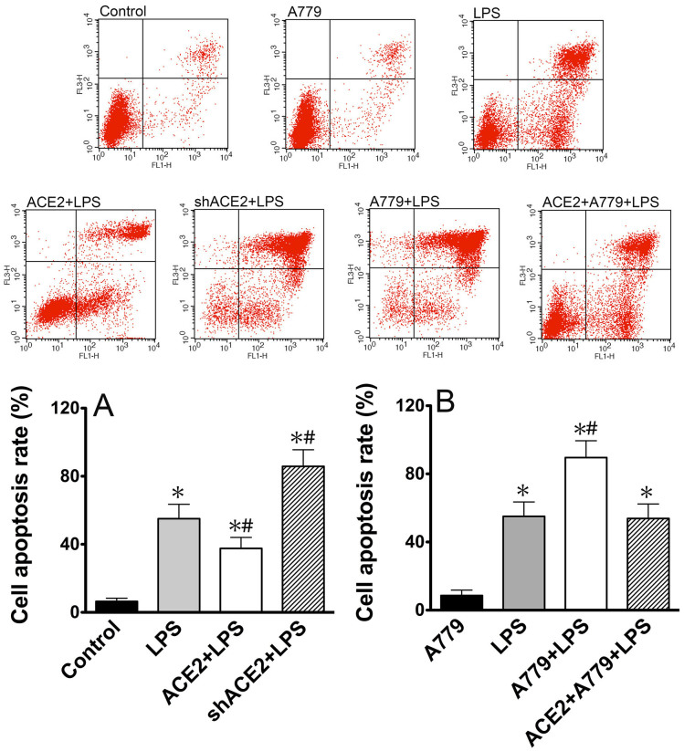
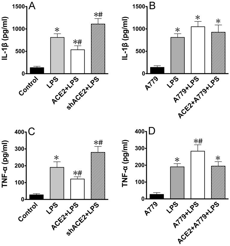
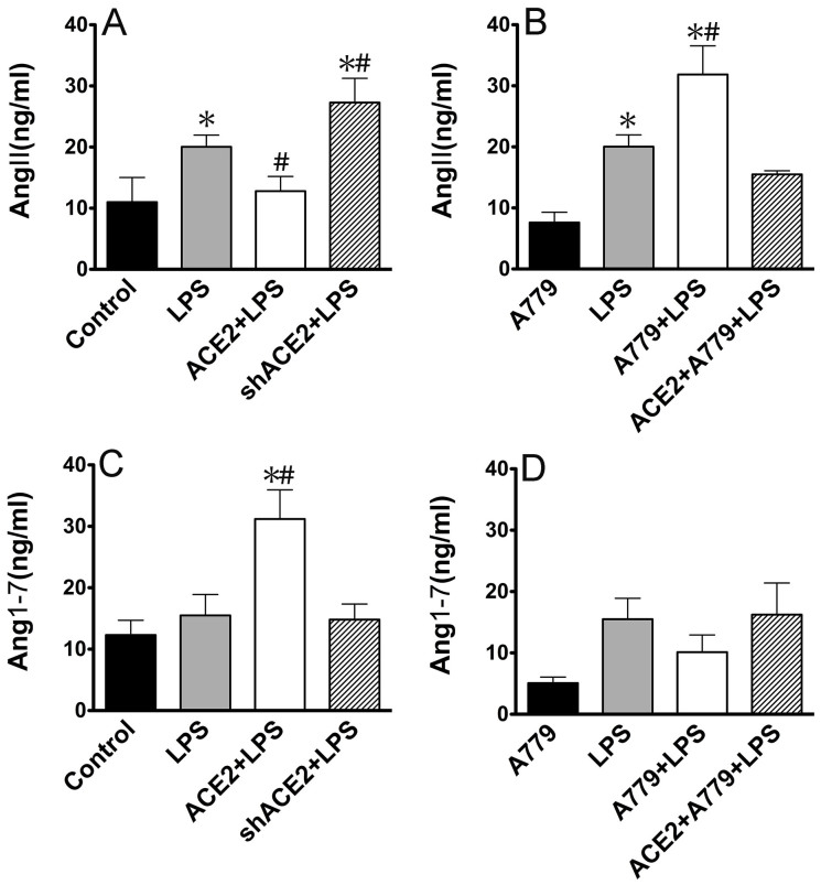
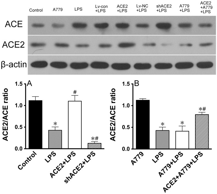
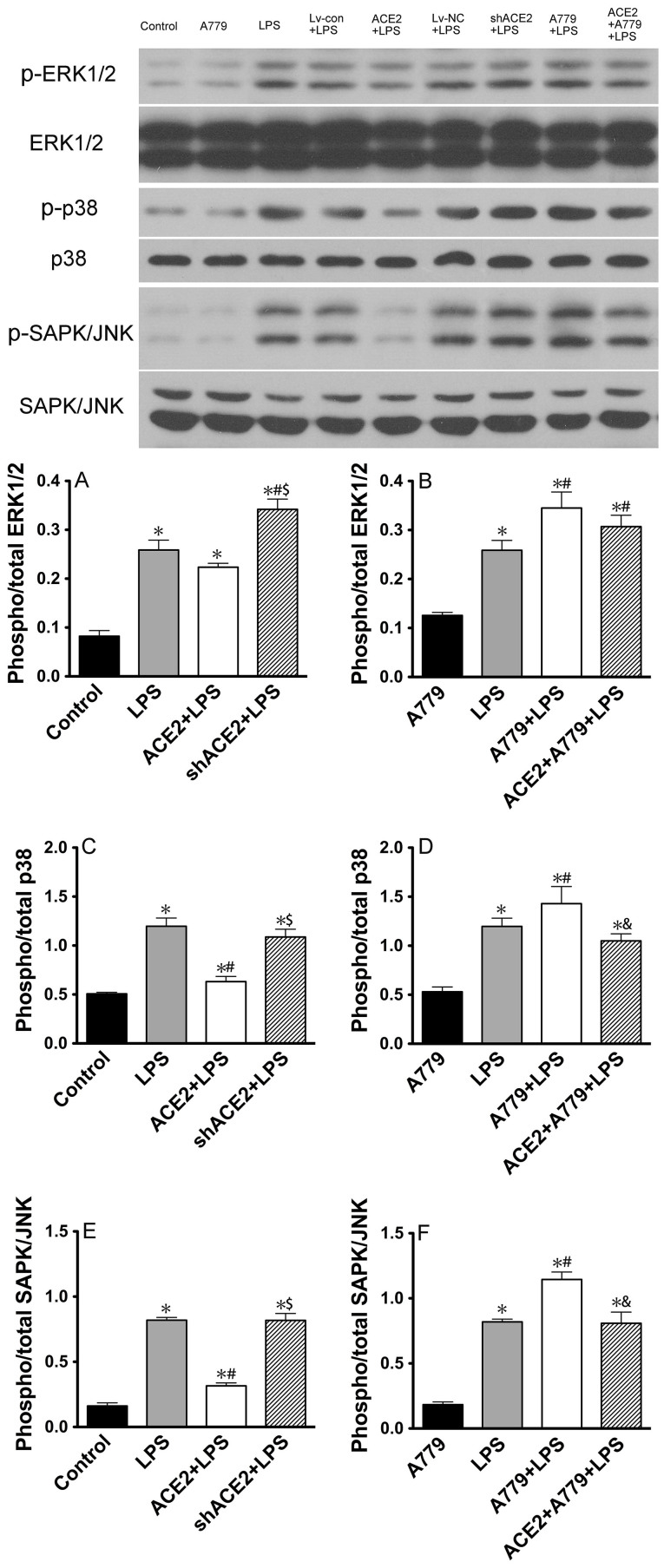
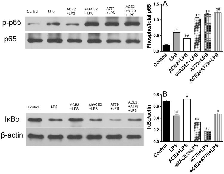
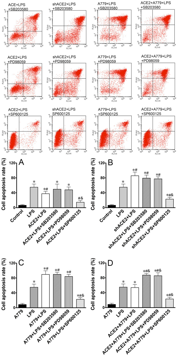
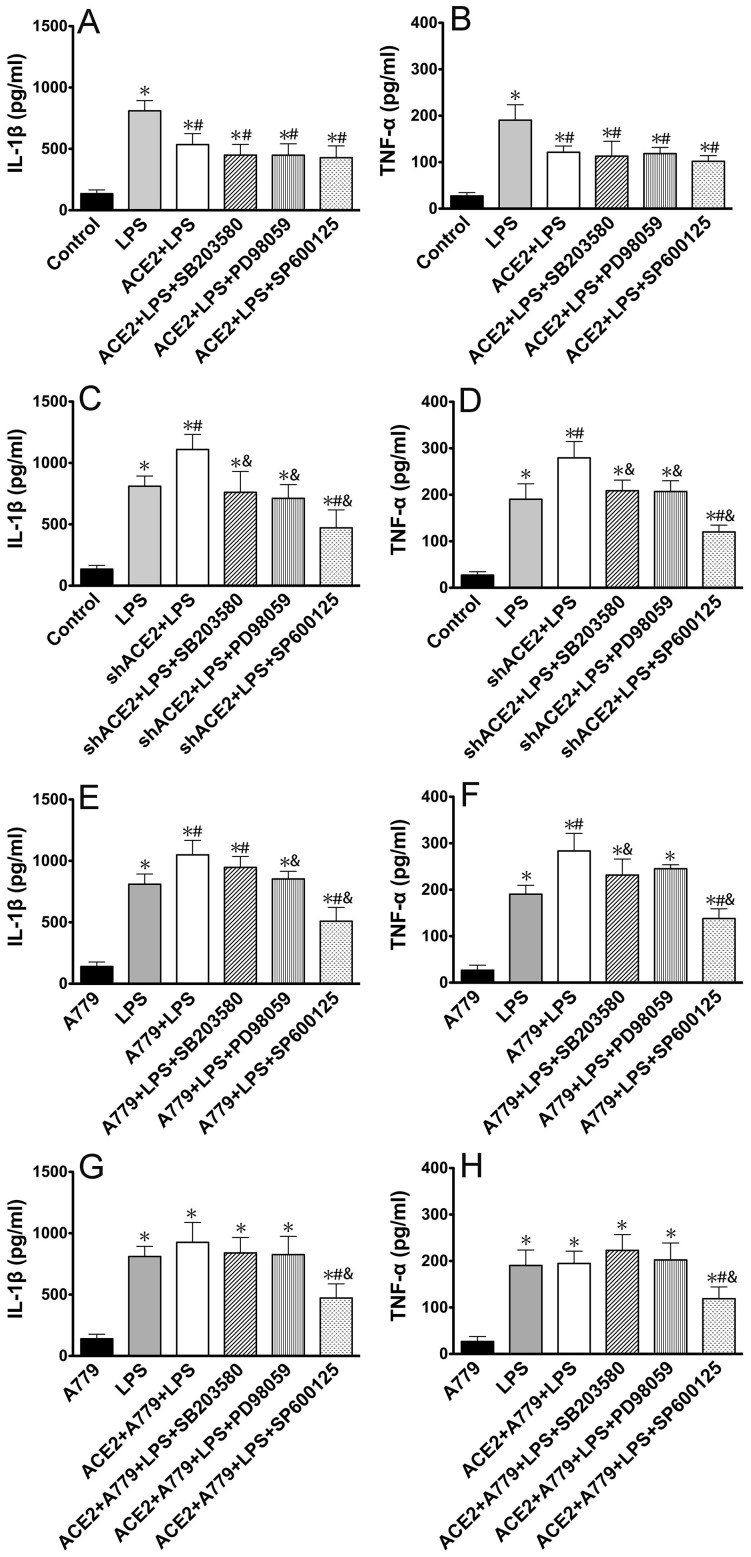
ACE2 and Ang-(1-7) have important roles in preventing acute lung injury. However, it is not clear whether upregulation of the ACE2/Ang-(1-7)/Mas axis prevents LPS-induced injury in pulmonary microvascular endothelial cells (PMVECs) by inhibiting the MAPKs/NF-κB pathways. Primary cultured rat PMVECs were transduced with lentiviral-borne Ace2 or shRNA-Ace2, and then treated or not with Mas receptor blocker (A779) before exposure to LPS. LPS stimulation resulted in the higher levels of AngII, Ang-(1-7), cytokine secretion, and apoptosis rates, and the lower ACE2/ACE ratio. Ace2 reversed the ACE2/ACE imbalance and increased Ang-(1-7) levels, thus reducing LPS-induced apoptosis and inflammation, while inhibition of Ace2 reversed all these effects. A779 abolished these protective effects of Ace2. LPS treatment was associated with activation of the ERK, p38, JNK, and NF-κB pathways, which were aggravated by A779. Pretreatment with A779 prevented the Ace2-induced blockade of p38, JNK, and NF-κB phosphorylation. However, only JNK inhibitor markedly reduced apoptosis and cytokine secretion in PMVECs with Ace2 deletion and A779 pretreatment. These results suggest that the ACE2/Ang-(1-7)/Mas axis has a crucial role in preventing LPS-induced apoptosis and inflammation of PMVECs, by inhibiting the JNK/NF-κB pathways.


Figures

Figure 1
Figure 1. Effects of ACE2 and Mas–receptor antagonist A779 on apoptosis of PMVECs treated with LPS for 48 h.
(A) LPS exposure caused a significant apoptosis in PMVECs but was attenuated by ACE2 overexpression and aggravated by ACE2 downregulation. (B) LPS–induced apoptosis was markedly increased by A779 pretreatment but was improved by ACE2 overexpression. There was no significant difference between the ACE2 + A779 + LPS group and LPS group in apoptosis. All data are expressed as mean ± SD. * P < 0.05 compared with PBS control group; #P < 0.05 compared with LPS group.
Figure 2
Figure 2. Effects of ACE2 and Mas–receptor antagonist A779 on cytokine secretion in PMVECs.
(A, C) LPS exposure for 48 h caused a significant increase of the levels of IL–1β and TNF–α in the supernatants of cultured PMVECs but was markedly attenuated by ACE2 overexpression and aggravated by ACE2 downregulation. (B, D) LPS–induced IL–1β and TNF–α secretions were markedly increased by A779 pretreatment, but were improved by ACE2 overexpression. There was no significant difference in cytokine secretion between the ACE2 + A779 + LPS and LPS groups. All data are expressed as mean ± SD. * P < 0.05 compared with control group; #P < 0.05 compared with LPS group.
Figure 3
Figure 3. Effects of ACE2 and Mas–receptor antagonist A779 on AngII/Ang–(1–7) secretion in PMVECs.
(A, C) LPS exposure for 48 h caused a significant increase of AngII and a slightly increase of Ang–(1–7). The increase of AngII was markedly attenuated by ACE2 overexpression and aggravated by ACE2 downregulation. The up–regulation of Ang–(1–7) was further increased by ACE2 overexpression, but reduced by ACE2 knockdown. (B, D) LPS–induced AngII secretion was markedly increased by A779 pretreatment, which was suppressed by ACE2 overexpression. But, Ang–(1–7) level was not affected by A779 pretreatment before LPS administration. All data are expressed as mean ± SD. * P < 0.05 compared with control group; #P < 0.05 compared with LPS group.
Figure 4
Figure 4. Effects of ACE2 and Mas–receptor antagonist A779 on the ACE2/ACE expression in PMVECs.
(A) LPS exposure for 48 h caused a significant decrease of the ACE2/ACE ratio in PMVECs that was markedly reversed by ACE2 overexpression and aggravated by ACE2 downregulation. (B) The ratio of ACE2/ACE was greatly reduced with A779 pretreatment prior to LPS stimulation that was reversed by ACE2 overexpression. There was no difference in the ACE2/ACE ratio between the A779 + LPS and LPS groups. All data are expressed as mean ± SD. * P < 0.05 compared with control group; #P < 0.05 compared with LPS group.
Figure 5
Figure 5. Effects of ACE2 and Mas–receptor antagonist A779 on the phosphorylation of MAPKs in PMVECs.
(A, C, E) LPS exposure for 48 h caused a significant increase in phosphorylation of ERK1/2, p38, and SAPK/JNK in PMVECs. ACE2 overexpression effectively reduced LPS–induced p38 and SAPK/JNK phosphorylation levels, but ERK1/2 phosphorylation was unaffected. In contrast, downregulation of ACE2 in LPS–exposed cells obviously enhanced ERK1/2 phosphorylation levels, but failed to change p38 and SAPK/JNK phosphorylation. (B, D, F) The phosphorylation levels of ERK1/2, p38, and SAPK/JNK were significantly enhanced by A779 pretreatment prior to LPS stimulation. ACE2 overexpression before A779 pretreatment obviously downregulated the levels of p38 and SAPK/JNK phosphorylation, but slightly decreased ERK1/2 phosphorylation level. All data are expressed as mean ± SD. * P < 0.05 compared with control group; #P < 0.05 compared with LPS group; $ P < 0.05 compared with ACE2 + LPS group; &P < 0.05 compared with A779 + LPS group.
Figure 6
Figure 6. Effects of ACE2 and Mas–receptor antagonist A779 on NF–κB p65 phosphorylation and IκBα expression in PMVECs.
(A, B) LPS exposure for 48 h caused a significant increase in phosphorylation of NF–κB p65 and decrease in levels of IκBα in PMVECs. ACE2 overexpression effectively reduced LPS–induced NF–κB p65 phosphorylation levels and reversed IκBα levels. In contrast, downregulation of ACE2 in LPS–exposed cells obviously enhanced NF–κB p65 phosphorylation levels and suppressed IκBα levels. The phosphorylation of NF–κB p65 and downregulation of IκBα were significantly enhanced by A779 pretreatment prior to LPS stimulation. A779 pretreatment markedly reversed the effect of ACE2 overexpression on NF–κB p65 phosphorylation and IκBα expression. All data are expressed as mean ± SD. * P < 0.05 compared with the control group; #P < 0.05 compared with the LPS group.
Figure 7
Figure 7. Effects of the specific inhibitors of MAPKs on LPS–induced apoptosis in PMVECs.
The protective effect of ACE2 overexpression on LPS–induced apoptosis was enhanced by SP600125 and deteriorated by SB203580 and PD98059. SP600125 markedly reduced apoptosis of PMVECs with Ace2 deletion and Mas receptor blocker pretreatment. Mas receptor blocker abolished the protective effect of ACE2, which was aggravated by SB203580 and PD98059 and also SP600125. All data are expressed as mean ± SD. * P < 0.05 compared with PBS control group; #P < 0.05 compared with LPS group; &P < 0.05 compared with the ACE2 + LPS (A), shACE2 + LPS (B), A779 + LPS (C), and ACE2 + A779 + LPS (D) groups.
Figure 8
Figure 8. Effects of the specific inhibitors of MAPKs on LPS–induced cytokine secretion in PMVECs.
(A, B) In the ACE2 + LPS group, SB203580, PD98059 and SP600125 pretreatment did not affect LPS–induced IL–1β and TNF–α secretions. (C, D) Pre–incubation with SB203580, PD98059, and especially SP600125, significantly suppressed the secretion of cytokines, in the shACE2 + LPS group. (E, F) In PMVECs exposed to LPS, pretreatment with A779 caused a significant increase in IL–1β and TNF–α secretions, which were slightly reduced by SB203580 and PD98059 as well as markedly attenuated by SP600125. (G, H) LPS–induced IL–1β and TNF–α secretions in the ACE2 + A779 + LPS group were significantly inhibited by SP600125 pre–incubation and were not affected by SB203580, or PD98059. All data are expressed as mean ± SD. * P < 0.05 compared with the PBS control group; #P < 0.05 compared with the LPS group; &P < 0.05 compared with the ACE2 + LPS (A), shACE2 + LPS (B), A779 + LPS (C), and ACE2 + A779 + LPS (D) groups.

References

    1. Rubenfeld G. D. et al. Incidence and Outcomes of Acute Lung Injury. N Engl J Med 353, 1685–93 (2005). - PubMed
    1. Phua J. et al. Has mortality from acute respiratory distress syndrome decreased over time? : A systematic review. Am J Respir Crit Care Med 179, 220–7 (2009). - PubMed
    1. Cheng C. et al. Lipopolysaccharide induces expression of SSeCKS in rat lung microvascular endothelial cell. Mol Cell Biochem 305, 1–8 (2007). - PubMed
    1. Weir M. R. & Dzau V. J. The renin–angiotensin–aldosterone system: a specific target for hypertension management. Am J Hypertens 12, 205S–213S (1999). - PubMed
    1. de Man F. S. et al. Dysregulated renin–angiotensin–aldosterone system contributes to pulmonary arterial hypertension. Am J Respir Crit Care Med 186, 780–9 (2012). - PMC - PubMed

Publication types

MeSH terms