Review
. 2015 Mar-Apr;4(2):135-50.
doi: 10.1002/wdev.170. Epub 2015 Jan 21.

Strength in numbers: quantitative single-molecule RNA detection assays

Affiliations
Review

Strength in numbers: quantitative single-molecule RNA detection assays

Imre Gaspar et al. Wiley Interdiscip Rev Dev Biol. 2015 Mar-Apr.

Abstract

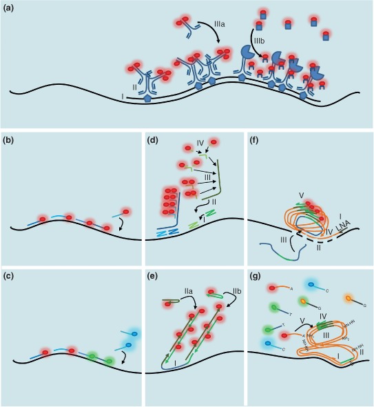
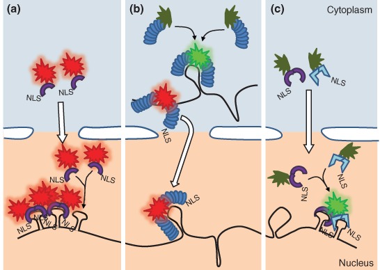
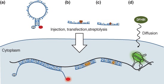
Gene expression is a fundamental process that underlies development, homeostasis, and behavior of organisms. The fact that it relies on nucleic acid intermediates, which can specifically interact with complementary probes, provides an excellent opportunity for studying the multiple steps--transcription, RNA processing, transport, translation, degradation, and so forth--through which gene function manifests. Over the past three decades, the toolbox of nucleic acid science has expanded tremendously, making high-precision in situ detection of DNA and RNA possible. This has revealed that many--probably the vast majority of--transcripts are distributed within the cytoplasm or the nucleus in a nonrandom fashion. With the development of microscopy techniques we have learned not only about the qualitative localization of these molecules but also about their absolute numbers with great precision. Single-molecule techniques for nucleic acid detection have been transforming our views of biology with elementary power: cells are not average members of their population but are highly distinct individuals with greatly and suddenly changing gene expression, and this behavior of theirs can be measured, modeled, and thus predicted and, finally, comprehended. For further resources related to this article, please visit the WIREs website.

Conflict of interest: The authors have declared no conflicts of interest for this article.


Figures

Figure 1
Figure 1
Single‐molecule RNA detection by hybridization. (a) During conventional fluorescent RNA in situ hybridization (FISH), a hapten‐labeled single‐stranded nucleic acid, a few hundred nucleotides long, is (I) hybridized to the target messenger RNA (mRNA). (II) The hapten molecules are subsequently detected by specific primary antibodies. Amplified fluorescent signal is created by detecting the primary antibodies (IIIa) with labeled secondary antibodies or (IIIb) by enzyme‐mediated catalytic conversion of a soluble substrate into stable deposits associated with the surrounding cellular matrix. (b) Single‐molecule FISH based on an array of singly labeled fluorescent probes, simultaneously hybridizing to target, usually in a tandem with a few nucleotide long gaps. (c) RNA bar‐coding based on single‐molecule FISH (smFISH). Smaller arrays of probes labeled with different colors hybridize the target to create one (spectral bar‐coding) or a spatial pattern of super‐localized spots (spatial bar‐coding) with unique color combination. (d) Branched DNA (bDNA) FISH, e.g., RNAscope. (I) Two target‐specific probes hybridize juxtaposed creating a landing platform for (II) the preamplifier, which (III) binds an array of identical amplifiers. The amplifiers gather (IV) multiple copies of singly labeled probes. (e) Hybridization chain reaction. (I) The initiator hybridizes to the target. (IIa and IIb) The two labeled hairpins bind the two overhangs of the initiator deploying fluorescence and regenerating the overhangs to maintain the chain reaction. (f) Rolling circle amplification (RCA) based on padlock probes. (I) Reverse transcription is carried out using a locked nucleic acid (LNA) primer. (II) The RNA of the RNA/cDNA (complementary DNA) duplex is digested with RNase H, allowing binding of the linear padlock probe to the LNA/RNA fixed cDNA. (III) The padlock probe is circularized via mismatch‐sensitive DNA ligation. (IV) Phi29 polymerase‐based RCA is initiated, creating a DNA nanoball that carries multiple covalently bound copies of the padlock probe complementer. (V) These are detected by a singly labeled probe specific to the padlock probe. (g) FISSEQ, sequencing RNA in situ. (I) Random hexamer‐based reverse transcription (RT). (II) The obtained cDNA is circularized by CircLigase and (III) this circular ss cDNA is amplified during RCA, creating a DNA nanoball. (IV) Multiple copies of the sequencing primer hybridize within the nanoball to the adapter introduced by the RT primer. (V) SOLiD sequencing‐by‐ligation is carried out on each spot.
Figure 2
Figure 2
Fluorescent protein‐based messenger RNA (mRNA) visualization. (a) Ribonucleoprotein complex (RNP) detection via phage coat protein–fluorescent protein (pCP–FP)–p‐loop system. Fluorescently labeled monomeric—or intramolecularly dimerized—coat protein molecules carrying nuclear localization signal (NLS) are sequestered to the nucleus, where they bind to the respective p‐loop of a transgenically encoded RNA target. (b) Pumilio Homology Domain (PUM‐HD) FP system. A full FP (red) or the two halves of a split FP (green) are fused to two PUM‐HDs with different sequence specificities. The two PUM‐HDs bind to adjacent segments of the target, restoring fluorescence in case of a split FP label (green). Target binding can take place both in the nucleus and in the cytoplasm. (c) Split FP modification of the pCF‐FP–p‐loop system. Nonfluorescent orthologous intramolecularly dimerized pCPs (e.g., MCP and PCP) fused to split Venus are targeted to the nuclei, where they bind to their respective p‐loops, which are arranged in an alternating fashion, restoring Venus fluorescence. Fluorescently labeled RNPs get exported to cytoplasm while nonassociated label molecules remain sequestered in the nucleus (a–c).
Figure 3
Figure 3
Fluorogenic approaches for RNA detection in situ and in vivo. (a) Molecular beacons. Upon hybridization the fluorophore goes from a quenched, dark state, into a nonquenched bright state. (b) Exciton‐controlled hybridization‐sensitive fluorescent oligonucleotide (ECHO) probes. One thymidine base is covalently coupled to a pair of thiazole orange (TO) dyes. During hybridization, the two TO dyes are displaced and locked by the surrounding stacking forces, resulting in an increase of fluorescence (bright state). (c) Forced intercalation (FIT) probes. One base is replaced by a single TO dye. When in duplex, the high viscosity arising from surrounding hydrogen bonds restricts rotational movement of the TO dye, resulting in an increased fluorescence (bright state). (d) Spinach and Spinach2. A transgenically encoded messenger RNA (mRNA) carrying the Spinach(2) aptamer binds to the nonfluorescent, cell‐permeable green fluorescent protein (GFP) fluorophore DFHBI, providing a protective environment that allows fluorescence to develop.

Similar articles

Cited by

References

    1. Gall JG, Pardue ML. Formation and detection of RNA‐DNA hybrid molecules in cytological preparations. Proc Natl Acad Sci USA 1969, 63:378–383. - PMC - PubMed
    1. Guesdon JL. Immunoenzymatic techniques applied to the specific detection of nucleic acids. A review. J Immunol Methods 1992, 150:33–49. - PubMed
    1. Weil TT, Parton RM, Davis I. Making the message clear: visualizing mRNA localization. Trends Cell Biol 2010, 20:380–390. - PMC - PubMed
    1. Singer RH, Ward DC. Actin gene expression visualized in chicken muscle tissue culture by using in situ hybridization with a biotinated nucleotide analog. Proc Natl Acad Sci USA 1982, 79:7331–7335. - PMC - PubMed
    1. Kosman D, Mizutani CM, Lemons D, Cox WG, McGinnis W, Bier E. Multiplex detection of RNA expression in Drosophila embryos. Science 2004, 305:846. - PubMed