. 2015 Jun;165(2):135-44.
doi: 10.1007/s12011-015-0234-6. Epub 2015 Feb 3.

The Effect of Cadmium on COX-1 and COX-2 Gene, Protein Expression, and Enzymatic Activity in THP-1 Macrophages

Affiliations

The Effect of Cadmium on COX-1 and COX-2 Gene, Protein Expression, and Enzymatic Activity in THP-1 Macrophages

Tomasz Olszowski et al. Biol Trace Elem Res. 2015 Jun.

Abstract

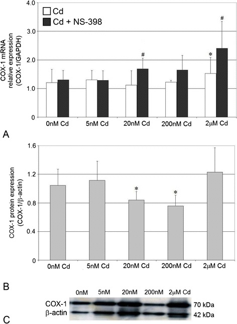
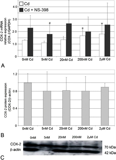
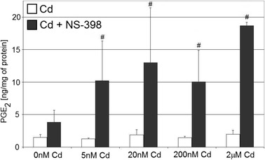
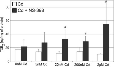
The aim of this study was to examine the effects of cadmium in concentrations relevant to those detected in human serum on cyclooxygenase-1 (COX-1) and cyclooxygenase-2 (COX-2) expression at mRNA, protein, and enzyme activity levels in THP-1 macrophages. Macrophages were incubated with various cadmium chloride (CdCl2) solutions for 48 h at final concentrations of 5 nM, 20 nM, 200 nM, and 2 μM CdCl2. The mRNA expression and protein levels of COXs were analyzed with RT-PCR and Western blotting, respectively. Prostaglandin E2 (PGE2) and stable metabolite of thromboxane B2 (TXB2) concentrations in culture media were determined using ELISA method. Our study demonstrates that cadmium at the highest tested concentrations modulates COX-1 and COX-2 at mRNA level in THP-1 macrophages; however, the lower tested cadmium concentrations appear to inhibit COX-1 protein expression. PGE2 and TXB2 production is not altered by all tested Cd concentrations; however, the significant stimulation of PGE2 and TXB2 production is observed when macrophages are exposed to both cadmium and COX-2 selective inhibitor, NS-398. The stimulatory effect of cadmium on COXs at mRNA level is not reflected at protein and enzymatic activity levels, suggesting the existence of some posttranscriptional, translational, and posttranslational events that result in silencing of those genes' expression.


Conflict of interest statement

The authors declare that there is no conflict of interests regarding the publication of this paper.

Figures

Fig. 1
Fig. 1
The effect of cadmium on COX-1 mRNA and protein expression in macrophages cultured with various cadmium solutions. a COX-1 mRNA expression following cadmium exposure without or with addition of COX-2 selective inhibitor, NS-398; b COX-1 protein expression (densitometric analysis of protein normalized to β-actin; c representative Western blot following cadmium exposure. Monocytes/macrophages were cultured with cadmium solutions for 48 h. After incubation, cells were harvested by scraping and mRNA was measured by using real-time PCR method (n = 4) and protein expression by using Western blotting method (n = 3). Asterisk, statistically significant as compared with 0 nM Cd—cells incubated in RPMI medium with 10 % FBS and with DMSO addition (Wilcoxon test). Number sign, statistically significant as compared with the experiment 0 nM Cd with NS-398 (Wilcoxon test)
Fig. 2
Fig. 2
Imaging of COX-1 enzyme by fluorescence microscopy in macrophages cultured with cadmium. Monocytes/macrophages were cultured with Cd solutions for 48 h. The immunohistochemistry was performed using specific primary antibody, mouse anti-COX-1 (the overnight incubation at 4 °C), and secondary antibodies conjugated with flouorochrome–anti-mouse IgG FITC (incubation for 45 min at room temperature). The nuclei of cells were DAPI stained. Image analysis was performed with a fluorescent microscope using filters 38 HE GFP for green fluorescence and 49 DAPI for blue fluorescence
Fig. 3
Fig. 3
The effect of cadmium on COX-2 mRNA and protein expression in macrophages cultured with various cadmium solutions. a COX-2 mRNA expression following cadmium exposure without or with addition of COX-2 selective inhibitor, NS-398; b COX-2 protein expression (densitometric analysis of protein normalized to β-actin; c representative Western blot following cadmium exposure. Monocytes/macrophages were cultured with cadmium solutions for 48 h. After incubation, cells were harvested by scraping and mRNA was measured by using real-time PCR method (n = 4) and protein expression by using Western blotting method (n = 3). Asterisk, statistically significant as compared with 0 nM Cd—cells incubated in RPMI medium with 10 % FBS and with DMSO addition (Wilcoxon test). Number sign, statistically significant as compared with the experiment 0 nM Cd with NS-398 (Wilcoxon test)
Fig. 4
Fig. 4
Imaging of COX-2 enzyme by fluorescence microscopy in macrophages cultured with cadmium. Monocytes/macrophages were cultured with Cd solutions for 48 h. The immunohistochemistry was performed using specific primary antibody, mouse anti-COX-2 (the overnight incubation at 4 °C), and secondary antibodies conjugated with flouorochrome–anti-mouse IgG FITC (incubation for 45 min at room temperature). The nuclei of cells were DAPI stained. Image analysis was performed with a fluorescent microscope using filters 38 HE GFP for green fluorescence and 49 DAPI for blue fluorescence
Fig. 5
Fig. 5
The effect of cadmium on quantity of PGE2 in culture supernatants of macrophages cultured with various cadmium solutions. Monocytes/macrophages were cultured with cadmium solutions for 48 h. After incubation, cells were harvested by scraping and PGE2 concentration was measured by ELISA method (n = 6). Number sign, statistically significant as compared with the experiment 0 nM Cd with NS-398 (Wilcoxon test)
Fig. 6
Fig. 6
The effect of cadmium on quantity of TXB2 in culture supernatants of macrophages cultured with various cadmium solutions. Monocytes/macrophages were cultured with cadmium solutions for 48 h. After incubation, cells were harvested by scraping and TXB2 concentration was measured by ELISA method (n = 6). Number sign, statistically significant as compared with the experiment 0 nM Cd with NS-398 (Wilcoxon test)

Similar articles

Cited by

References

    1. Kurumbail RG, Kiefer JR, Marnett LJ. Cyclooxygenase enzymes: catalysis and inhibition. Curr Opin Struct Biol. 2001;11:752–760. doi: 10.1016/S0959-440X(01)00277-9. - DOI - PubMed
    1. Smith CJ, Zhang Y, Koboldt CM, Muhammad J, Zweifel BS, Shaffer A, Talley JJ, Masferrer JL, Seibert K, Isakson PC. Pharmacological analysis of cyclooxygenase-1 in inflammation. Proc Natl Acad Sci USA. 1998;95:13313–13318. doi: 10.1073/pnas.95.22.13313. - DOI - PMC - PubMed
    1. Rouzer CA, Marnett LJ. Cyclooxygenases: structural and functional insights. J Lipid Res. 2009;50:S29–S34. doi: 10.1194/jlr.R800042-JLR200. - DOI - PMC - PubMed
    1. Morita I. Distinct functions of COX-1 and COX-2. Prostaglandins Other Lipid Mediat. 2002;68–69:165–175. doi: 10.1016/S0090-6980(02)00029-1. - DOI - PubMed
    1. Gilroy DW, Colville-Nash PR. New insights into the role of COX-2 in inflammation. J Mol Med (Berl) 2000;78:121–129. doi: 10.1007/s001090000094. - DOI - PubMed

Publication types

MeSH terms

LinkOut - more resources