. 2015 Feb 3:5:8216.
doi: 10.1038/srep08216.

Structural basis of natural promoter recognition by the retinoid X nuclear receptor

Affiliations

Structural basis of natural promoter recognition by the retinoid X nuclear receptor

Judit Osz et al. Sci Rep. .

Abstract

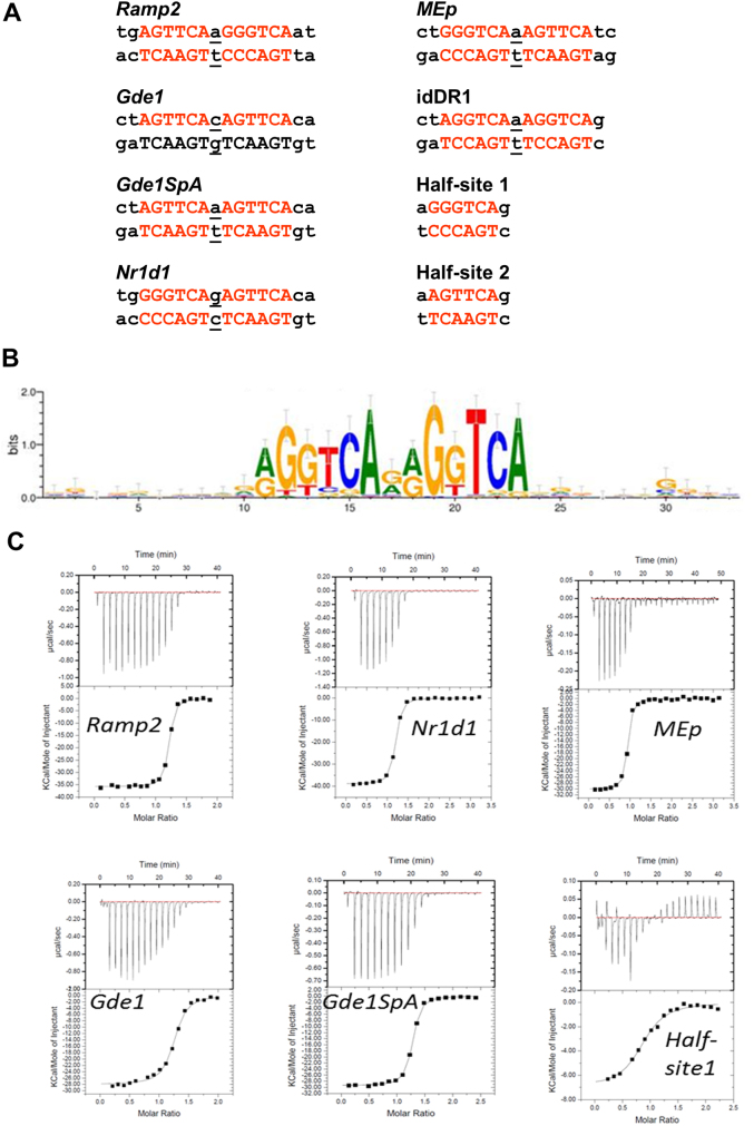
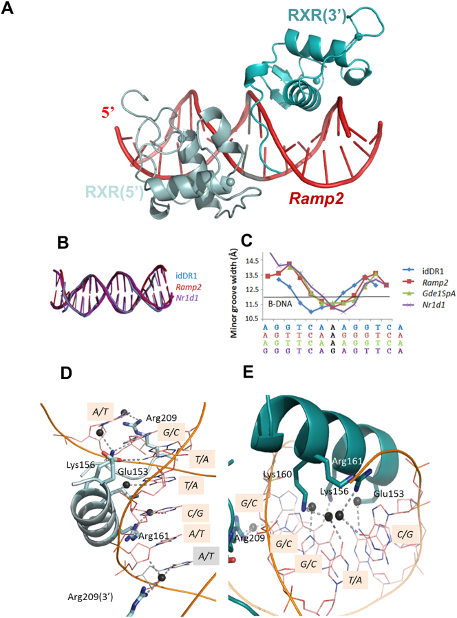
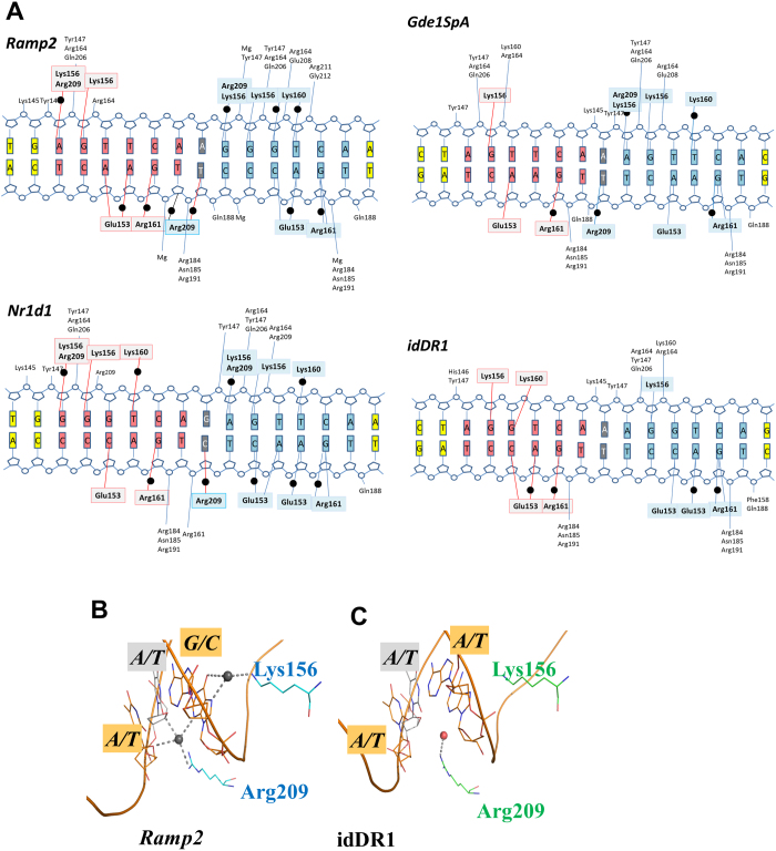
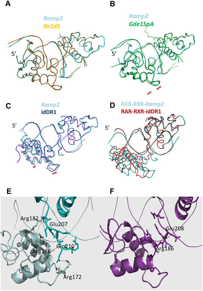
Retinoid X receptors (RXRs) act as homodimers or heterodimerisation partners of class II nuclear receptors. RXR homo- and heterodimers bind direct repeats of the half-site (A/G)G(G/T)TCA separated by 1 nucleotide (DR1). We present a structural characterization of RXR-DNA binding domain (DBD) homodimers on several natural DR1s and an idealized symmetric DR1. Homodimers displayed asymmetric binding, with critical high-affinity interactions accounting for the 3' positioning of RXR in heterodimers on DR1s. Differing half-site and spacer DNA sequence induce changes in RXR-DBD homodimer conformation notably in the dimerization interface such that natural DR1s are bound with higher affinity than an idealized symmetric DR1. Subtle changes in the consensus DR1 DNA sequence therefore specify binding affinity through altered RXR-DBD-DNA contacts and changes in DBD conformation suggesting a general model whereby preferential half-site recognition determines polarity of heterodimer binding to response elements.


Figures

Figure 1
Figure 1
(a) Natural DR1 response element double strand nucleotide (ds) sequences of DNA DR1 response elements. Hexanucleotide half-site motifs are shown in red. (b) RXR binding motif identified by RXR-ChIP sequencing from Ref. . (c) Quantification of the interaction between RXR and DR1s by ITC. Representative ITC isotherms for the binding of the DR1 duplex (Ramp2, Nr1d1, MEp, Gde1, Gde1SpA and half-site 1) to the RXR-DBD. The top panels show the raw ITC data expressed as the change in thermal power with respect to time over the period of titration. Lower panels: change in molar heat is expressed as a function of molar ratio of corresponding DR1 to dimer-equivalent RXR or half-site to monomer RXR. The solid lines in the lower panels represent the fit of data to a one-site model using the ORIGIN software. Standard free energies of binding and entropic contributions were obtained, respectively, as ΔG = −RT ln(Ka) and TΔS = ΔH − ΔG, from the Ka and ΔH values derived from ITC curve fitting.
Figure 2
Figure 2
(a) Overall structure of RXR DBD-Ramp2. The upstream RXR (in light cyan) and downstream RXR (in cyan) bound to their hexanucleotide motifs shown in red. The spheres indicate the Zn molecules. (b) Comparison of DNA bending of the DR1 elements. The ds oligonucleotides used in the crystallographic structures of RXR-DBD homodimers complexed with DR1, show similar deformation. (c) Plot of the minor groove widths of the DR1 ds oligonucleotides. The values were derived using the 3DNA software. The solid black line represents standard values for B-DNA. (d-e) RXR homodimers exhibit specific interactions and polarity on natural DR1s. Ramp2 DNA sequence recognition by the upstream RXR subunit (d). View along the DNA-recognition helix (α1) of RXR showing residues Glu153, Lys156, Arg161 and Arg209 and their direct and water-mediated base contacts. Hydrogen-bonds and water molecules are shown as dotted blue lines and dark spheres, respectively. The interspacer nucleotide is highlighted in grey. The corresponding view of Ramp2 DNA sequence recognition by the downstream RXR subunit (e).
Figure 3
Figure 3. Polarity of the bound RXR homodimers revealed by asymmetric DNA recognition of natural DR1s.
The RXR-DBDs establish unique interactions to recognize natural asymmetric DR1s that are more numerous than in the complex with symmetric idDR1 as revealed by the schematic view of the protein/DNA contacts calculated with NUCPLOT with a 3.9 Å distance cutoff (a). Note that all crystal structures have comparable resolution (between 2 Å and 2.35 Å). For the three natural DR1s studied, the 3′ half-site is more tightly bound with more observed interactions with RXR. Bridging water molecules are shown as black circles. The residues forming hydrogen-bond interactions with the 5′ subunit and 3′subunit are highlighted in light grey and cyan, respectively. The first hexanucleotide is shown in salmon, the second one in blue and the interspacer nucleotide in grey. The gray circles indicate the DNA phosphates and the labeled residues their contacts with RXR. (b-c) Comparison of the interactions around the interspacer nucleotides of the natural Ramp2 and the idealized DR1 complexes. The interspacer is highlighted in grey and the surrounding base pairs in orange. Only the RXR DBD complex with natural DR1 forms specific H-bonds with the interspacer nucleotide. For Ramp2, the downstream DBD extends its interactions upstream of the 3′ half-site to reach the backbone sugar of the last nucleotide of the 5′ site. H-bonds are shown by dashed lines and the water molecules as dark spheres for Ramp2 and red sphere for the idDR1.
Figure 4
Figure 4. Structural changes of RXR-DBDs induced by the half-site sequence.
Significant differences in RXR-DBD positioning and in the dimerization interface are observed for the natural DR1s. (a–c) Superimposed crystal structures of RXR-DBD-Ramp2 (cyan) with RXR-DBD-Nr1d1 (a; orange), RXR-DBD-Gde1SpA (b; green) and RXR-DBD-idDR1 (c; blue). Superposition was performed on the 3′ bound RXR-DBD. The largest differences are observed for the idDR1 as indicated by red arrows. Differences are also observed for the complex with the natural Gde1SpA DR1. (d) Superimposed crystal structures of RXR DBD-Ramp2 (cyan) with RAR-RXR DBD-idDR1 (PDB ID: 1XDK, red). Superposition was performed on the 3′ bound RXR-DBD. (e–f) Dimerization interface that involves the DNA minor groove, hydrogen-bonding between atoms of the 2 subunits (residues highlighted) and Van der Waals interactions for RXR-DBD-Ramp2 (e) and RXR-DBD-idDR1 (f).

References

    1. Mangelsdorf D. J. et al. The nuclear receptor superfamily: the second decade. Cell 83, 835–9 (1995). - PMC - PubMed
    1. Altucci L. & Gronemeyer H. Nuclear receptors in cell life and death. Trends Endocrinol Metab 12, 460–8 (2001). - PubMed
    1. Aagaard M. M., Siersbaek R. & Mandrup S. Molecular basis for gene-specific transactivation by nuclear receptors. Biochim Biophys Acta 1812, 824–35 (2011). - PubMed
    1. Huang P., Chandra V. & Rastinejad F. Structural overview of the nuclear receptor superfamily: insights into physiology and therapeutics. Annu Rev Physiol 72, 247–72 (2010). - PMC - PubMed
    1. Claessens F. & Gewirth D. T. DNA recognition by nuclear receptors. Essays Biochem 40, 59–72 (2004). - PubMed

Publication types

Substances

LinkOut - more resources