. 2015 Feb 3:13:6.
doi: 10.1186/s12951-015-0067-7.

Investigating the immunomodulatory nature of zinc oxide nanoparticles at sub-cytotoxic levels in vitro and after intranasal instillation in vivo

Affiliations

Investigating the immunomodulatory nature of zinc oxide nanoparticles at sub-cytotoxic levels in vitro and after intranasal instillation in vivo

Shruti R Saptarshi et al. J Nanobiotechnology. .

Abstract

Background: This study evaluates the time-dependent pro-inflammatory response of the model human lung epithelial cells (A549) to industrially relevant zinc oxide nanoparticles (ZnO NPs). In terms of toxicity, ZnO-NPs are categorised into the group of high toxicity nanomaterials. However information on pro-inflammatory potential of these NPs at sub-toxic concentrations is limited. Understanding how cellular defense mechanisms function in the presence of sub-cytotoxic concentrations of these NPs is vital. Moreover, there is an urgent need for additional in vivo studies addressing pulmonary toxicity due to accidental inhalation of ZnO NPs.

Results: Exposure to sub-cytotoxic ZnO NP concentrations (20 μg/mL) induced significant up-regulation of mRNA for the pro-inflammatory cytokine IL-8 and redox stress marker heme oxygenase-1, along with increased release of IL-8. The highest pro-inflammatory response was recorded after 4 to 6 hr exposure to ZnO NPs over a 24 hr period. Pre-treatment of A549 cells with the sulfhydryl antioxidant N-acetyl cysteine (at 5 mM) resulted in significant reduction of the up-regulation of inflammatory markers, confirming the role of reactive oxygen species in the observed immunomodulatory effects, independent of cytotoxicity. Furthermore, we report for the first time that, intranasal instillation of a single dose (5 mg/kg) of pristine or surfactant-dispersed ZnO NPs can cause pulmonary inflammation, already after 24 hr in a murine model. This was confirmed by up-regulation of eotaxin mRNA in the lung tissue and release of pro-inflammatory cytokines in the sera of mice exposed to ZnO NPs.

Conclusion: Our study highlights that even at sub-cytotoxic doses ZnO NPs can stimulate a strong inflammatory and antioxidant response in A549 cells. ZnO NP mediated cytotoxicity may be the outcome of failure of cellular redox machinery to contain excessive ROS formation. Moreover exposure to a single but relatively high dose of ZnO NPs via intranasal instillation may provoke acute pulmonary inflammatory reactions in vivo.


Figures

Figure 1
Figure 1
Cytotoxicity profile of pristine and surfactant-dispersed ZnO NPs after 24 hr exposure of human lung epithelial A549 cells, using the (A) MTS assay and (B) lactate dehydrogenase release assays. Cytotoxicity was dependent on NP size and dispersal state. Concentrations at or below the dotted line (MTS assay) and beyond the dotted line (LDH release assay) were significantly different from untreated control cells. Values are expressed as mean ± SEM (n = 5 separate experiments).
Figure 2
Figure 2
ZnO NP mediated pro-inflammation in A549 cells exposed to the sub-cytotoxic dose (20 μg/mL) over 24 hr. (A) ZnO NP induced up-regulation of HO-1 mRNA indicative of a strong antioxidant response in A549 cells. Highest HO-1 expression was seen at 6hr which was a 30-fold increase in gene expression compared to untreated cells at 6hr (p<0.001). Block bars compare the statistical significance between pristine or surfactant-dispersed solutions for each ZnO NP size. (B) mRNA over expression of the pro-inflammatory cytokine IL-8 in A549 cells stimulated with 20 μg/mL of ZnO NPs at 1, 2, 4, 6 or 24hr. Maximum expression of IL-8 gene was recorded at 4hr. (C) Release of pro-inflammatory cytokine IL-8 by ZnO NP-exposed A549 cells at 1, 2, 4, 6 or 24 hr. Statistical significance is shown in (B) and (C) compared to untreated cells at each time point. *p<0.05,**<0.01,***p<0.001, ****p<0.0001. Data are expressed as mean ±SEM of the fold levels compared to untreated controls at each time point (n=3) separate experiments, each performed with triplicates).
Figure 3
Figure 3
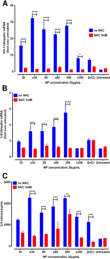
Mitigation of the up-regulation of (A) HO-1 mRNA expression and (B) IL-8 mRNA expression (C) IL-8 cytokine release in antioxidant treated A549 cells after exposure to ZnO NPs (20 μg/mL) for 6 hr. Statistical significance after comparing no NAC or 5 mM NAC cells where *p < 0.05, ** < 0.01, ***p < 0.001, ****p < 0.0001. Data are expressed as mean ± SEM of the fold levels compared to untreated controls at each time point (n = 3) separate experiments, each performed with triplicates).
Figure 4
Figure 4
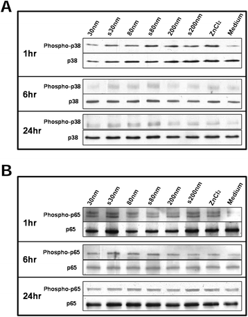
Immunoblotting showing (A) phosphorylation of p38 protein, belonging to the redox-sensitive p38 Mitogen activated protein kinase (MAPK) pathway and (B) p65 protein, involved in NFκB activation, after exposing A549 cells to 20 μg/mL dose of ZnO NPs.
Figure 5
Figure 5
H&E staining of lung sections of mice treated with (A) vehicle control, (B) LPS, (C) pristine 30 nm ZnO NPs and (D) surfactant-dispersed 30 nm ZnO NPs via intratracheal instillation. AV indicates the alveolar region of the lung. Red arrows indicate haemorrhage and black arrows demonstrate thickened bronchial walls.
Figure 6
Figure 6
qPCR analysis of mRNA levels for inflammatory markers (A) eotaxin, (B) MCP-1 and (C) TNFα in the lung tissues of treated mice. (D) Proteome profile array of pro-inflammatory cytokines quantified in pooled sera from each experimental group. Statistical significance is compared to vehicle control. Pink bars for A,B,C and dotted line for D represent the vehicle control. Data are expressed as mean ± SEM (n = 12) from two separate experiments.

Similar articles

Cited by

References

    1. Wang ZL. Zinc oxide nanostructures: growth, properties and applications. J Phys Condens Matter. 2004;16:R829–58. doi: 10.1088/0953-8984/16/25/R01. - DOI
    1. Saptarshi S, Duschl A, Lopata A. Interaction of nanoparticles with proteins: relation to bio-reactivity of the nanoparticle. J Nanobiotechnol. 2013;11:26. doi: 10.1186/1477-3155-11-26. - DOI - PMC - PubMed
    1. Wu W, Samet JM, Peden DB, Bromberg PA. Phosphorylation of p65 is required for zinc oxide nanoparticle-induced interleukin 8 expression in human bronchial epithelial cells. Environ Health Perspect. 2010;118:982–7. doi: 10.1289/ehp.0901635. - DOI - PMC - PubMed
    1. Karlsson HL, Cronholm P, Gustafsson J, Möller L. Copper oxide nanoparticles are highly toxic: a comparison between metal oxide nanoparticles and carbon nanotubes. Chem Res Toxicol. 2008;21:1726–32. doi: 10.1021/tx800064j. - DOI - PubMed
    1. Lin W, Xu Y, Huang C-C, Ma Y, Shannon K, Chen D-R, et al. Toxicity of nano- and micro-sized ZnO particles in human lung epithelial cells. J Nanoparticle Res. 2009;11:25–39. doi: 10.1007/s11051-008-9419-7. - DOI

Publication types

MeSH terms

LinkOut - more resources