

A 2D-DIGE-based proteomic analysis reveals differences in the platelet releasate composition when comparing thrombin and collagen stimulations
- PMID: 25645904
- PMCID: PMC4316189
- DOI: 10.1038/srep08198
A 2D-DIGE-based proteomic analysis reveals differences in the platelet releasate composition when comparing thrombin and collagen stimulations
Abstract
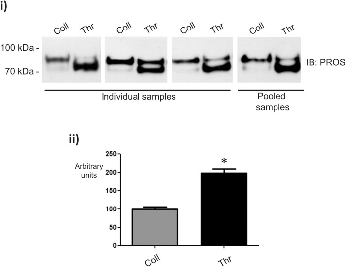
Upon stimulation, platelets release a high number of proteins (the releasate). There are clear indications that these proteins are involved in the pathogenesis of several diseases, such as atherosclerosis. In the present study we compared the platelet releasate following platelet activation with two major endogenous agonists: thrombin and collagen. Proteome analysis was based on 2D-DIGE and LC-MS/MS. Firstly, we showed the primary role of thrombin and collagen receptors in platelet secretion by these agonists; moreover, we demonstrated that GPVI is the primary responsible for collagen-induced platelet activation/aggregation. Proteomic analysis allowed the detection of 122 protein spots differentially regulated between both conditions. After excluding fibrinogen spots, down-regulated in the releasate of thrombin-activated platelets, 84 differences remained. From those, we successfully identified 42, corresponding to 37 open-reading frames. Many of the differences identified correspond to post-translational modifications, primarily, proteolysis induced by thrombin. Among others, we show vitamin K-dependent protein S, an anticoagulant plasma protein, is up-regulated in thrombin samples. Our results could have pathological implications given that platelets might be playing a differential role in various diseases and biological processes through the secretion of different subsets of granule proteins and microvesicles following a predominant activation of certain receptors.
Figures
Similar articles
-
Proteomics of the TRAP-induced platelet releasate.J Proteomics. 2009 Feb 15;72(1):91-109. doi: 10.1016/j.jprot.2008.10.009. Epub 2008 Nov 8. J Proteomics. 2009. PMID: 19049909
-
High precision platelet releasate definition by quantitative reversed protein profiling--brief report.Arterioscler Thromb Vasc Biol. 2013 Jul;33(7):1635-8. doi: 10.1161/ATVBAHA.113.301147. Epub 2013 May 2. Arterioscler Thromb Vasc Biol. 2013. PMID: 23640497
-
Application of 2-dimensional difference gel electrophoresis (2D-DIGE) to the study of thrombin-activated human platelet secretome.Platelets. 2008 Feb;19(1):43-50. doi: 10.1080/09537100701609035. Platelets. 2008. PMID: 18231937
-
Taking the stock of granule cargo: Platelet releasate proteomics.Platelets. 2017 Mar;28(2):119-128. doi: 10.1080/09537104.2016.1254762. Epub 2016 Dec 8. Platelets. 2017. PMID: 27928935 Review.
-
Platelet proteomics.J Thromb Haemost. 2003 Jul;1(7):1593-601. doi: 10.1046/j.1538-7836.2003.00311.x. J Thromb Haemost. 2003. PMID: 12871296 Review.
Cited by
-
Is autologous platelet activation the key step in ovarian therapy for fertility recovery and menopause reversal?Biomedicine (Taipei). 2022 Dec 1;12(4):1-8. doi: 10.37796/2211-8039.1380. eCollection 2022. Biomedicine (Taipei). 2022. PMID: 36816178 Free PMC article. Review.
-
Platelet-released extracellular vesicles: the effects of thrombin activation.Cell Mol Life Sci. 2022 Mar 14;79(3):190. doi: 10.1007/s00018-022-04222-4. Cell Mol Life Sci. 2022. PMID: 35288766 Free PMC article.
-
Platelets mediate trained immunity against bone and joint infections in a mouse model.Bone Joint Res. 2022 Feb;11(2):73-81. doi: 10.1302/2046-3758.112.BJR-2021-0279.R1. Bone Joint Res. 2022. PMID: 35118873 Free PMC article.
-
Autologous platelet-rich plasma for healing chronic venous leg ulcers: Clinical efficacy and potential mechanisms.Int Wound J. 2019 Jun;16(3):788-792. doi: 10.1111/iwj.13098. Epub 2019 Mar 12. Int Wound J. 2019. PMID: 30864220 Free PMC article.
-
Alteration of platelet GPVI signaling in ST-elevation myocardial infarction patients demonstrated by a combination of proteomic, biochemical, and functional approaches.Sci Rep. 2016 Dec 22;6:39603. doi: 10.1038/srep39603. Sci Rep. 2016. PMID: 28004756 Free PMC article.
References
-
- Parguiña A. F., Rosa I. & García A. Proteomics applied to the study of platelet-related diseases: aiding the discovery of novel platelet biomarkers and drug targets. J. Proteomics. 76 Spec No 275–286 (2012). - PubMed
-
- Leslie M. Beyond clotting: the powers of platelets. Science 328, 562–564 (2010). - PubMed
-
- Coppinger J. A. et al. Characterization of the proteins released from activated platelets leads to localization of novel platelet proteins in human atherosclerotic lesions. Blood 103, 2096–2104 (2004). - PubMed
-
- Coppinger J. A. et al. Moderation of the platelet releasate response by aspirin. Blood 109, 4786–4792 (2007). - PubMed
-
- Della Corta A. et al. Application of 2-dimensional difference gel electrophoresis (2D-DIGE) to the study of thrombin-activated human platelet secretome. Platelets 19, 43–50 (2008). - PubMed
Publication types
MeSH terms
Substances
LinkOut - more resources
Full Text Sources
Other Literature Sources
