. 2014 Jul:112:116-41.

Steroid differentiation: the safety profile of various steroids on retinal cells in vitro and their implications for clinical use (an American Ophthalmological Society thesis)

Affiliations

Steroid differentiation: the safety profile of various steroids on retinal cells in vitro and their implications for clinical use (an American Ophthalmological Society thesis)

Baruch D Kuppermann et al. Trans Am Ophthalmol Soc. 2014 Jul.

Abstract

Purpose: To determine if potentially viable alternatives to the clinical use of intravitreal triamcinolone acetonide should be considered based on a comparative assessment of the in vitro effects of five commercially available corticosteroids. We hypothesized that dexamethasone, betamethasone, methylprednisolone, loteprednol etabonate, and fluocinolone acetonide, at clinically relevant doses, may show different levels of in vitro cytotoxicity to retinal cells.

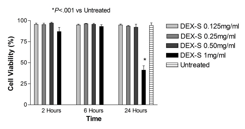
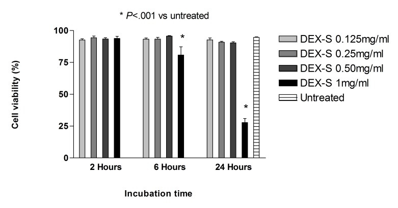
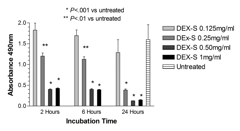
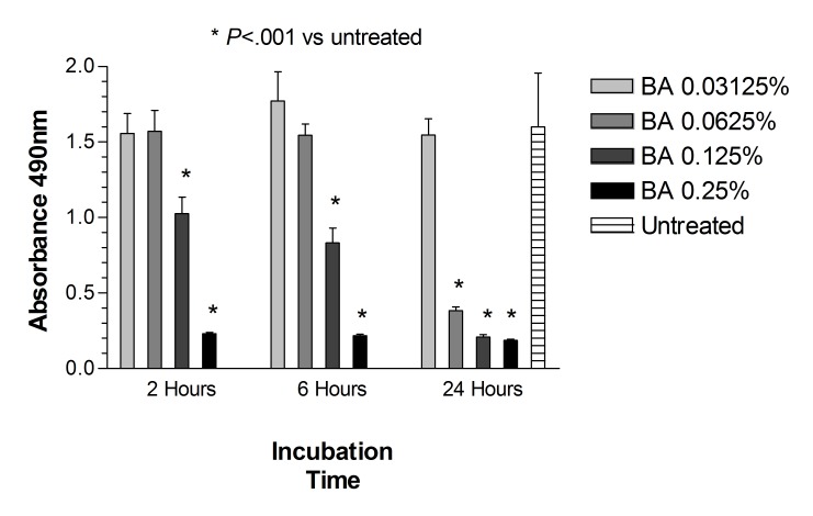
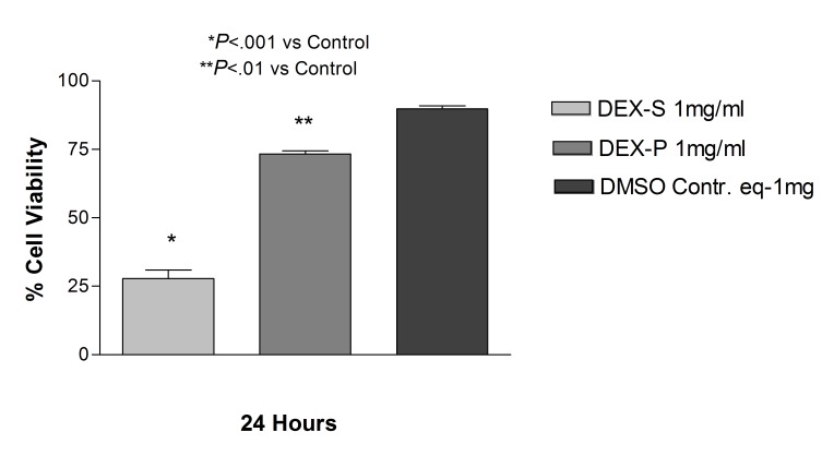
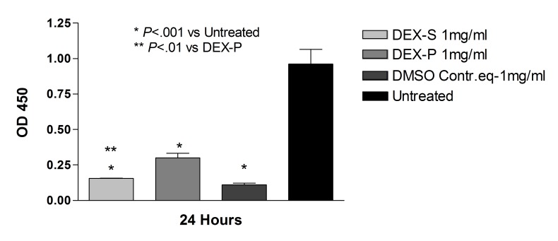
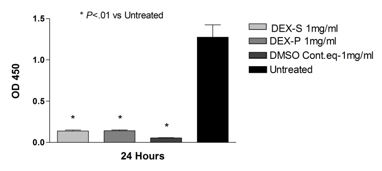
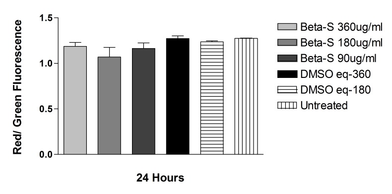
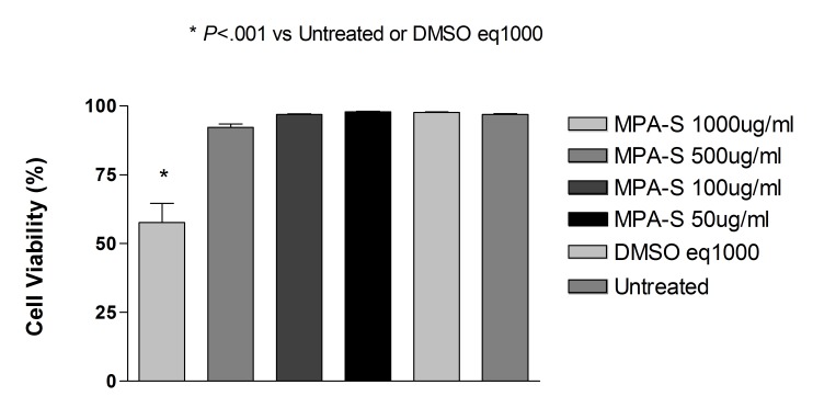
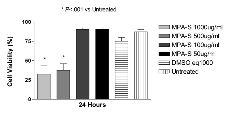
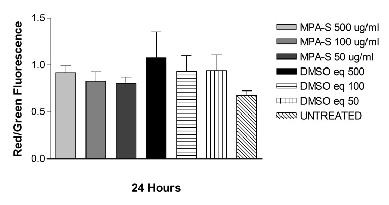
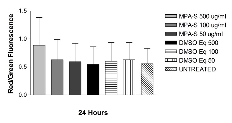
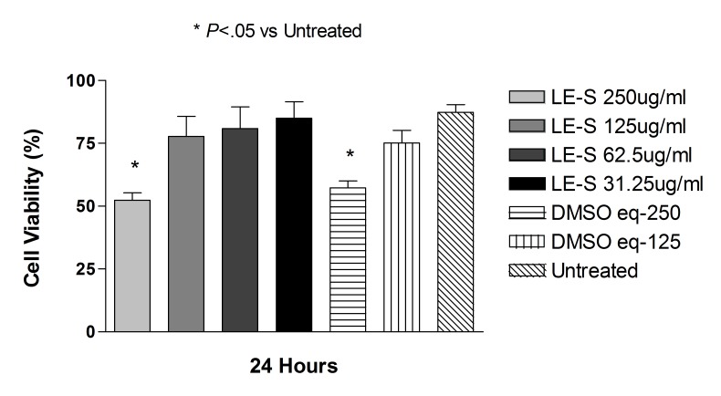
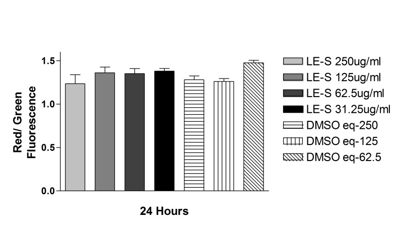
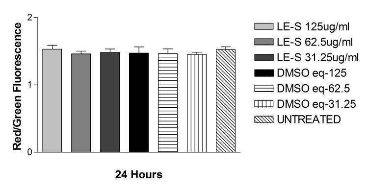
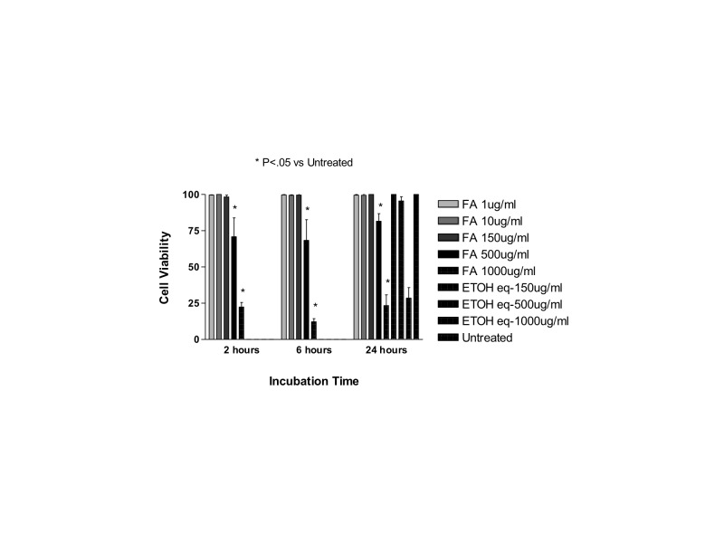
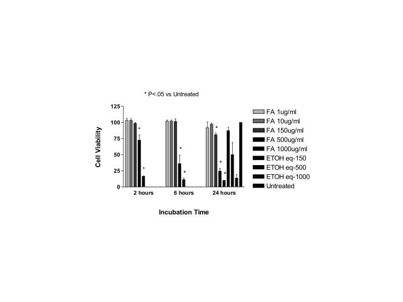
Methods: Cultures of human retinal pigment epithelial cells (ARPE-19) and rat embryonal neurosensory precursor retinal cells (R28) were treated with dexamethasone, betamethasone, methylprednisolone, loteprednol, or fluocinolone acetonide. Cell viability as a measure of cell death was determined by trypan blue dye exclusion assay. The mechanical effect of drug crystals was evaluated by solubilizing the steroid formulations. Mitochondrial dehydrogenase and membrane potential were assessed to measure cell damage.

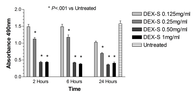
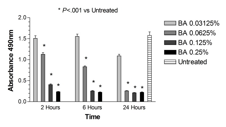
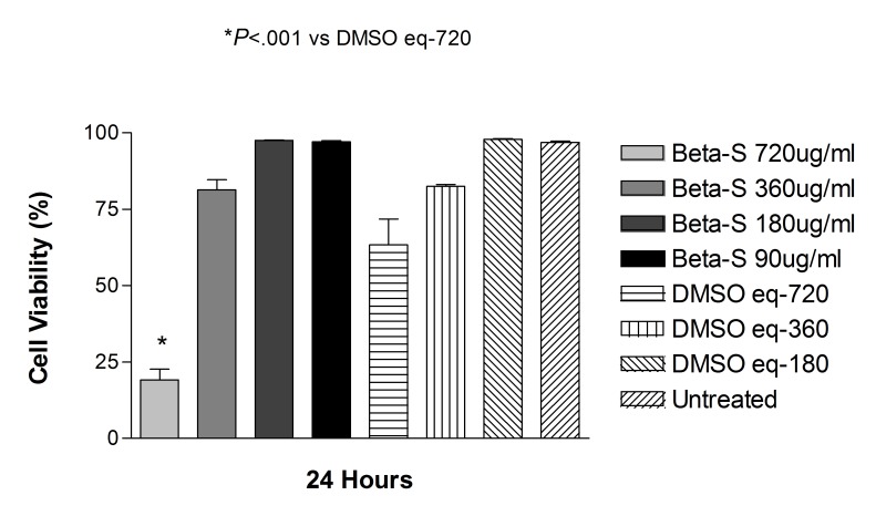
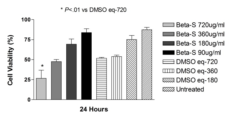
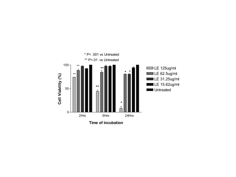
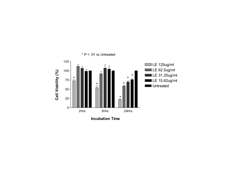
Results: Betamethasone, loteprednol, and methylprednisolone, in commercially available forms, caused significant cytotoxic changes to retinal cells in vitro at clinically relevant doses. This effect was less pronounced with solubilized betamethasone. Dexamethasone at concentrations up to 5 times the clinical dose of free drug injections and 1000 times greater than a drug implant did not cause decreased cell viability. Fluocinolone acetonide at doses 1000 times higher than observed with drug delivery systems showed no cytotoxic effect.

Conclusions: Betamethasone, loteprednol, and methylprednisolone exhibited cytotoxicity at clinically relevant doses and do not appear to be good therapeutic options for intravitreal use. In comparison, dexamethasone and fluocinolone acetonide, which exhibited fewer cytotoxic effects than other steroids, may be potentially viable alternatives to triamcinolone acetonide for clinical use.


Figures

FIGURE 1
FIGURE 1
Percentage of cell viability for human retinal pigment epithelial cells exposed to commercially available dexamethasone solution (Dex-S). There was a significant decrease in cell viability after 24 hours for cells exposed to 1 mg/mL Dex-S when compared to untreated controls (P<.001).
FIGURE 2
FIGURE 2
Percentage of cell viability for human retinal pigment epithelial cells exposed to the preservative in dexamethasone solution, benzyl alcohol (BA). After 24 hours, cells exposed to the highest concentration tested (equivalent to the amount contained in 1 mg/mL dexamethasone solution) showed a significant decrease in cell viability when compared to controls (P<.001).
FIGURE 3
FIGURE 3
Percentage of cell viability for rat embryonal neurosensory precursor retinal cells exposed to commercially available dexamethasone solution (Dex-S). There was a significant decrease in cell viability after 6 or 24 hours of exposure for cells treated with 1 mg/mL Dex-S when compared to untreated controls (P<.001).
FIGURE 4
FIGURE 4
Percentage of cell viability for rat embryonal neurosensory precursor retinal cells exposed to the preservative benzyl alcohol (BA). After 24 hours, cells exposed to 1 mg/mL dexamethasone solution showed a significant decrease in cell viability when compared to controls (P<.001).
FIGURE 5
FIGURE 5
Mitochondrial dehydrogenase (WST-1) assay for human retinal pigment epithelial cells exposed to dexamethasone solution (Dex-S). After 2, 6, or 24 hours, cells exposed to 0.25, 0.5, or 1 mg/mL had a significant reduction in mitochondrial activity when compared to untreated controls (P<.001 for 0.5 or 1.0 mg/mL; P<.01 for 0.25 mg/mL).
FIGURE 6
FIGURE 6
Mitochondrial dehydrogenase (WST-1) assay for human retinal pigment epithelial cells exposed to benzyl alcohol (BA). After 2, 6, or 24 hours, cells exposed to BA at concentrations equivalent to 0.5 or 1.0 mg/mL had a significant reduction in mitochondrial activity when compared to untreated controls (P<.01 for 2 or 6 hours of exposure; P<.001 for 24 hours of exposure). Similar findings were observed after 24 hours of cell exposure to BA at equivalent concentration of 0.25 mg/mL (P<.001).
FIGURE 7
FIGURE 7
Mitochondrial dehydrogenase (WST-1) assay for human retinal pigment epithelial cells exposed to dexamethasone solution (Dex-S). After 2, 6, or 24 hours, cells exposed to 0.25, 0.5, or 1 mg/mL had a significant reduction in mitochondrial activity when compared to untreated controls.
FIGURE 8
FIGURE 8
Mitochondrial dehydrogenase (WST-1) assay for rat embryonal neurosensory precursor retinal cells exposed to benzyl alcohol (BA). After 2, 6, or 24 hours, cells exposed to BA at concentrations equivalent to 0.25, 0.5, or 1.0 mg/mL had a significant reduction in mitochondrial activity when compared to untreated controls (P<.001).
FIGURE 9
FIGURE 9
Comparison between human retinal pigment epithelial cells treated with 1 mg/mL dexamethasone solution (Dex-S), 1.0 mg/mL dexamethasone powder (Dex-P), or DMSO controls. There was no significant difference in cell viability between cells treated with dexamethasone powder or the commercially available solution, and both showed a decrease in cell viability when compared to controls (P<.001).
FIGURE 10
FIGURE 10
Comparison between rat embryonal neurosensory precursor retinal cells treated with 1.0 mg/mL dexamethasone solution (Dex-S), 1.0 mg/mL dexamethasone powder (Dex-P), or DMSO controls. Cells exposed to 1.0 mg/mL dexamethasone powder showed a significant decrease in cell viability when compared to controls, but less toxicity than cells treated with commercially available dexamethasone (P<.01).
FIGURE 11
FIGURE 11
Mitochondrial dehydrogenase (WST-1) assay for human retinal pigment epithelial cells after 24-hour exposure to dexamethasone solution (Dex-S), dexamethasone powder solubilized in DMSO (Dex-P), DMSO at an equivalent dose to solubilize the powder, and untreated controls. There was a reduction in mitochondrial dehydrogenase in all groups compared to untreated controls (P<.001). Cells treated with 1.0 mg/mL Dex-P had significantly higher mitochondrial dehydrogenase activity when compared to cells treated with Dex-S at the same concentration (P<.01).
FIGURE 12
FIGURE 12
Mitochondrial dehydrogenase (WST-1) assay for rat embryonal neurosensory precursor retinal cells after 24-hour exposure to dexamethasone solution (Dex-S), dexamethasone powder solubilized in DMSO (Dex-P), DMSO at an equivalent dose used to solubilize the powder, and untreated controls. There was a reduction in mitochondrial dehydrogenase in all groups compared to untreated controls (P<.01). No difference was found between cells treated with Dex-S and Dex-P.
FIGURE 13
FIGURE 13
Cell viability of human retinal pigment epithelial cells exposed to betamethasone solubilized in DMSO (Beta-S). Only the highest concentration tested (720 μg/mL) showed a significant decrease in cell viability when compared to untreated cells or DMSO-treated controls (P<.001).
FIGURE 14
FIGURE 14
Cell viability of rat embryonal neurosensory precursor retinal cells exposed to betamethasone solubilized in DMSO (Beta-S). Only the highest concentration tested (720 μg/mL) showed a significant decrease in cell viability when compared to untreated cells or DMSO-treated controls (P<.01).
FIGURE 15
FIGURE 15
Mitochondrial membrane potential (ΔΨm) of human retinal pigment epithelial cells exposed to betamethasone solubilized in DMSO (Beta-S). No significant difference in mitochondrial potential was found between cells treated with solubilized betamethasone, DMSO-treated, or untreated controls.
FIGURE 16
FIGURE 16
Mitochondrial membrane potential (ΔΨm) of rat embryonal neurosensory precursor retinal cells exposed to Beta-S. No significant difference in mitochondrial potential was found between cells treated with solubilized betamethasone, DMSO-treated, or untreated controls.
FIGURE 17
FIGURE 17
Cell viability of human retinal pigment epithelial cells exposed to methyl prednisolone solubilized in DMSO. Only the highest concentration tested (1000 μg/mL) showed a significant decrease in cell viability when compared to untreated cells or DMSO-treated controls (P<.001).
FIGURE 18
FIGURE 18
Cell viability of rat embryonal neurosensory precursor retinal cells exposed to methylprednisolone solubilized in DMSO. The concentration of 500 or 1000 μg/mL showed a significant decrease in cell viability when compared to untreated cells or DMSO-treated controls (P<.001).
FIGURE 19
FIGURE 19
Mitochondrial membrane potential (ΔΨm) of human retinal pigment epithelial cells exposed to methylprednisolone solubilized in DMSO. No significant difference in mitochondrial potential was found between cells treated with solubilized methylprednisolone, DMSO-treated, or untreated controls.
FIGURE 20
FIGURE 20
Mitochondrial membrane potential (ΔΨm) of rat embryonal neurosensory precursor retinal cells exposed to methylprednisolone solubilized in DMSO. No significant difference in mitochondrial potential was found between cells treated with solubilized methylprednisolone, DMSO-treated, or untreated controls.
FIGURE 21
FIGURE 21
Cell viability of human retinal pigment epithelial cells exposed to commercially available loteprednol etabonate (LE). After 24 hours, there was a significant decrease in cell viability for cells exposed to 125, 62.5, or 31.25 μg/mL compared to untreated controls (P<.001). After 2 or 6 hours of exposure, cells treated with 125 or 62.5 μg/mL also showed a reduction in cell viability when compared to untreated controls (P<.01).
FIGURE 22
FIGURE 22
Cell viability of rat embryonal neurosensory precursor retinal cells exposed to commercially available loteprednol etabonate (LE). After 24 hours, there was a significant decrease in cell viability for cells exposed to 125, 62.5, or 31.25 μg/mL compared to untreated controls (P<.01). After 2 or 6 hours, only the highest concentration tested (125 μg/mL) showed a reduction in cell viability when compared to untreated controls (P<.01).
FIGURE 23
FIGURE 23
Cell viability of human retinal pigment epithelial cells exposed for 24 hours to loteprednol etabonate solubilized in DMSO (LE-S). Cells treated with the highest concentration tested of LE-S (250 μg/mL) had a significant decrease in viability when compared to untreated controls or cells incubated with the same amount of DMSO (P<.001).
FIGURE 24
FIGURE 24
Cell viability of rat embryonal neurosensory precursor retinal cells exposed for 24 hours to loteprednol etabonate solubilized in DMSO (LE-S). No significant decrease in cell viability was noted when any concentration of LE-S was compared to its respective DMSO controls; cells treated with the highest dose tested of LE-S (250 μg/mL) and its respective DMSO controls had a significant reduction in cell viability when compared to untreated controls (P<.05).
FIGURE 25
FIGURE 25
Mitochondrial membrane potential (ΔΨm) of human retinal pigment epithelial cells exposed to loteprednol etabonate solubilized in DMSO (LE-S). No significant difference in mitochondrial potential was found between cells treated with solubilized loteprednol, DMSO-treated, or untreated controls.
FIGURE 26
FIGURE 26
Mitochondrial membrane potential (ΔΨm) of rat embryonal neurosensory precursor retinal cells exposed to loteprednol etabonate solubilized in DMSO (LE-S). No significant difference in mitochondrial potential was found between cells treated with solubilized loteprednol, DMSO-treated, or untreated controls.
FIGURE 27
FIGURE 27
Cell viability of human retinal pigment epithelial cells exposed to fluocinolone acetonide (FA). Cells treated with 500 or 1000 μg/mL FA showed a significant reduction in cell viability after 2, 6, or 24 hours of exposure (P<.05) when compared to ethyl alcohol (EtOH) or untreated controls.
FIGURE 28
FIGURE 28
Cell viability of rat embryonal neurosensory precursor retinal cells exposed to fluocinolone acetonide (FA). Cells treated with 500 or 1000 μg/mL FA showed a significant reduction in cell viability after 2, 6, or 24 hours of exposure (P<.05). Cells exposed to 150 μg/mL FA showed a significant reduction in cell viability after 24 hours of exposure, when compared to ethyl alcohol (EtOH) or untreated controls (P<.05).

Similar articles

Cited by

References

    1. Leopold IH. Nonsteroidal and steroidal anti-inflammatory agents. In: Sears ML, Tarkkanen A, editors. Surgical Pharmacology of the Eye. New York: Raven Press; 1985.
    1. Pagano G, Bruno A, Cavallo-Perin P, Cesco L, Imbimbo B. Glucose intolerance after short-term administration of corticosteroids in healthy subjects. Prednisone, deflazacort, and betamethasone. Arch Intern Med. 1989;149(5):1098–1101. - PubMed
    1. Saag KG. Glucocorticosteroid-induced osteoporosis. Endocrinol Metab Clin North Am. 2003;32(1):135–157. - PubMed
    1. Stanbury RM, Graham EM. Systemic corticosteroid therapy—side effects and their management. Br J Ophthalmol. 1998;82(6):704–708. - PMC - PubMed
    1. Weijtens O, van der Sluijs FA, Schoemaker RC, et al. Peribulbar corticosteroid injection: vitreal and serum concentration after dexamethasone disodium phosphate injection. Am J Ophthalmol. 1997;123(3):358–363. - PubMed

Publication types

MeSH terms