. 2015 Apr;56(4):871-87.
doi: 10.1194/jlr.M056614. Epub 2015 Feb 2.

Source and role of intestinally derived lysophosphatidic acid in dyslipidemia and atherosclerosis

Affiliations

Source and role of intestinally derived lysophosphatidic acid in dyslipidemia and atherosclerosis

Mohamad Navab et al. J Lipid Res. 2015 Apr.

Abstract

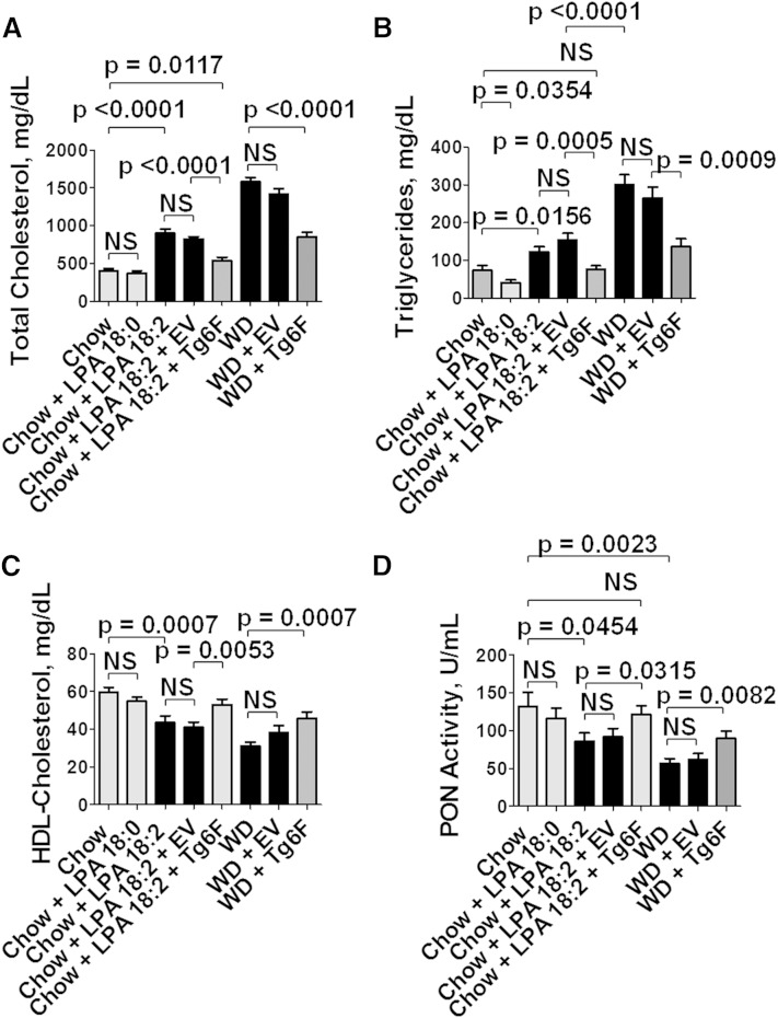
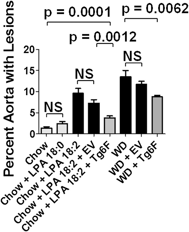
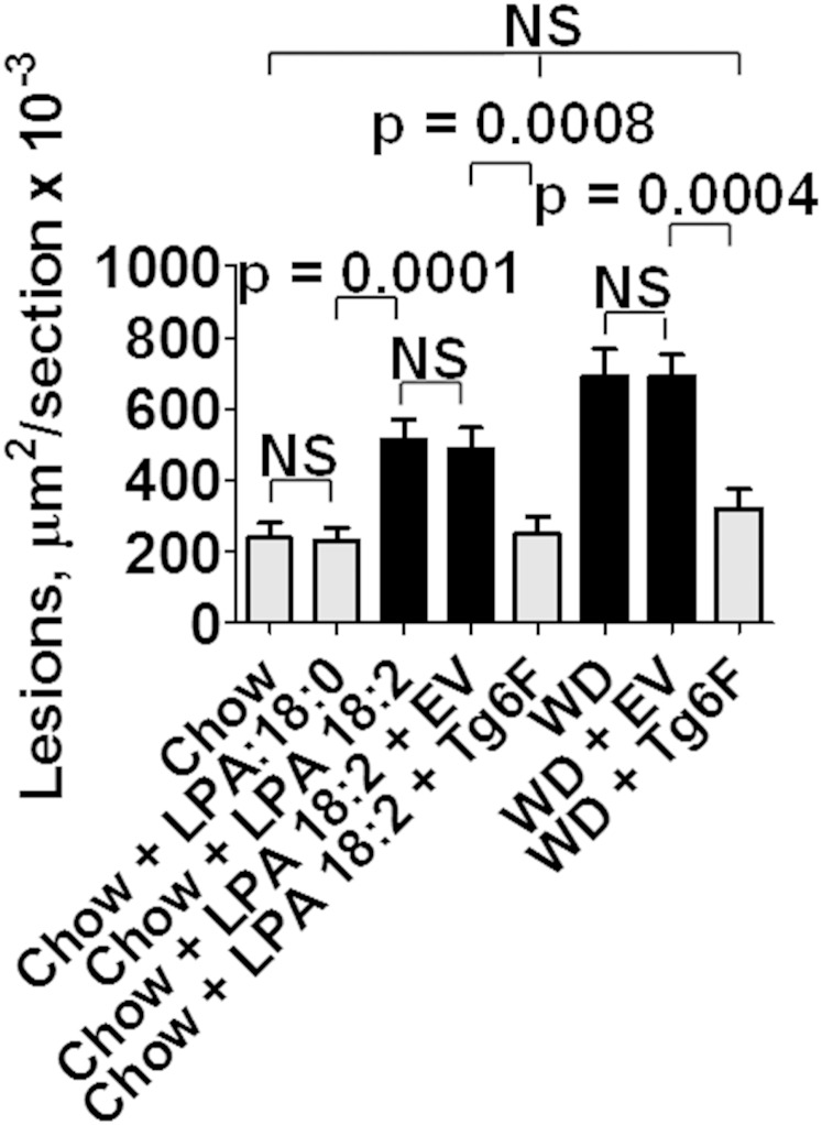
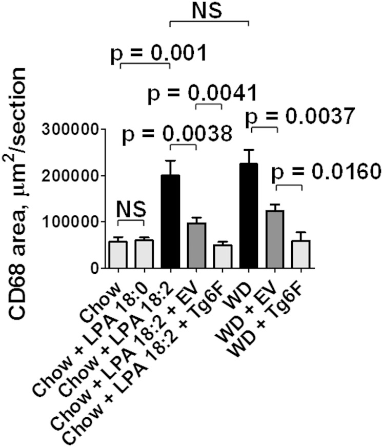
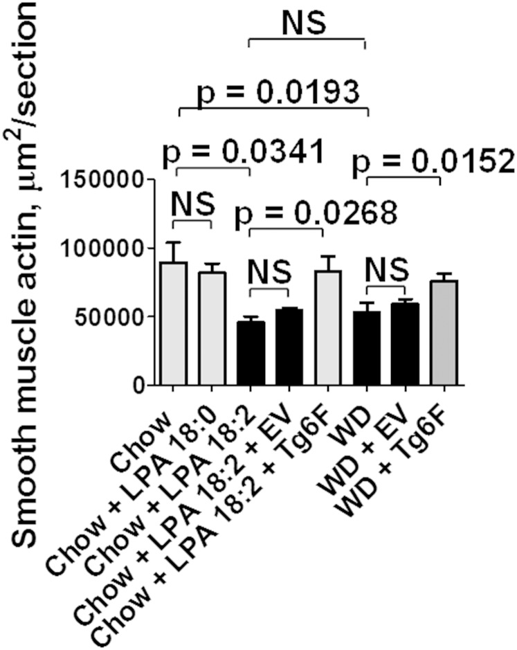
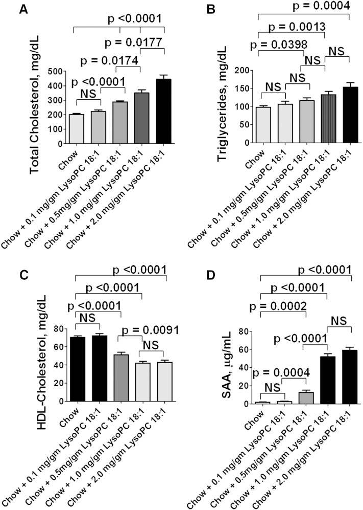
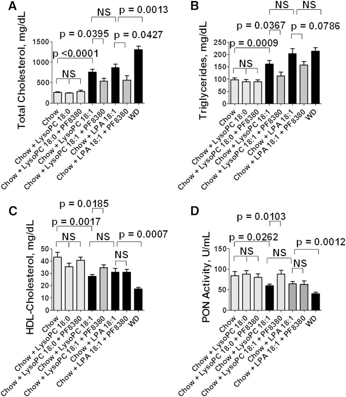
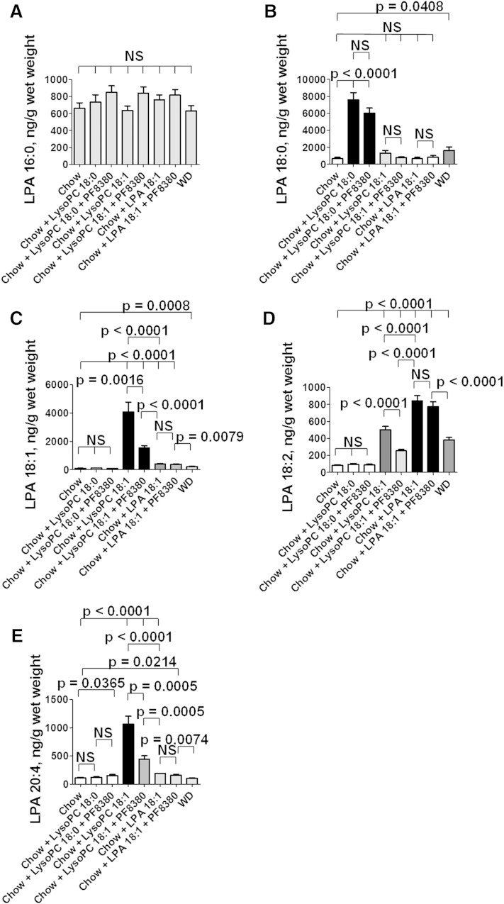
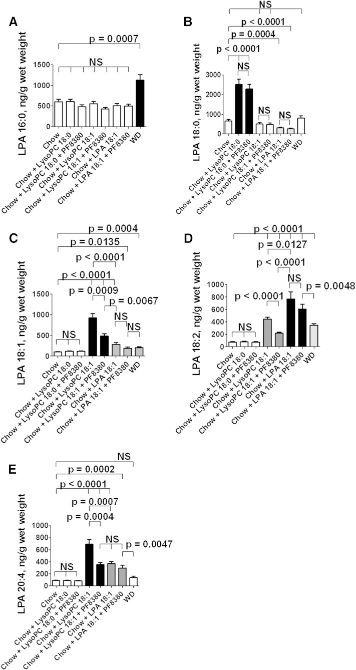
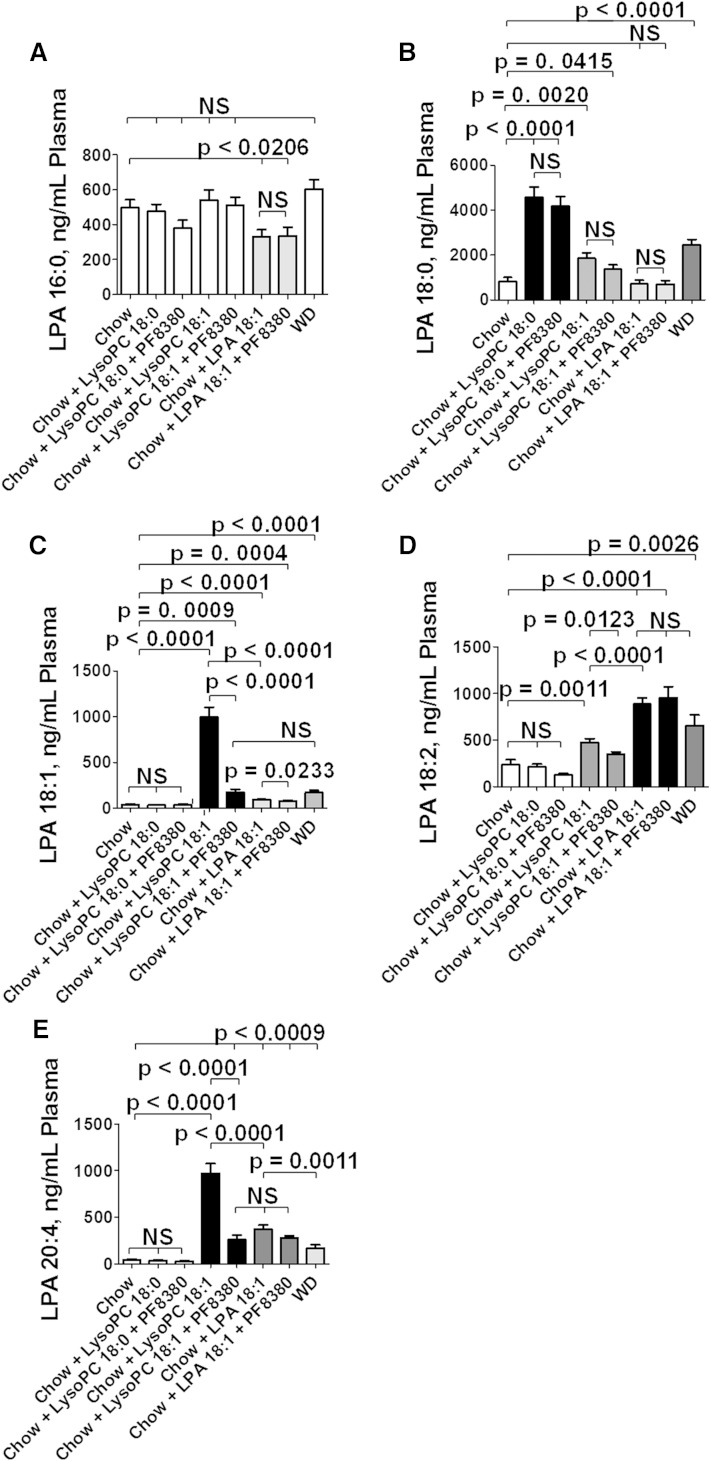
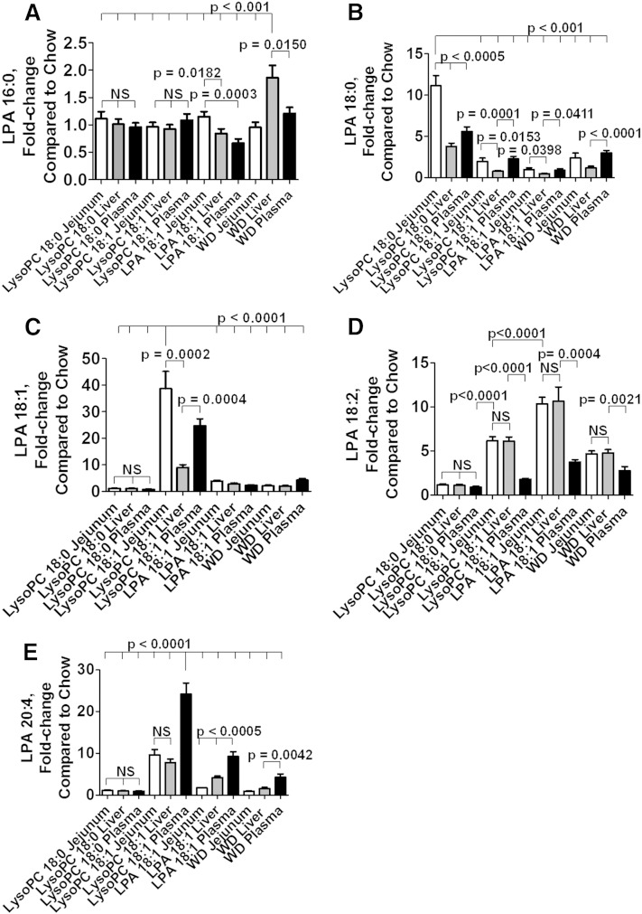
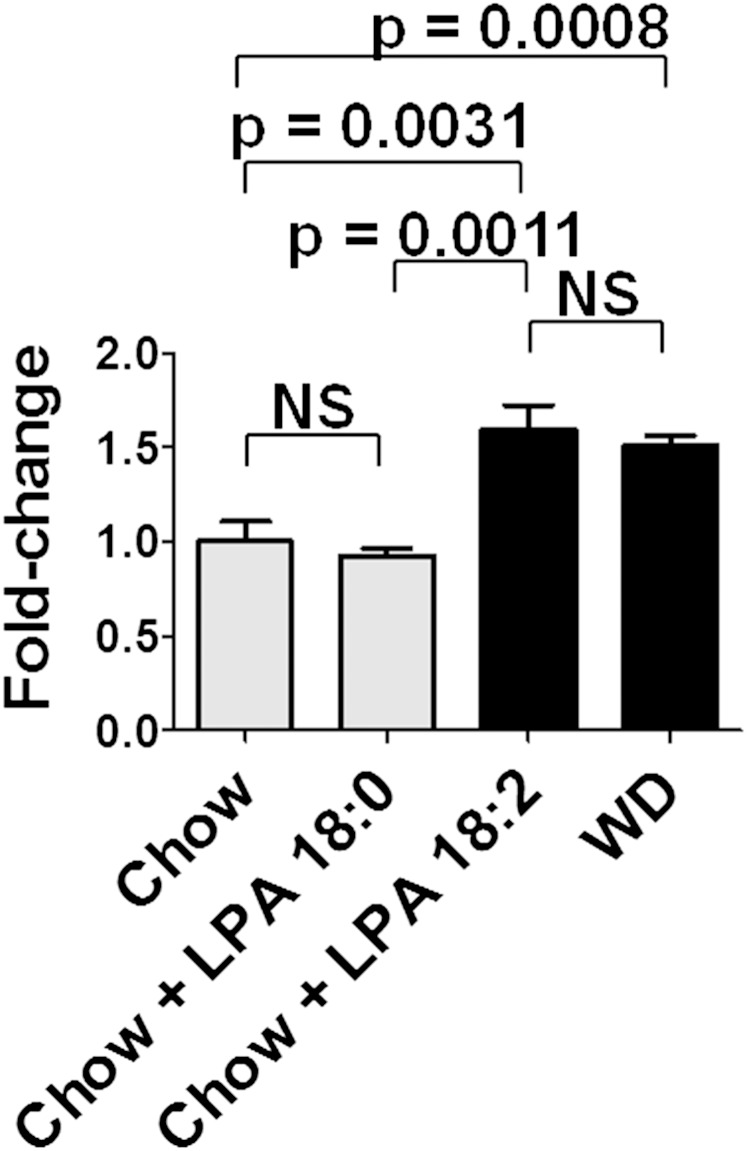
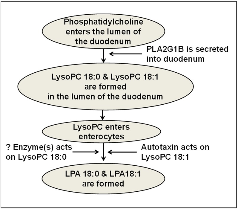
We previously reported that i) a Western diet increased levels of unsaturated lysophosphatidic acid (LPA) in small intestine and plasma of LDL receptor null (LDLR(-/-)) mice, and ii) supplementing standard mouse chow with unsaturated (but not saturated) LPA produced dyslipidemia and inflammation. Here we report that supplementing chow with unsaturated (but not saturated) LPA resulted in aortic atherosclerosis, which was ameliorated by adding transgenic 6F tomatoes. Supplementing chow with lysophosphatidylcholine (LysoPC) 18:1 (but not LysoPC 18:0) resulted in dyslipidemia similar to that seen on adding LPA 18:1 to chow. PF8380 (a specific inhibitor of autotaxin) significantly ameliorated the LysoPC 18:1-induced dyslipidemia. Supplementing chow with LysoPC 18:1 dramatically increased the levels of unsaturated LPA species in small intestine, liver, and plasma, and the increase was significantly ameliorated by PF8380 indicating that the conversion of LysoPC 18:1 to LPA 18:1 was autotaxin dependent. Adding LysoPC 18:0 to chow increased levels of LPA 18:0 in small intestine, liver, and plasma but was not altered by PF8380 indicating that conversion of LysoPC 18:0 to LPA 18:0 was autotaxin independent. We conclude that i) intestinally derived unsaturated (but not saturated) LPA can cause atherosclerosis in LDLR(-/-) mice, and ii) autotaxin mediates the conversion of unsaturated (but not saturated) LysoPC to LPA.

Keywords: 6F peptide; apolipoprotein A-I mimetic peptides; genetically engineered tomato plants; lysophosphatidylcholine.


Figures

Fig. 1.
Fig. 1.
Addition of unsaturated (but not saturated) LPA to standard mouse chow results in dyslipidemia and decreased PON activity. Female LDLR−/− mice 4–6 months of age (20–22 per group) were fed standard mouse chow, or standard mouse chow supplemented with 1 μg per gram chow of LPA 18:0 or LPA 18:2, or the mice were fed a WD. Some of the mice also received 2.2% by weight of freeze-dried EV tomatoes or Tg6F tomatoes. After 13 weeks, the mice were fasted overnight, and 20 mice from each group were bled to determine plasma lipids and PON activity as described in Materials and Methods. A: Plasma total cholesterol levels. B: Plasma triglyceride levels. C: Plasma HDL-cholesterol levels. D: Plasma PON activity. The data shown are mean ± SEM. NS, not significant.
Fig. 2.
Fig. 2.
Addition of unsaturated (but not saturated) LPA to standard mouse chow results in aortic atherosclerosis as determined by en face analysis. The percent of aorta with atherosclerosis as determined by en face analysis for the mice described in Fig. 1 was determined as described in Materials and Methods. The data shown are mean ± SEM. NS, not significant.
Fig. 3.
Fig. 3.
Addition of unsaturated (but not saturated) LPA to standard mouse chow results in aortic atherosclerosis as determined by aortic root analysis. The area containing atherosclerotic lesions in aortic root sections of 15–19 randomly selected mice in each group described in Fig. 1 was determined as described in Materials and Methods. The data shown are mean ± SEM. NS, not significant. If chow is included in multiple comparisons, each comparison is to chow.
Fig. 4.
Fig. 4.
Addition of unsaturated (but not saturated) LPA to standard mouse chow results in macrophage-rich aortic atherosclerosis. Aortic root sections from the mice described in Fig. 3 were randomly selected from 4 to 11 mice in each group for determination of macrophage area by staining for CD68 as described in Materials and Methods. The data shown are mean ± SEM. NS, not significant.
Fig. 5.
Fig. 5.
Addition of unsaturated (but not saturated) LPA to mouse chow results in a significant reduction of aortic α smooth muscle cell actin. Aortic root sections from the mice described in Fig. 3 were randomly selected from 6 to 17 mice in each group for determination of aortic lesion α smooth muscle cell actin as described in Materials and Methods. The data shown are mean ± SEM. NS, not significant.
Fig. 6.
Fig. 6.
Adding LysoPC 18:1 to standard mouse chow dose-dependently produces dyslipidemia and inflammation in LDLR−/− mice. Female LDLR−/− mice 4–6 months of age (20 per group) were fed mouse chow or mouse chow supplemented with LysoPC 18:1 at a dose of 0.1 mg/g chow, 0.5 mg/g chow, 1.0 mg/g chow, or 2 mg/g chow. After 2 weeks, the mice were fasted overnight, and blood was collected for determination of plasma lipids and SAA as described in Materials and Methods. A: Plasma total cholesterol levels. B: Plasma triglyceride levels. C: Plasma HDL-cholesterol levels. D: Plasma SAA levels. The data shown are mean ± SEM. NS, not significant. If chow is included in multiple comparisons, each comparison is to chow.
Fig. 7.
Fig. 7.
The dyslipidemia induced by supplementing standard mouse chow with LysoPC 18:1 is significantly ameliorated by adding a specific autotaxin inhibitor (PF8380). Male LDLR−/− mice 9–10 months of age (20 per group) were fed standard mouse chow or standard mouse chow plus LysoPC 18:0 or LysoPC 18:1 at 1 mg per gram chow without or with 30 mg/kg of the specific oral autotaxin inhibitor PF8380, or the mice were fed standard mouse chow supplemented with LPA 18:1 at 1 μg per gram chow without or with 30 mg/kg of PF8380, or the mice were fed a WD. After 2 weeks, the mice were fasted overnight, bled, and plasma lipids and PON activity were determined as described in Materials and Methods. A: Plasma total cholesterol levels. B: Plasma triglyceride levels. C: HDL-cholesterol levels. D: PON activity. The data shown are mean ± SEM. NS, not significant. If chow is included in multiple comparisons, each comparison is to chow.
Fig. 8.
Fig. 8.
Adding LysoPC 18:0 or LysoPC 18:1 or LPA 18:1 to standard mouse chow produces complex changes in the levels of LPA species in the tissue of the jejunum. Male LDLR−/− mice 5–7 months of age (20 per group) were fed standard mouse chow or standard mouse chow plus LysoPC 18:0 or LysoPC 18:1 at 1 mg per gram chow without or with 30 mg/kg of the specific oral autotaxin inhibitor PF8380, or the mice were fed standard mouse chow supplemented with LPA 18:1 at 1 μg per gram chow without or with 30 mg/kg of PF8380, or the mice were fed a WD. After 2 weeks, the mice were fasted overnight; under anesthesia, the mice were bled and perfused to remove all blood as described in Materials and Methods; and the jejunum was harvested and processed to determine LPA tissue levels as described in Materials and Methods. A: The levels of LPA 16:0. B: The levels of LPA 18:0. C: The levels of LPA 18:1. D: The levels of LPA 18:2. E: The levels of LPA 20:4. The data shown are mean ± SEM. NS, not significant. If chow is included in multiple comparisons, each comparison is to chow.
Fig. 9.
Fig. 9.
Adding LysoPC 18:0 or LysoPC 18:1 or LPA 18:1 to standard mouse chow produces directional changes in the liver similar to those seen in the jejunum for most LPA species. After perfusion to remove all blood from the tissues, the liver was harvested from the mice described in Fig. 8, and hepatic LPA tissue levels were determined as described in Materials and Methods. A: The levels of LPA 16:0. B: The levels of LPA 18:0. C: The levels of LPA 18:1. D: The levels of LPA 18:2. E: The levels of LPA 20:4. The data shown are mean ± SEM. NS, not significant. If chow is included in multiple comparisons, each comparison is to chow.
Fig. 10.
Fig. 10.
Adding LysoPC 18:0 or LysoPC 18:1 or LPA 18:1 to standard mouse chow produces directional changes in the plasma similar to those seen in the jejunum and liver for most LPA species. Plasma LPA levels in the mice described in Fig. 8 were determined as described in Materials and Methods. A: The levels of LPA 16:0. B: The levels of LPA 18:0. C: The levels of LPA 18:1. D: The levels of LPA 18:2. E: The levels of LPA 20:4. The data shown are mean ± SEM. NS, not significant. If chow is included in multiple comparisons, each comparison is to chow.
Fig. 11.
Fig. 11.
Directly comparing changes in jejunum, liver, and plasma. An average value was calculated for each LPA species in each tissue in mice receiving standard chow without supplements in Figs. 8–10. The fold-change compared with the average value on standard mouse chow without supplements was then calculated for each mouse on each diet (except for those that received PF8380), and for each LPA species in the mice described in Figs. 8–10. In each panel, the fold-change in the jejunum compared with standard mouse chow without supplements is shown in white bars. In each panel the fold-change in the liver compared with standard mouse chow without supplements is shown in the gray bars. In each panel the fold-change in the plasma compared with standard mouse chow without supplements is shown in the black bars. Comparisons for mice receiving PF8380 were omitted to reduce the number of bars in each panel. A: The fold-change in levels of LPA 16:0. B: The fold-change in levels of LPA 18:0. C: The fold-change in levels of LPA 18:1. D: The fold-change in levels of LPA 18:2. E: The fold-change in levels of LPA 20:4. The data shown are mean ± SEM. NS, not significant. In the case of multiple comparisons, the index condition to which the others are compared is indicated by a long vertical line (e.g., in A, the multiple comparisons shown at the top of the figure were to the values for WD liver).
Fig. 12.
Fig. 12.
Adding unsaturated (but not saturated LPA) to standard mouse chow induces gene expression of Lpcat3 in the jejunum to the same level as seen after feeding the mice a WD. Female LDLR−/− mice age 7–8 months (7–8 per group) were fed standard mouse chow or standard mouse chow supplemented with 1 μg LPA 18:0 or LPA 18:2 per gram chow, or the mice were fed WD. After 2 weeks, the jejunum was harvested, and mRNA levels for Lpcat3 were determined by RT-qPCR as described in Materials and Methods. The data shown are mean ± SEM. NS, not significant.
Fig. 13.
Fig. 13.
A schematic representation of the formation of LPA 18:0 and LPA 18:1 in the small intestine. It is hypothesized that phosphatidylcholine in the lumen of the small intestine is acted on by pancreatic PLA2G1B to produce saturated LysoPC (e.g., LysoPC 18:0) or unsaturated LysoPC (e.g., LysoPC 18:1) in the lumen of the small intestine. The resulting LysoPC enters the enterocytes of the small intestine where the saturated LysoPC is converted to saturated LPA (e.g., LPA 18:0) by unknown (?) enzyme(s), while unsaturated LysoPC is converted to unsaturated LPA (e.g., LPA 18:1) by lysophospholipase D (autotaxin).

References

    1. Reddy S. T., Navab M., Anantharamaiah G. M., Fogelman A. M. 2014. Searching for a successful HDL-based treatment strategy. Biochim. Biophys. Acta. 1841: 162–167. - PubMed
    1. Navab M., Shechter I., Anantharamaiah G. M., Reddy S. T., Van Lenten B. J., Fogelman A. M. 2010. Structure and function of HDL mimetics. Arterioscler. Thromb. Vasc. Biol. 30: 164–168. - PMC - PubMed
    1. Bloedon L. T., Dunbar R., Duffy D., Pinell-Salles P., Norris R., DeGroot B. J., Movva R., Navab M., Fogelman A. M., Rader D. J. 2008. Safety, pharmacokinetics, and pharmacodynamics of oral apoA-I mimetic peptide D-4F in high-risk cardiovascular patients. J. Lipid Res. 49: 1344–1352. - PMC - PubMed
    1. Dunbar R.L., Bloedon L.T., Duffy D., Norris R.B., Movva R., Navab M., Fogelman A.M., Rader D.J. 2007. Daily oral administration of the apolipoprotein A-I mimetic peptide D-4F in patients with coronary heart disease or equivalent risk improves high-density lipoprotein anti-inflammatory function (Abstract). J. Am. Coll. Cardiol. 49: 366A.
    1. Watson C. E., Weissbach N., Kjems L., Ayalasomayajula S., Zhang Y., Chang I., Navab M., Hama S., Hough G., Reddy S. T., et al. 2011. Treatment of patients with cardiovascular disease with L-4F, an apoA-1 mimetic, did not improve select biomarkers of HDL function. J. Lipid Res. 52: 361–373. - PMC - PubMed

Publication types

MeSH terms