Review
. 2015 Feb 2;7(2):a006023.
doi: 10.1101/cshperspect.a006023.

Signaling in muscle contraction

Affiliations
Review

Signaling in muscle contraction

Ivana Y Kuo et al. Cold Spring Harb Perspect Biol. .

Abstract

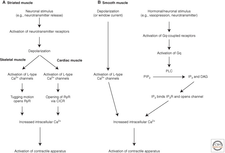
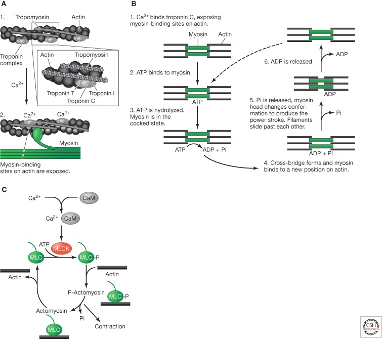
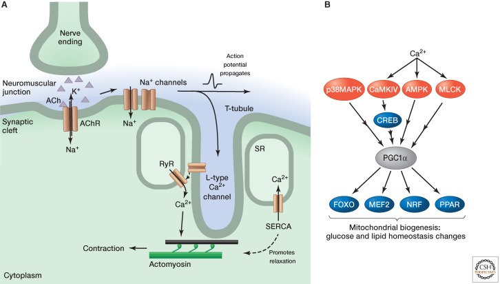
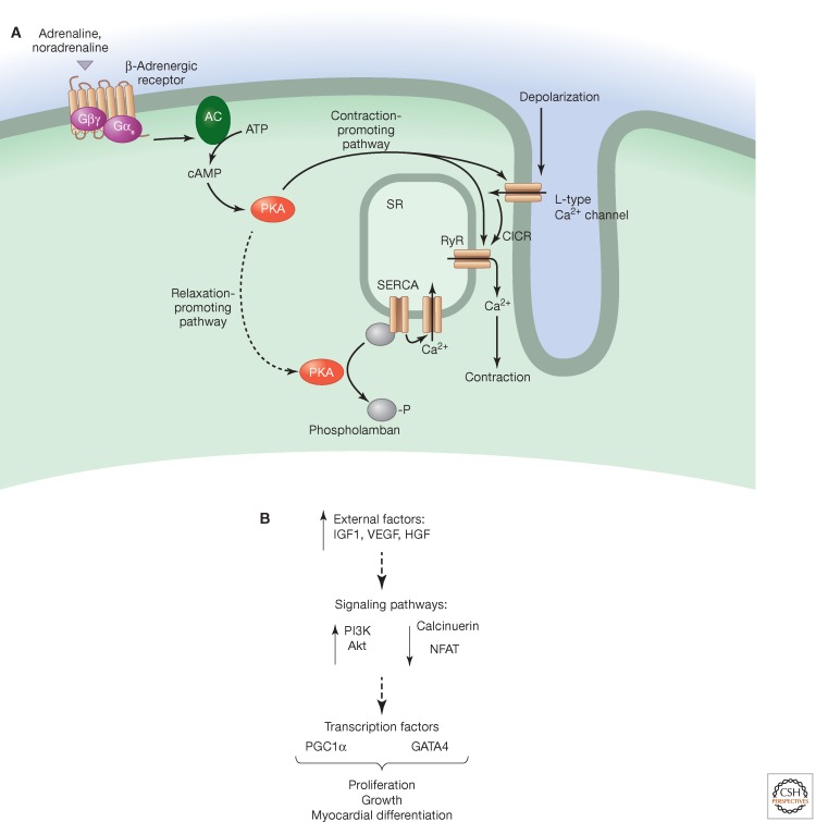
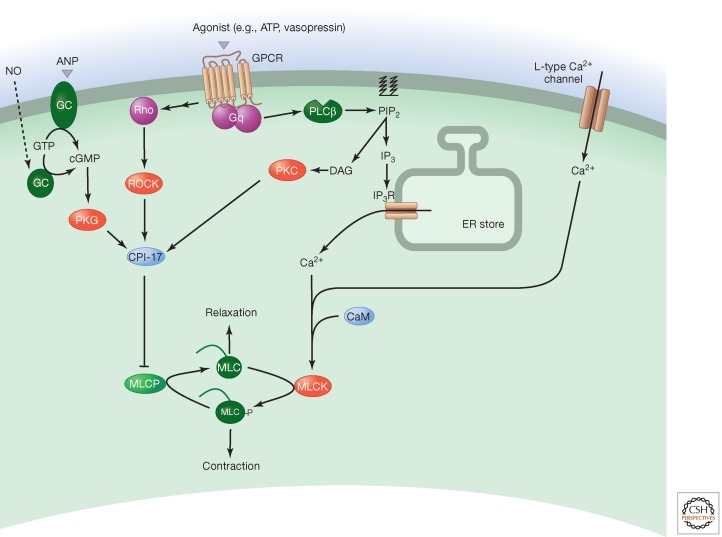
Signaling pathways regulate contraction of striated (skeletal and cardiac) and smooth muscle. Although these are similar, there are striking differences in the pathways that can be attributed to the distinct functional roles of the different muscle types. Muscles contract in response to depolarization, activation of G-protein-coupled receptors and other stimuli. The actomyosin fibers responsible for contraction require an increase in the cytosolic levels of calcium, which signaling pathways induce by promoting influx from extracellular sources or release from intracellular stores. Rises in cytosolic calcium stimulate numerous downstream calcium-dependent signaling pathways, which can also regulate contraction. Alterations to the signaling pathways that initiate and sustain contraction and relaxation occur as a consequence of exercise and pathophysiological conditions.


Figures

Figure 1.
Figure 1.
Overview of muscle contraction signals in striated (A), and smooth (B) muscle.
Figure 2.
Figure 2.
Calcium triggers contraction in striated muscle. (A) Actomyosin in striated muscle. (1) Striated muscle in the relaxed state has tropomyosin covering myosin-binding sites on actin. (2) Calcium binds to troponin C, which induces a conformational change in the troponin complex. This causes tropomyosin to move deeper into the actin groove, revealing the myosin-binding sites. (B) Cross-bridge cycle in striated muscle. (1) Calcium binds to troponin C, causing the conformational shift in tropomyosin that reveals myosin-binding sites on actin. (2) ATP then binds to myosin. (3) ATP is then hydrolyzed. (4) A cross-bridge forms and myosin binds to a new position on actin. (5) Pi is released and myosin changes conformation, resulting in the power stroke that causes the filaments to slide past each other. (6) ADP is then released. (C) Contraction in smooth muscle. In smooth muscle, calcium binds to calmodulin and causes the activation of myosin light chain (MLC) kinase (MLCK). This phosphorylates MLC, which then binds to actin to form phosphorylated actomyosin, enabling the cross-bridge cycle to start.
Figure 3.
Figure 3.
Skeletal muscle contraction and changes with exercise. (A) Neurotransmitter (acetylcholine, ACh) released from nerve endings binds to receptors (AChRs) on the muscle surface. The ensuing depolarization causes sodium channels to open, which elicits an action potential that propagates along the cell. The action potential invades T-tubules and causes the L-type calcium channels to open, which in turn causes ryanodine receptors (RyRs) in the SR to open and release calcium, which stimulates contraction. Calcium is pumped back into the SR by (SR/ER calcium ATPase SERCA) pumps. The decreasing cytosolic calcium levels cause calcium to disassociate from troponin C and, consequently, tropomyosin reverts to a conformation that covers the myosin-binding sites. (B) Signaling in exercised skeletal muscle. Both calcium and calcium-independent signals stimulate the transcriptional coactivator PGC1α. This activates a number of transcription factors that regulate genes associated with mitochondrial biogenesis, glucose, and lipid homeostasis.
Figure 4.
Figure 4.
Cardiac muscle contraction and changes with exercise. (A) Cardiac muscle contraction can occur as a consequence of calcium entry through L-type calcium channels, which activate ryanodine receptor (RyR) channels in the SR. Alternatively, β-adrenergic receptors on the cell membrane lead to activation of adenylyl cyclase (AC), which stimulates PKA. This can promote contraction by phosphorylating RyR and L-type calcium channels or relaxation by phosphorylating the SERCA pump inhibitor phospholamban. (B) Changes with exercise lead to an activation of the PI3K/Akt pathway, and a down-regulation of NFAT and calcinurin.
Figure 5.
Figure 5.
Smooth muscle contraction. Calcium released by L-type calcium channels or IP3Rs downstream from Gq-coupled cell-surface receptors causes smooth muscle contraction. It binds to calmodulin (CaM) and the resulting complex stimulates myosin light-chain (MLC) kinase (MLCK). This phosphorylates MLC to promote contraction. A RhoA/ROCK pathway and a diacylglycerol (DAG) pathway contribute to calcium sensitization by altering the phosphorylation status of myosin light-chain phosphatase (MLCP). Relaxation is mediated through the cGMP/PKG pathway downstream from nitric oxide (NO) and agonists such as atrial natriuretic peptide (ANP).

References

    1. Andersson DC, Marks AR 2010. Fixing ryanodine receptor Ca leak—A novel therapeutic strategy for contractile failure in heart and skeletal muscle. Drug Discov Today 7: e151–e157. - PMC - PubMed
    1. Balakumar P, Jagadeesh G 2010. Multifarious molecular signaling cascades of cardiac hypertrophy: Can the muddy waters be cleared? Pharmacol Res 62: 365–383. - PubMed
    1. Baryshnikov SG, Pulina MV, Zulian A, Linde CI, Golovina VA 2009. Orai1, a critical component of store-operated Ca2+ entry, is functionally associated with Na+/Ca2+ exchanger and plasma membrane Ca2+ pump in proliferating human arterial myocytes. Am J Physiol Cell Physiol 297: C1103–C1112. - PMC - PubMed
    1. Berra-Romani R, Mazzocco-Spezzia A, Pulina MV, Golovina VA 2008. Ca2+ handling is altered when arterial myocytes progress from a contractile to a proliferative phenotype in culture. Am J Physiol Cell Physiol 295: C779–C790. - PMC - PubMed
    1. Bezprozvanny I, Watras J, Ehrlich BE 1991. Bell-shaped calcium-response curves of Ins(1,4,5)P3- and calcium-gated channels from endoplasmic reticulum of cerebellum. Nature 351: 751–754. - PubMed

Publication types

MeSH terms