Review
. 2015 Feb 2;7(2):a015800.
doi: 10.1101/cshperspect.a015800.

The centrosome and its duplication cycle

Affiliations
Review

The centrosome and its duplication cycle

Jingyan Fu et al. Cold Spring Harb Perspect Biol. .

Abstract

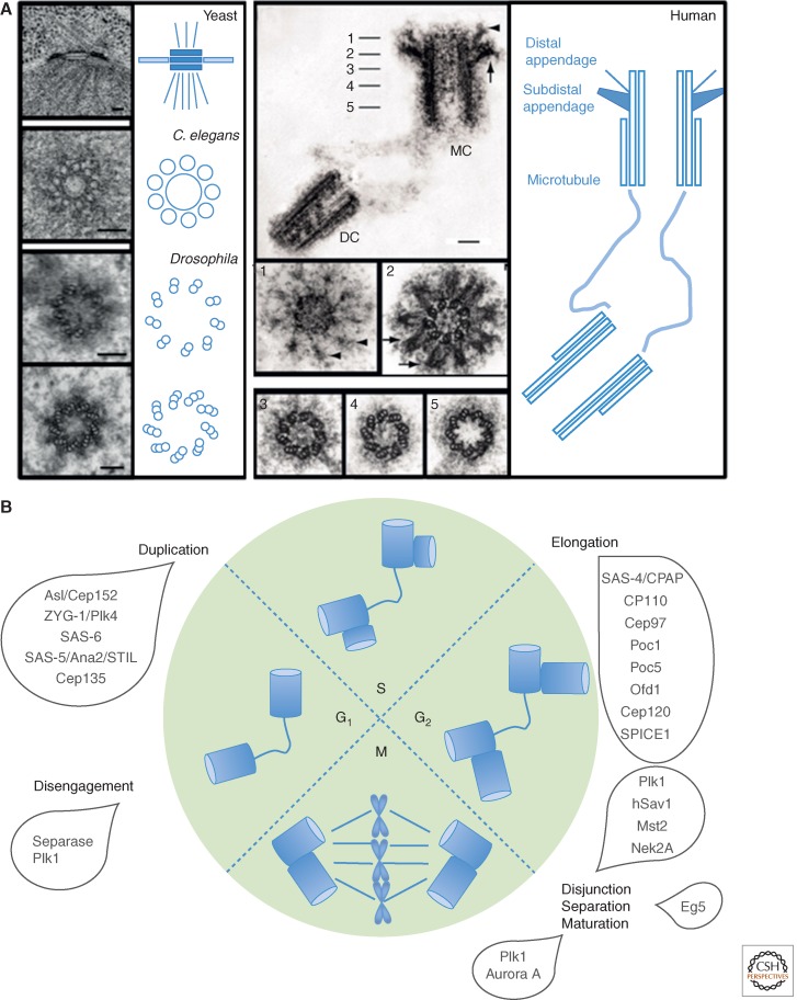
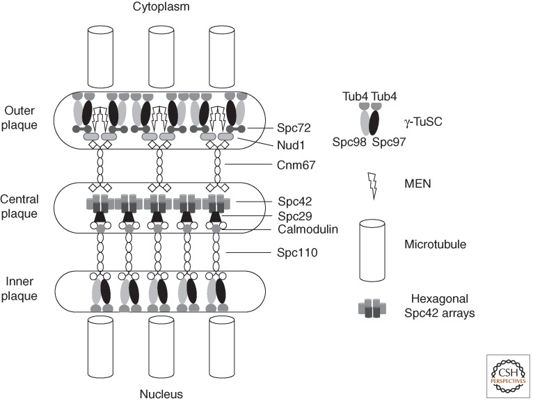
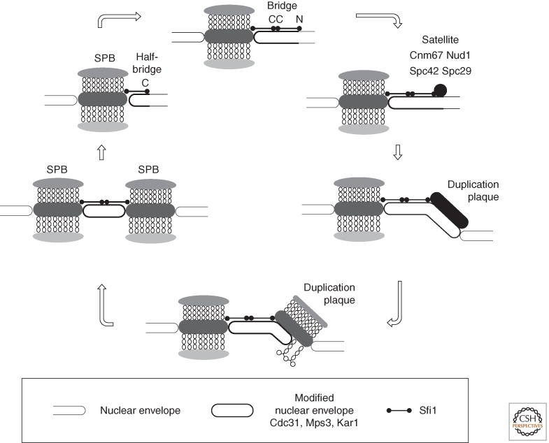
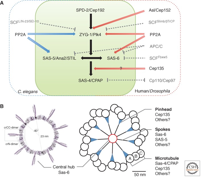
The centrosome was discovered in the late 19th century when mitosis was first described. Long recognized as a key organelle of the spindle pole, its core component, the centriole, was realized more than 50 or so years later also to comprise the basal body of the cilium. Here, we chart the more recent acquisition of a molecular understanding of centrosome structure and function. The strategies for gaining such knowledge were quickly developed in the yeasts to decipher the structure and function of their distinctive spindle pole bodies. Only within the past decade have studies with model eukaryotes and cultured cells brought a similar degree of sophistication to our understanding of the centrosome duplication cycle and the multiple roles of this organelle and its component parts in cell division and signaling. Now as we begin to understand these functions in the context of development, the way is being opened up for studies of the roles of centrosomes in human disease.


Figures

Figure 1.
Figure 1.
The structure and duplication cycle of centrosomes. (A) Electron microscopy reveals the structures of the spindle pole body (SPB) centrosome with ninefold symmetrical centriole as its core. Scale bars, 100 nm. (B) C. elegans, Caenorhabditis elegans; DC, daughter centriole; MC, mother centriole. The centrosome duplication cycle occurs in concert with the cell-division cycle. Key events and players in the centrosome cycle are indicated.
Figure 2.
Figure 2.
A highly schematic representation of molecular architecture of the budding yeast spindle pole body (SPB). A hexagonal crystalline array of Spc42 units associate with Spc29/Spc110 complexes on the nuclear side and cnm67 dimers on the cytoplasmic side of the SPB. These spacer proteins separate the central Spc42 plaque from the γ-TuSC microtubule-nucleating centers at the inner and outer plaques. At the inner plaque the interaction between the spacer Spc110 is direct with one Spc110 dimer associating with a single γ-TuSC (Erlemann et al. 2012). It is estimated that a functional microtubule nucleation unit comprises seven γ-TuSCs, two additional Spc98, and three extra γ-tubulins (Erlemann et al. 2012). This estimate agrees well with the reconstitution of 13-fold symmetric γ-tubulin microtubule-nucleating units in vitro (Kollman et al. 2008, 2010). At the cytoplasmic outer plaque, the association between the spacer and the γ-TuSC is mediated through the association of Nud1 with Spc72. Despite the fact that Spc72 interacts with both Spc97 and Spc98 in two hybrid assays (Knop and Schiebel 1998), in vivo measurements suggest that one Spc72 dimer interacts with a single γ-TuSC (Erlemann et al. 2012). Nud1 also acts as a scaffolding molecule for the mitotic exit network (MEN) that couples the SPB position with cell-cycle control. The stoichiometries of other associations remain to be established. The representation of Spc29 in between Spc110 and Spc42 is highly schematic, as the exact nature of its function as part of the Spc110 complex remains to be established.
Figure 3.
Figure 3.
Budding yeast spindle pole body (SPB) duplication. A highly schematic representation of SPB duplication in budding yeast. The key role played by Sfi1 C-C homotypic dimerization in establishing a point for the formation of the satellite that expands to form the duplication plaque is shown in the top panel. Immunoelectron microscopy indicates that this satellite contains at least Cnm67, Nud1, Spc42, and Spc29. (For full details, see Adams and Kilmartin 1999, ; Kilmartin 2014.)
Figure 4.
Figure 4.
Centriole assembly. (A) Comparison of Caenorhabditis elegans (C. elegans) and Human/Drosophila pathways. Common elements are in the green box. (B) Structure organization of nine Sas-6 dimers (left) (Kitagawa et al. 2011c), and the relationship of cartwheel to centriole wall/microtubules (right). Molecular components are indicated.
Figure 5.
Figure 5.
Centrosome disjunction and centriole disengagement. (A) Series of upstream events trigger the dissociation of C-Nap1 and rootletin from the centrosome leading to loss of centrosome cohesion. Main players are depicted. (B) Roles of Plk1 and separase in disjoining mother and daughter centrioles.
Figure 6.
Figure 6.
Pericentriolar material (PCM) assembly. (A) The zones of the Drosophila centrosome (Fu and Glover 2012). (B) Expansion of the PCM in mitosis. Comparison of human and Drosophila components. (C) Pathway of PCM assembly deduced from studies in Drosophila (Fu and Glover 2012; Conduit et al. 2014a,b). tub, Tubulin.
Figure 7.
Figure 7.
Cilia assembly and disassembly cycle. Role of Plk1 and Aurora A in activating tubulin deacetylation for cilia disassembly (left). Cilia assembly pathway (right).
Figure 8.
Figure 8.
Key landmarks in the control of asymmetric spindle pole body (SPB) function in budding yeast. The figure shows the key landmarks that are referred to in the text imposed on depictions of bud growth from a mother yeast cell. See text for full details on how each of these molecules/features contributes to asymmetries of SPB function that ensure each daughter cell will inherit one genome.
Figure 9.
Figure 9.
Inheritance of centrosomes in asymmetric cell division in Drosophila. The stem cells of the male germline inherit the mother centrosome (A) in contrast to the stem cell of the neuroblasts that inherit the daughter (B). GMC, ganglion mother cell; MT, microtubule; PCM, pericentriolar material.

References

    1. Abrieu A, Brassac T, Galas S, Fisher D, Labbe JC, Doree M 1998. The Polo-like kinase Plx1 is a component of the MPF amplification loop at the G2/M-phase transition of the cell cycle in Xenopus eggs. J Cell Sci 111: 1751–1757. - PubMed
    1. Adames NR, Cooper JA 2000. Microtubule interactions with the cell cortex causing nuclear movements in Saccharomyces cerevisiae. J Cell Biol 149: 863–874. - PMC - PubMed
    1. Adams IR, Kilmartin JV 1999. Localization of core spindle pole body (SPB) components during SPB duplication in Saccharomyces cerevisiae. J Cell Biol 145: 809–823. - PMC - PubMed
    1. Adams IR, Kilmartin JV 2000. Spindle pole body duplication: A model for centrosome duplication? Trend Cell Biol 10: 329–335. - PubMed
    1. Alfa CE, Ducommun B, Beach D, Hyams JS 1990. Distinct nuclear and spindle pole body populations of cyclin-cdc2 in fission yeast. Nature 347: 680–682. - PubMed

MeSH terms

Substances

LinkOut - more resources