Review
. 2015 Feb 2;7(2):a021808.
doi: 10.1101/cshperspect.a021808.

Place cells, grid cells, and memory

Affiliations
Review

Place cells, grid cells, and memory

May-Britt Moser et al. Cold Spring Harb Perspect Biol. .

Abstract

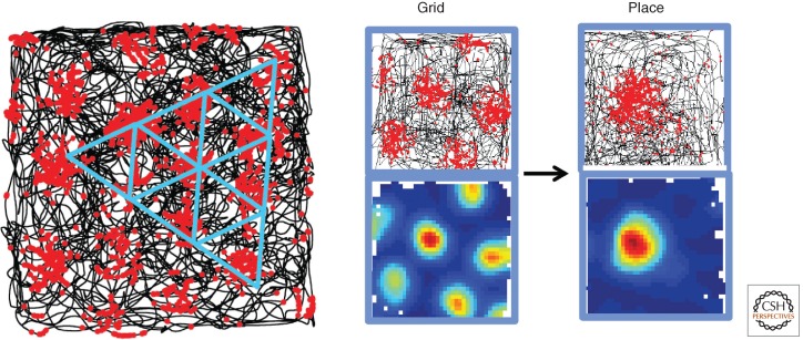
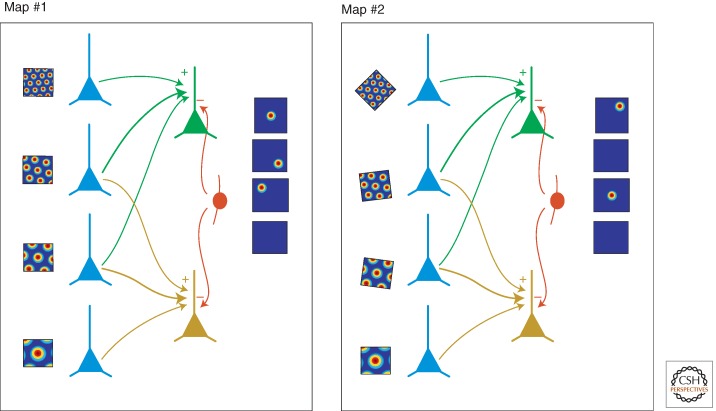
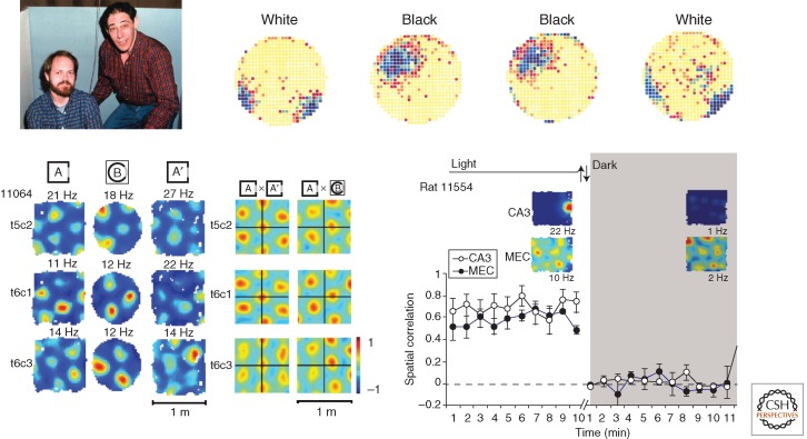
The hippocampal system is critical for storage and retrieval of declarative memories, including memories for locations and events that take place at those locations. Spatial memories place high demands on capacity. Memories must be distinct to be recalled without interference and encoding must be fast. Recent studies have indicated that hippocampal networks allow for fast storage of large quantities of uncorrelated spatial information. The aim of the this article is to review and discuss some of this work, taking as a starting point the discovery of multiple functionally specialized cell types of the hippocampal-entorhinal circuit, such as place, grid, and border cells. We will show that grid cells provide the hippocampus with a metric, as well as a putative mechanism for decorrelation of representations, that the formation of environment-specific place maps depends on mechanisms for long-term plasticity in the hippocampus, and that long-term spatiotemporal memory storage may depend on offline consolidation processes related to sharp-wave ripple activity in the hippocampus. The multitude of representations generated through interactions between a variety of functionally specialized cell types in the entorhinal-hippocampal circuit may be at the heart of the mechanism for declarative memory formation.


Figures

Figure 1.
Figure 1.
Grid cells and place cells. (Left) A grid cell from the entorhinal cortex of the rat brain. The black trace shows the trajectory of a foraging rat in part of a 1.5-m-diameter-wide square enclosure. Spike locations of the grid cell are superimposed in red on the trajectory. Each red dot corresponds to one spike. Blue equilateral triangles have been drawn on top of the spike distribution to illustrate the regular hexagonal structure of the grid pattern. (Right) Grid cell and place cell. (Top) Trajectory with spike locations, as in the left part. (Bottom) Color-coded rate map with red showing high activity and blue showing low activity. Grid cells are thought to provide much, but not all, of the entorhinal spatial input to place cells.
Figure 2.
Figure 2.
Schematic illustration of how periodic grid cells could be transformed to nonperiodic place cells by linear summation of output from grid cells with overlapping firing fields, but different spacing and orientation, and how differential responses among modules of grid cells might give rise to remapping in the hippocampus. (Left) Map 1, grid cells with different spacing converge to generate place cells in a subset of the hippocampal place-cell population. Each grid cell belongs to a different grid module. (Right) Map 2, differential realignment of each of the grid maps induces recruitment of a new subset of place cells. (From images in Solstad et al. 2006 and Fyhn et al. 2007; modified, with permission, from the authors and Nature Publishing Group © 2006 and 2007, respectively.)
Figure 3.
Figure 3.
Remapping in place cells and grid cells. (Top left) John Kubie and Bob Muller in 1983. (Top right) Color-coded firing rate map for a hippocampal place cell from an early remapping experiment (purple, high rate; yellow, low rate). The cell fired at different locations in different versions of the recording cylinder, one with a black cue card and one with a white cue card. (Bottom left) Realignment of entorhinal grid cells under conditions that generate global remapping in the hippocampus. The rat was tested in boxes with square or circular surfaces. The left panel shows color-coded rate maps for three grid cells (t5c2, t6c1, and t6c3) (color coded as in Fig. 1). The right panel shows cross-correlation maps for pairs of rate maps (same grid cells as in the left panel; repeated trials in A or one trial in A and one trial in B). The cross-correlation maps are color-coded, with red corresponding to high correlation and blue to low (negative) correlation. Note that the center of the cross-correlation map is shifted in the same direction and at a similar distance from the origin in all three grid cells, suggesting that all grid cells in an ensemble respond coherently to changes in the environment very much unlike the remapping that is observed in the hippocampus. (Bottom right) Response to a change in the environment (darkness) in a simultaneously recorded pair of grid and place cells. (Top left photo courteously provided by John Kubie; top right image is modified from data in Bostock et al. 1991; bottom image from Fyhn et al. 2007; reprinted, with permission, from the authors and Nature Publishing Group © 2007.)

References

    1. Abbott LF, Blum KI 1996. Functional significance of long-term potentiation for sequence learning and prediction. Cereb Cortex 6: 406–416. - PubMed
    1. Barnes CA, McNaughton BL, Mizumori SJ, Leonard BW, Lin LH 1990. Comparison of spatial and temporal characteristics of neuronal activity in sequential stages of hippocampal processing. Prog Brain Res 83: 287–300. - PubMed
    1. Barry C, Lever C, Hayman R, Hartley T, Burton S, O’Keefe J, Jeffery K, Burgess N 2006. The boundary vector corpcell model of place cell firing and spatial memory. Rev Neurosci 17: 71–97. - PMC - PubMed
    1. Barry C, Hayman R, Burgess N, Jeffery KJ 2007. Experience-dependent rescaling of entorhinal grids. Nat Neurosci 10: 682–684. - PubMed
    1. Bi GQ, Poo MM 1998. Synaptic modifications in cultured hippocampal neurons: Dependence on spike timing, synaptic strength, and postsynaptic cell type. J Neurosci 18: 10464–10472. - PMC - PubMed

LinkOut - more resources