. 2015 Feb 17;112(7):2192-7.
doi: 10.1073/pnas.1500265112. Epub 2015 Feb 2.

Characterization of nonprimate hepacivirus and construction of a functional molecular clone

Affiliations

Characterization of nonprimate hepacivirus and construction of a functional molecular clone

Troels K H Scheel et al. Proc Natl Acad Sci U S A. .

Abstract

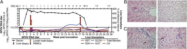
Nonprimate hepacivirus (NPHV) is the closest known relative of hepatitis C virus (HCV) and its study could enrich our understanding of HCV evolution, immunity, and pathogenesis. High seropositivity is found in horses worldwide with ∼ 3% viremic. NPHV natural history and molecular virology remain largely unexplored, however. Here, we show that NPHV, like HCV, can cause persistent infection for over a decade, with high titers and negative strand RNA in the liver. NPHV is a near-universal contaminant of commercial horse sera for cell culture. The complete NPHV 3'-UTR was determined and consists of interspersed homopolymer tracts and an HCV-like 3'-terminal poly(U)-X-tail. NPHV translation is stimulated by miR-122 and the 3'-UTR and, similar to HCV, the NPHV NS3-4A protease can cleave mitochondrial antiviral-signaling protein to inactivate the retinoic acid-inducible gene I pathway. Using an NPHV consensus cDNA clone, replication was not observed in primary equine fetal liver cultures or after electroporation of selectable replicons. However, intrahepatic RNA inoculation of a horse initiated infection, yielding high RNA titers in the serum and liver. Delayed seroconversion, slightly elevated circulating liver enzymes and mild hepatitis was observed, followed by viral clearance. This establishes the molecular components of a functional NPHV genome. Thus, NPHV appears to resemble HCV not only in genome structure but also in its ability to establish chronic infection with delayed seroconversion and hepatitis. This NPHV infectious clone and resulting acute phase sera will facilitate more detailed studies on the natural history, pathogenesis, and immunity of this novel hepacivirus in its natural host.

Keywords: 3′-untranslated region; animal model; equine liver disease; hepatitis C virus; infectious cDNA clone.


Conflict of interest statement

The authors declare no conflict of interest.

Figures

Fig. 1.
Fig. 1.
NPHV RNA titers in individual animal and commercial horse sera. NPHV RNA genome equivalents (GE) per milliliter were measured by qRT-PCR.
Fig. 2.
Fig. 2.
Structure of the NPHV 3′-UTR and constructed consensus clones. (A) Elements and predicted structure of the complete NPHV 3′-UTR. Homopolymer tracts are shown in red. Lengths in nucleotides are indicated. (B) The 3′-UTR regions according to A included in versions of translation reporters. The dotted line indicates the HCV 3′-terminal stem loop. (C) Schematic of the full-length NPHV sequence, as inserted downstream of a T7 promoter in the NZP1 consensus clone. (D) Schematic of the NZP1 subgenomic replicon.
Fig. 3.
Fig. 3.
Translation of NPHV and detection of viral proteins. (A) Representative images of Clone8 cells 1 d posttransfection with RNA from NZP1, NZP1-GNN, or the protease-deficient control, NS3pro(−). (B) Translation from monocistronic translation reporters in Huh-7.5 or E.Derm cells. Relative Renilla (Rluc) to firefly (Fluc) luciferase values are shown after normalization for RNA amounts. Mean and SD are shown. Differences were evaluated by ANOVA. For P values, **P < 0.01, ***P < 0.001, and ****P < 0.0001. N/A, not applicable. (C) Western blot (WB) of lysates from T7-expressing HEK293 cells. Crude horse serum (lot 8211574) was used as a primary antibody. (D) WB of recombinant NS5A(ΔAAH) and the HEK293 lysates from B, using purified polyclonal NPHV NS5A8211574 antibody. The size difference for NS5A is due to absence of the amphipathic α-helix (AAH) in the recombinant protein. (E) NPHV and HCV NS5A antibodies do not cross-react. Lysates from T7-expressing HEK293 cells transfected with pNZP1 (NPHV) or pJc1 (HCV) were used for WB, using NPHV NS5A8211574 antibody or HCV NS5A9E10 antibody. (F) Immunostaining of T7-expressing HEK293 or BHK-J cells with and without NPHV (pNZP1) or HCV (pJ6/JFH1-GNN) detected with NPHV NS5A8211574 or HCV NS5A9E10 antibody.
Fig. 4.
Fig. 4.
Characterization of cell lines and primary EFLCs. (A) miR-122 levels in unmodified cell lines and in cell lines transduced with lentiviruses to express miR-122. (B) miR-122 levels in processed equine fetal liver tissue. Large cell (EFLCs, red) and small cell (blue) preparations before plating and EFLC cultures postplating (days 5–22, orange) were compared with Huh-7.5 cells (white). (C) Equine albumin (eAlb) concentration in EFLCs compared with horse serum and typical human albumin (hAlb) for HFLCs (23). (D) Representative phase-contrast and fluorescence images of EFLC cultures on day 6 postplating. EFLCs were transduced with lentiviruses expressing RFP-NLS-MAVS driven from the human albumin promoter. Error bars in A and B represent SD.
Fig. 5.
Fig. 5.
Course of infection after intrahepatic inoculation of NPHV RNA. (A) In vitro transcribed RNA from the NZP1 consensus clone was inoculated directly into the liver of a horse (Movie S1), and viral RNA, NS3 antibodies, and liver biomarkers were quantified over time. Liver biomarkers are plotted as percentage of reference interval (AST, 199–374 units/L; SDH, 0–11 units/L; GLDH, 1–8 units/L; GGT, 8–29 units/L; bilirubin, 0.5–2.5 mg/dL (total) and 0.1–0.3 mg/dL (direct); creatine kinase, 142–548 units/L). n.d., not done. (B and C) Representative sections of liver portal tract regions (Left) and lobules (Right) before (B) and 17 wk post (C)-NPHV RNA inoculation. Portal lymphocytic infiltrates (blue nuclei) breach the limiting plate (C, Left). Individual necrotic hepatocytes in the limiting plate indicative of piecemeal necrosis are indicated by the arrow. Individual necrotic hepatocytes are dispersed in the lobules (C, Right, thick arrow). Small foci of lymphocytic inflammation are highlighted (C, Right, thin arrow). The asterisk denotes a bile duct.

References

    1. Scheel TK, Simmonds P, Kapoor A. Surveying the global virome: Identification and characterization of HCV-related animal hepaciviruses. Antiviral Res. 2014 doi: 10.1016/j.antiviral.2014.12.014. - DOI - PMC - PubMed
    1. Kapoor A, et al. Characterization of a canine homolog of hepatitis C virus. Proc Natl Acad Sci USA. 2011;108(28):11608–11613. - PMC - PubMed
    1. Lyons S, et al. Viraemic frequencies and seroprevalence of non-primate hepacivirus and equine pegiviruses in horses and other mammalian species. J Gen Virol. 2014;95(Pt 8):1701–1711. - PubMed
    1. Burbelo PD, et al. Serology-enabled discovery of genetically diverse hepaciviruses in a new host. J Virol. 2012;86(11):6171–6178. - PMC - PubMed
    1. Lyons S, et al. Nonprimate hepaciviruses in domestic horses, United Kingdom. Emerg Infect Dis. 2012;18(12):1976–1982. - PMC - PubMed

Publication types

Associated data

LinkOut - more resources