

Excessive growth hormone expression in male GH transgenic mice adversely alters bone architecture and mechanical strength
- PMID: 25646711
- PMCID: PMC4399323
- DOI: 10.1210/en.2014-1572
Excessive growth hormone expression in male GH transgenic mice adversely alters bone architecture and mechanical strength
Abstract
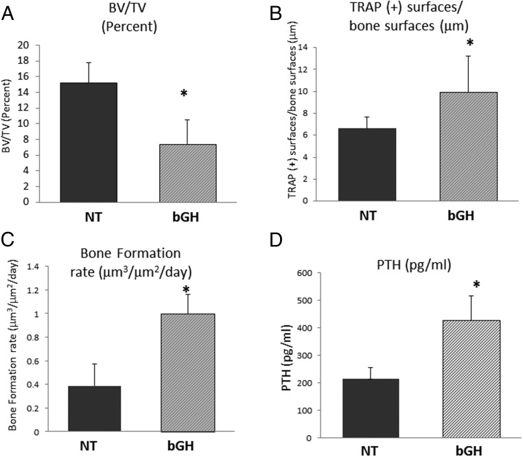
Patients with acromegaly have a higher prevalence of vertebral fractures despite normal bone mineral density (BMD), suggesting that GH overexpression has adverse effects on skeletal architecture and strength. We used giant bovine GH (bGH) transgenic mice to analyze the effects of high serum GH levels on BMD, architecture, and mechanical strength. Five-month-old hemizygous male bGH mice were compared with age- and sex-matched nontransgenic littermates controls (NT; n=16/group). Bone architecture and BMD were analyzed in tibia and lumbar vertebrae using microcomputed tomography. Femora were tested to failure using three-point bending and bone cellular activity determined by bone histomorphometry. bGH transgenic mice displayed significant increases in body weight and bone lengths. bGH tibia showed decreases in trabecular bone volume fraction, thickness, and number compared with NT ones, whereas trabecular pattern factor and structure model index were significantly increased, indicating deterioration in bone structure. Although cortical tissue perimeter was increased in transgenic mice, cortical thickness was reduced. bGH mice showed similar trabecular BMD but reduced trabecular thickness in lumbar vertebra relative to controls. Cortical BMD and thickness were significantly reduced in bGH lumbar vertebra. Mechanical testing of femora confirmed that bGH femora have decreased intrinsic mechanical properties compared with NT ones. Bone turnover is increased in favor of bone resorption in bGH tibia and vertebra compared with controls, and serum PTH levels is also enhanced in bGH mice. These data collectively suggest that high serum GH levels negatively affect bone architecture and quality at multiple skeletal sites.
Figures


Similar articles
-
Lifelong Excess in GH Elicits Sexually Dimorphic Effects on Skeletal Morphology and Bone Mechanical Properties.J Bone Miner Res. 2022 Nov;37(11):2201-2214. doi: 10.1002/jbmr.4699. Epub 2022 Sep 16. J Bone Miner Res. 2022. PMID: 36069368 Free PMC article.
-
Body composition, bone mass and microstructural analysis in GH-transgenic mice reveals that skeletal changes are specific to bone compartment and gender.Growth Horm IGF Res. 2002 Apr;12(2):116-25. doi: 10.1054/ghir.2002.0272. Growth Horm IGF Res. 2002. PMID: 12175649
-
Longitudinal in vivo effects of growth hormone overexpression on bone in transgenic mice.J Bone Miner Res. 2004 May;19(5):802-10. doi: 10.1359/JBMR.040308. Epub 2004 Mar 15. J Bone Miner Res. 2004. PMID: 15068504
-
Administration of growth hormone in selectively protein-deprived rats decreases BMD and bone strength.Bone. 2010 Jun;46(6):1574-81. doi: 10.1016/j.bone.2010.02.015. Epub 2010 Feb 21. Bone. 2010. PMID: 20178866
-
Transgenic over-expression of plasminogen activator inhibitor-1 results in age-dependent and gender-specific increases in bone strength and mineralization.Bone. 2007 Dec;41(6):995-1004. doi: 10.1016/j.bone.2007.08.020. Epub 2007 Aug 17. Bone. 2007. PMID: 17888748 Free PMC article.
Cited by
-
High-resolution-cone beam tomography analysis of bone microarchitecture in patients with acromegaly and radiological vertebral fractures.Endocrine. 2016 Nov;54(2):532-542. doi: 10.1007/s12020-016-1078-3. Epub 2016 Sep 6. Endocrine. 2016. PMID: 27601020
-
Skeletal disorders associated with the growth hormone-insulin-like growth factor 1 axis.Nat Rev Endocrinol. 2022 Jun;18(6):353-365. doi: 10.1038/s41574-022-00649-8. Epub 2022 Mar 14. Nat Rev Endocrinol. 2022. PMID: 35288658 Review.
-
Bone histomorphometry in acromegaly patients with fragility vertebral fractures.Pituitary. 2018 Feb;21(1):56-64. doi: 10.1007/s11102-017-0847-1. Pituitary. 2018. PMID: 29214508
-
Ablation of Hepatic Production of the Acid-Labile Subunit in Bovine-GH Transgenic Mice: Effects on Organ and Skeletal Growth.Endocrinology. 2017 Aug 1;158(8):2556-2571. doi: 10.1210/en.2016-1952. Endocrinology. 2017. PMID: 28475811 Free PMC article.
-
Effects of GH/IGF axis on bone and cartilage.Mol Cell Endocrinol. 2021 Jan 1;519:111052. doi: 10.1016/j.mce.2020.111052. Epub 2020 Oct 14. Mol Cell Endocrinol. 2021. PMID: 33068640 Free PMC article. Review.
References
-
- Ohlsson C, Bengtsson BA, Isaksson OG, Andreassen TT, Slootweg MC. Growth hormone and bone. Endocr Rev. 1998;19:55–79. - PubMed
-
- Ohlsson C, Nilsson A, Isaksson OG, Lindahl A. Effect of growth hormone and insulin-like growth factor-I on DNA synthesis and matrix production in rat epiphyseal chondrocytes in monolayer culture. J Endocrinol. 1992;133:291–300. - PubMed
-
- Tritos NA, Biller BM. Growth hormone and bone. Curr Opin Endocrinol Diabetes Obes. 2009;16:415–422. - PubMed
Publication types
MeSH terms
Substances
LinkOut - more resources
Full Text Sources
Other Literature Sources
Medical
Molecular Biology Databases
Research Materials
