. 2015 Feb 3;10(2):e0117225.
doi: 10.1371/journal.pone.0117225. eCollection 2015.

Nanoscale imaging of caveolin-1 membrane domains in vivo

Affiliations

Nanoscale imaging of caveolin-1 membrane domains in vivo

Kristin A Gabor et al. PLoS One. .

Abstract

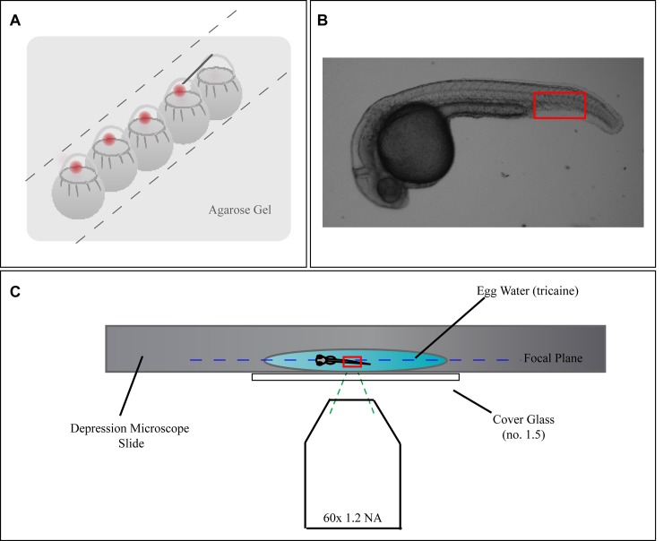
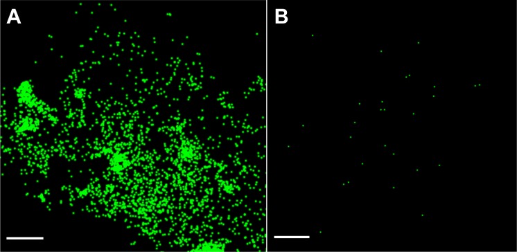
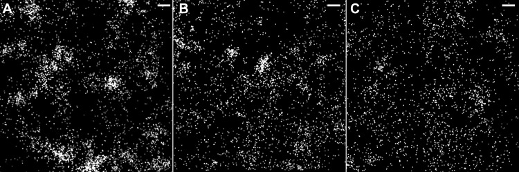
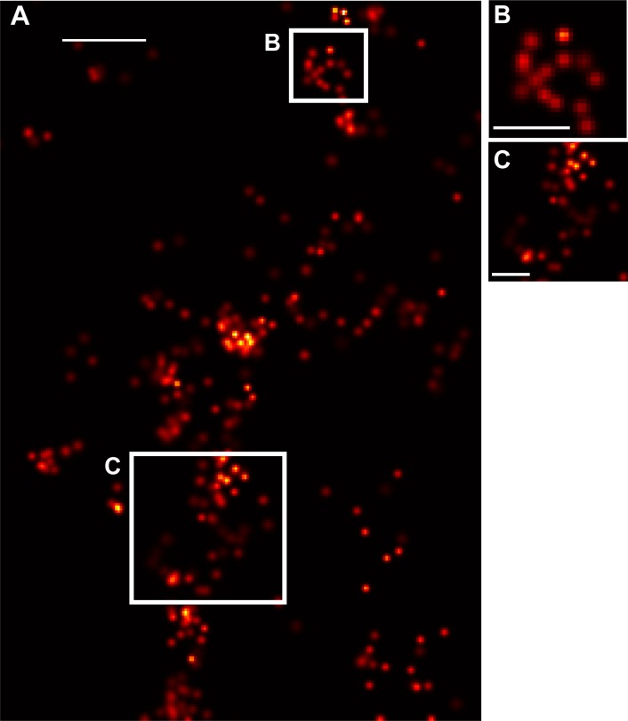
Light microscopy enables noninvasive imaging of fluorescent species in biological specimens, but resolution is generally limited by diffraction to ~200-250 nm. Many biological processes occur on smaller length scales, highlighting the importance of techniques that can image below the diffraction limit and provide valuable single-molecule information. In recent years, imaging techniques have been developed which can achieve resolution below the diffraction limit. Utilizing one such technique, fluorescence photoactivation localization microscopy (FPALM), we demonstrated its ability to construct super-resolution images from single molecules in a living zebrafish embryo, expanding the realm of previous super-resolution imaging to a living vertebrate organism. We imaged caveolin-1 in vivo, in living zebrafish embryos. Our results demonstrate the successful image acquisition of super-resolution images in a living vertebrate organism, opening several opportunities to answer more dynamic biological questions in vivo at the previously inaccessible nanoscale.


Conflict of interest statement

Competing Interests: The authors have declared that no competing interests exist.

Figures

Figure 1
Figure 1. Widefield FPALM Enables Penetration Into Sample.
Schematic of experimental setup. A) Diagram demonstrating how Casper fms embryos were injected at the 1-cell stage with linearized DNA plus phenol red indicator. B) A widefield image of a 24 hpf zebrafish embryo. Outlined in red is the region of the tail that FPALM images were acquired at, as it is the thinnest part of the fish and also has minimum autofluorescence. C) A 24 hpf embryo was placed in a microscope slide with a single shallow depression, immersed in a drop of non-lethal tricaine (solubilized in egg water). A cover slide (#1.5) was placed over the slide, covering the embryo and the coverslip was mounted on a microscopy setup suitable for FPALM.
Figure 2
Figure 2. Identification of Single Molecules in vivo.
The image demonstrates the ability to identify and localize single molecules above background in a living zebrafish embryo. Each image shows a single frame during image acquisition of a cell in an embryo during localization analysis. Casper fms zebrafish were injected at the 1-cell stage with plasmid DNA and images were captures 24 h post injection (i.e. 24 hpf). A single molecule is shown over consecutive frames, where the molecule is not present, turned on during the frame in which it is activated and localized, and then presumably has photobleached during acquisition of the subsequent frames. Note that the single molecule image appears to be approximately the width expected from the diffraction limit. Brightness and contrast were linearly adjusted in ImageJ for all frames for presentation purposes. Scale bar, 500 nm.
Figure 3
Figure 3. Cav-1 Specificity of Localized Molecules Demonstrates In vivo Visualization of Caveolae.
Casper fms zebrafish were injected at the 1-cell stage with plasmid DNA and images were captured 24 h post injection (i.e. 24 hpf). A) Control MO + Cav-1b dendra2 demonstrates single cav1 molecules are localized. B) Image rendered from Cav1b MO+Cav-1b dendra2 embryos with knockdown of Cav1b expression demonstrates that the molecules seen in control are in fact Cav1b molecules. Scale bar, 1 μm.
Figure 4
Figure 4. Widefield FPALM Enables Penetration Into Sample.
Single molecule localization with FPALM has greater Z-depth into the sample than TIRF microscopy. Images show a region of a zebrafish cell after localization of single molecules. Casper fms zebrafish were injected at the 1-cell stage with plasmid DNA and images were captured 24 h post injection (i.e. 24 hpf). (A) Image at focal plane of fish with high dendra2 expression (0 μm/10,046 molecules); (B) Image shown 5 μm above the focal plane depicted in A (9319 molecules); (C) Image shown 5 μm above the focal plane depicted in B, or 10 μm above the focal plane depicted in A (4434 molecules). Scale bar, 500 nm.
Figure 5
Figure 5. Caveolae-like structures evident at cell membrane.
(A) Shown are molecules along the membrane of one cell representative of the experiment, where clusters of Cav-1 molecules can be seen. Magnifications (B, C) of the regions marked by the white boxes in A show structures indicative of caveolae. Scale bar for A, 250 nm. Scale bar for B-C, 125 nm.

Similar articles

Cited by

References

    1. Rust MJ, Bates M, Zhuang X (2006) Sub-diffraction-limit imaging by stochastic optical reconstruction microscopy (STORM). Nat Methods 3: 793–795. - PMC - PubMed
    1. Hess ST, Girirajan TP, Mason MD (2006) Ultra-high resolution imaging by fluorescence photoactivation localization microscopy. Biophys J 91: 4258–4272. - PMC - PubMed
    1. Betzig E, Patterson GH, Sougrat R, Lindwasser OW, Olenych S, et al. (2006) Imaging intracellular fluorescent proteins at nanometer resolution. Science 313: 1642–1645. - PubMed
    1. Hell SW, Wichmann J (1994) Breaking the diffraction resolution limit by stimulated emission: stimulated-emission-depletion fluorescence microscopy. Opt Lett 19: 780–782. - PubMed
    1. Hess ST, Gould TJ, Gudheti MV, Maas SA, Mills KD, et al. (2007) Dynamic clustered distribution of hemagglutinin resolved at 40 nm in living cell membranes discriminates between raft theories. Proc Natl Acad Sci U S A 104: 17370–17375. - PMC - PubMed

Publication types

LinkOut - more resources