. 2015 Feb 3;13(2):e1002053.
doi: 10.1371/journal.pbio.1002053. eCollection 2015 Feb.

FAX1, a novel membrane protein mediating plastid fatty acid export

Affiliations

FAX1, a novel membrane protein mediating plastid fatty acid export

Nannan Li et al. PLoS Biol. .

Abstract

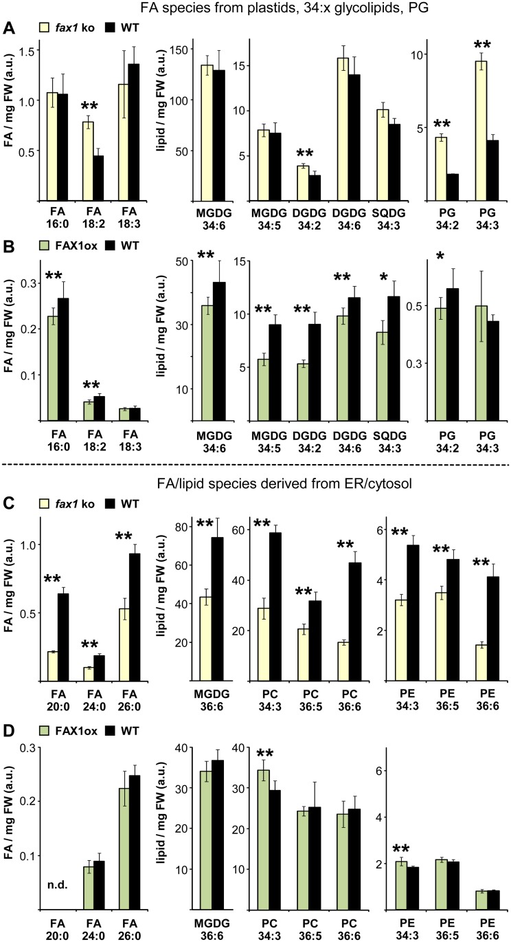
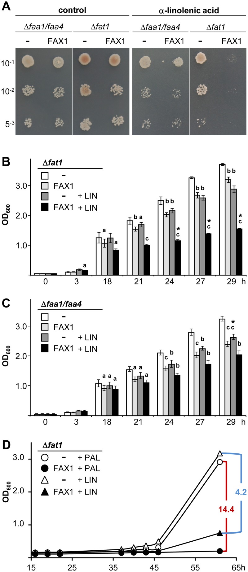
Fatty acid synthesis in plants occurs in plastids, and thus, export for subsequent acyl editing and lipid assembly in the cytosol and endoplasmatic reticulum is required. Yet, the transport mechanism for plastid fatty acids still remains enigmatic. We isolated FAX1 (fatty acid export 1), a novel protein, which inserts into the chloroplast inner envelope by α-helical membrane-spanning domains. Detailed phenotypic and ultrastructural analyses of FAX1 mutants in Arabidopsis thaliana showed that FAX1 function is crucial for biomass production, male fertility and synthesis of fatty acid-derived compounds such as lipids, ketone waxes, or pollen cell wall material. Determination of lipid, fatty acid, and wax contents by mass spectrometry revealed that endoplasmatic reticulum (ER)-derived lipids decreased when FAX1 was missing, but levels of several plastid-produced species increased. FAX1 over-expressing lines showed the opposite behavior, including a pronounced increase of triacyglycerol oils in flowers and leaves. Furthermore, the cuticular layer of stems from fax1 knockout lines was specifically reduced in C29 ketone wax compounds. Differential gene expression in FAX1 mutants as determined by DNA microarray analysis confirmed phenotypes and metabolic imbalances. Since in yeast FAX1 could complement for fatty acid transport, we concluded that FAX1 mediates fatty acid export from plastids. In vertebrates, FAX1 relatives are structurally related, mitochondrial membrane proteins of so-far unknown function. Therefore, this protein family might represent a powerful tool not only to increase lipid/biofuel production in plants but also to explore novel transport systems involved in vertebrate fatty acid and lipid metabolism.


Conflict of interest statement

The authors have declared that no competing interests exist.

Figures

Fig 1
Fig 1. Plant FAX and human Tmemb_14 proteins.
(A) Arabidopsis At-FAX1 (At3g57280, 226 amino acids [aa]) is accessible at the ARAMEMNON database [18], the sequence for pea Ps-FAX1 (232 aa) was deposited at National Center for Biotechnology Information (NCBI), GenBank acc. no. KF981436. Predicted chloroplast targeting peptides (ChloroP; http://www.cbs.dtu.dk/services/ChloroP), with 33 aa and 39 aa for At-FAX1 and Ps-FAX1, respectively, are marked with red triangles. The Tmemb_14 domain (Pfam|PF03647) of At-FAX1, including the four conserved hydrophobic domains, is indicated. Identical amino acids (49%) are shaded in black, hydrophobic α-helices (ARAMEMNON) are boxed in green, and peptides used for generation of antisera are indicated by red lines. (B) Members of the FAX/Tmemb_14 family in Arabidopsis. Whereas At-FAX1-At-FAX4 are predicted to be in plastids, At-FAX5-At-FAX7 most likely localize to membranes of the secretory pathway. Hydrophobic α-helices (black squares) and subcellular localization are depicted according to ARAMEMNON. Predicted chloroplast targeting peptides (ChloroP) are marked with red triangles. (C) At-FAX1 and Ps-FAX1 [sequence starting with Tmemb_14 domain, see (A)] in comparison to At-FAX6 (At3g20510) and human proteins of the Tmemb_14 superfamily: TMEM14A (Q9Y6G1) and TMEM14C (Q9POS9). Arabidopsis genome initiative (AGI) codes and InterPro accession numbers in brackets. Whereas At-FAX1 and Ps-FAX1 are slightly more similar to TMEM14C (17% identical, 35% similar aa), At-FAX6 shares 28% identical aa with both proteins TMEM14A and 14C. Predicted hydrophobic α-helices (ARAMEMNON) are boxed in green; α-helices in TMEM14A, 14C [14] are boxed in blue.
Fig 2
Fig 2. FAX1, a chloroplast IE protein of the Tmemb_14 family.
(A, B) Structural models and alignment (right) of the mature At-FAX1 / human TMEM14C (A) and of At-FAX6 / human TMEM14A (B) proteins. Membrane-spanning and amphiphilic α-helices of FAX and of TMEM14 are depicted in green/yellow-green and blue/light blue, respectively. Please note that At-FAX1 contains an additional N-terminal stretch that most likely folds into another α-helix (gray). First and last amino acid residues are indicated. (C) In vivo green fluorescent protein (GFP)-targeting. Arabidopsis leaf protoplasts were transiently transformed with constructs for FAX1- and NiCo-GFP (chloroplast IE marker; [19]). Images show GFP and chlorophyll fluorescence, as well as an overlay of both (bar = 5 μm). (D) Immunoblot analysis of FAX1 in chloroplast subfractions. Equal protein amounts (5μg) of pea chloroplast outer envelope (OE), inner envelope (IE), stroma (str), thylakoids (thy), as well as 2.5μg protein of Arabidopsis microsomal membranes (mm) and chloroplast envelopes (env) were separated by SDS-PAGE and subjected to immunoblot analysis using antibodies directed against Ps-FAX1 and At-FAX1. Antisera against marker proteins LSU (str), LHCP (thy), NiCo (IE), OEP16.1 (OE), and TPR7 (mm, see [20]) were used as controls. For LSU and LHCP less protein (1μg, 0.2μg, respectively) was loaded. Numbers indicate molecular mass of proteins in kDa.
Fig 3
Fig 3. Mutation of FAX1 in Arabidopsis affects plant growth.
(A) 30-day-old plants of FAX1 mutants and wild-type lines. fax1–1, fax1–2: homozygous knockout lines for FAX1; Col-0, WT2: wild-type FAX1 alleles, WT2 represents FAX1 wild-type progeny, segregated from heterozygous fax1–2; Co#7, Co#54: fax1–2 knockout complementation lines; ox#2, ox#4: FAX1 over-expressing lines in Col-0 background. (B) 7-week-old flowering plants of FAX1 mutants and wild type as specified in (A). (Inset) Comparison of primary inflorescence stalks (bottom parts of 2nd internode) from fax1–2, Col-0 and ox#2 plants. (C) Siliques produced by FAX1 mutants and wild-type lines as depicted in (B).
Fig 4
Fig 4. FAX1 function is essential for pollen cell wall assembly.
Pictures of flowers, anthers, and mature pollen of 5-week-old fax1–2 knockout and Col-0 wild-type plants (left and right panels, respectively). (A) Development of flower buds and young siliques. (B) Close-up of opened flowers. Arrowheads: non-pollinated stigma in fax1–2; arrows: anthers with released pollen in Col-0. (C) Cross sections of mature, dehisced anthers (light microscopy, bar = 50 μm). Black and white arrowheads in fax1–2: fully opened pollen sacks, and dark material covering endothecium/locule boundary, respectively. en: endothecium cells of anthers. (D), (E), (F) Transmission electron microscopic (TEM) pictures of anther cell/pollen grain intersections (D, bar = 5 μm; E, bar = 1μm) and pollen cell wall (F, bar = 500 nm) at mature tricellular pollen stages. White arrowheads in fax1–2: debris material sticking to pollen grains. en: endothecium cell; e: exine layer with eb: bacula, et: tectum structures; en: nexine layer (black arrowheads), i: intine layer; po: cytosol of pollen grain; pl: plastid; try: tryphine pollen coat.
Fig 5
Fig 5. FAX1 affects cell wall size and cuticular wax composition.
Stem tissue (1 cm at the bottom of the second internode) of the primary inflorescence stalk of 5-week-old fax1–1 knockout, Col-0 wild-type, and FAX1 over-expressor line ox#2 (left, middle, and right panels, respectively). (A) Light microscopic pictures of stem epidermal cells (bar = 10μm). (B) Transmission electron microscopic pictures of cell walls from stem epidermal cells (bar = 500 nm). (C) Close-ups of cell wall / cuticular layer boundary from cells in (B) (TEM, bar = 200 nm). cut: cuticular layer; cw: cell wall; cyt: cytosol. (D) C29 ketone wax coverage in μg per cm2 of stem surface from FAX1 mutant and wild-type lines (n = 3–7 ± SD; n = 12 for Col-0). For each replicate, three to four stem sections between internode 2–4 of 7-week-old, mature flowering plants were pooled. Asterisks indicate highly significant different contents (**: p < 0.001, Student’s t-test) when compared with Col-0 (for numerical values, see S1A Data).
Fig 6
Fig 6. Plastid FAX1 impacts cellular FA/lipid homeostasis in leaves.
Free fatty acid (FA) and polar lipid species were determined in leaf tissue of 7-week-old, mature flowering plants. Data (arbitrary units) are expressed relative to the internal standard (PC 34:0) and normalized to mg fresh weight (FW). For overview, we depict representatives of the most abundant species and those significantly different in FAX1 mutants. A complete dataset with details on analysis is given in S1 Table; values in mol % are listed in S2 Table. Levels in FAX1 mutants significantly different to wild type are indicated by asterisks (Student’s t-test, *: p < 0.05, **: p < 0.01). For a better resolution of differential patterns, y-axes are scaled differently. C16–18 FAs are exclusively and glycolipids in (A) and (B) are mainly synthesized in plastids (for details, see Discussion). Please note that the diacylglycerol backbone for the “34”-glycolipids can originate from prokaryotic (from plastids) and eukaryotic (from the ER) phospholipid precursors, respectively. C20–26 FAs and phospholipids in (C) and (D), as well as precursors for “36”- glycolipids, are only produced outside plastids in the cytosol and/or ER. (A), (C) Free FA and lipid levels in caulinary leaves of fax1–1, fax1–2 knockout and Col-0, WT2 wild-type lines (yellow and black bars, respectively) were determined by UPLC-Orbitrap MS [23]. Mean values (n = 6 ± SD), averaged over both fax1 knockouts and both wild types, respectively, are shown. (B), (D) Free FA and lipid content (n = 6–12 ± SD) in caulinary leaves of the FAX1 over-expressing line ox#4 and Col-0 wild type (green and black bars, respectively) was measured by UPLC-qTOF MS [24]. Please note that in comparison to the dataset in (A) and (C), usage of a different mass spectrometer results in different scaling of the relative values. DGDG: digalactosyl-diacylglycerol; FA: fatty acid; MGDG: monogalactosyl-diacylglycerol; n.d.: not determined; PC: phosphatidyl-choline; PE: phosphatidyl-ethanolamine; PG: phosphatidyl-glycerol; SQDG: sulphoquinovosyl-diacylglycerol.
Fig 7
Fig 7. Plastid FAX1 impacts TAG storage lipid homeostasis.
Triacylglycerol (TAG) oils were determined in tissues of 7-week-old, mature flowering plants (compare Fig. 6). Data (arbitrary units) are expressed relative to the internal standard (PC 34:0) and normalized to mg fresh weight (FW). For overview, we selected representatives of the most abundant species and those significantly different in FAX1 mutants. A complete dataset with details on analysis is given in S1 Table; values in mol % are listed in S4 Table. Levels in FAX1 mutants significantly different to wild type are indicated (Student’s t-test, *: p < 0.05, **: p < 0.01). We show high and low abundant TAGs (left and right graphs, respectively); thus for better resolution of differential patterns, y-axes are scaled differently. (A) TAG levels in caulinary leaves of fax1–1, fax1–2 knockout and Col-0, WT2 wild-type lines (yellow and black bars, respectively). Mean values (n = 4–6 ± SD), averaged over both fax1 knockouts and both wild types, respectively, are shown. (B) TAG content in caulinary leaves of the FAX1 over-expressing line ox#4 (green bars; n = 6–12 ± SD) and Col-0 wild type (black bars, n = 5–10 ± SD). (C) TAG levels in flowers of fax1–1, fax1–2 knockout and Col-0, WT2 wild-type lines (yellow and black bars, respectively). Mean values (n = 6 ± SD), averaged over both fax1 knockouts and both wild types, respectively, are shown. (D) TAG content (n = 7–12 ± SD, for TAG 58:6: n = 5) in flowers of the FAX1 over-expressing line ox#4 and Col-0 wild type (green and black bars, respectively). Please note that in comparison to the dataset for fax1 knockouts in (A) and (C), usage of a different mass spectrometer for FAX1ox data in (B) and (D) results in different scaling of the relative values (compare Fig. 6).
Fig 8
Fig 8. FAX1 mediates FA-transport in yeast.
The empty plasmid pDR195 (-) and the mature At-FAX1 cDNA in pDR195 (FAX1) were introduced into faa1/faa4 and fat1 yeast mutants, respectively. (A) Serial dilutions (OD600 of 10–1, 10–2, and 5–3) of rapidly growing yeast cells on SD-ura plates (0.1% glucose, 1% tergitol). Control plates (left) in comparison to plates with 3.6 mM α-linolenic acid (C18:3; right). (B), (C) Growth of fat1 (B) and faa1/faa4 (C) cells in liquid SD-ura [see (A)]. The OD600 was monitored within 29 h incubation at 30°C. White and light gray bars: growth of pDR195 (-) and matFAX1/pDR195 (FAX1) at control conditions. Gray and black bars: growth of (-) and FAX1 in presence of 3.6 mM α-linolenic acid (LIN). Error bars depict SD (n = 4), for numerical values, see S1B Data. a (p < 0.05), b (p < 0.005), c (p < 0.0005), * (p < 0.00005) indicate significantly different values (Student’s t-test) compared to (-) cells without and with LIN, respectively. (D) Representative growth curves of fat1 cells in liquid SD-ura (2% glucose, 0.5% Brij 58, 0.7% KH2PO4) in the presence of 10μM cerulenin (CER, inhibitor of FA-biosynthesis) and 100 μM palmitic acid (PAL, C16:0) or 100 μM α-linolenic acid (LIN, C18:3). Assays were performed according to [26,27]. For comparison with stearic (C18:0) and oleic acid (C18:1), see S4 Fig. White and black circles and triangles: growth of pDR195 (-) and matFAX1/pDR195 (FAX1) cells supplemented with PAL and LIN, respectively. Red/blue lines and numbers indicate maximal difference of cell density ratios for (-) versus FAX1 (compare S1B Data).

Comment in

  • Flagging the fatty acid ferryman.
    Roberts RG. Roberts RG. PLoS Biol. 2015 Feb 3;13(2):e1002054. doi: 10.1371/journal.pbio.1002054. eCollection 2015 Feb. PLoS Biol. 2015. PMID: 25646782 Free PMC article.

Similar articles

Cited by

References

    1. Li-Beisson Y, Shorrosh B, Beisson F, Andersson MX, Arondel V, et al. (2013) Acyl-lipid metabolism Arabidopsis Book 11: e0161 10.1199/tab.0161 - DOI - PMC - PubMed
    1. Ariizumi T, Toriyama K (2011) Genetic Regulation of Sporopollenin Synthesis and Pollen Exine Development. Annu. Rev. Plant Biol. 62: 437–460. 10.1146/annurev-arplant-042809-112312 - DOI - PubMed
    1. Wang Z, Benning C (2012) Chloroplast lipid synthesis and lipid trafficking through ER-plastid membrane contact sites. Biochem Soc Trans 40: 457–463. 10.1042/BST20110752 - DOI - PubMed
    1. Koo AJK, Ohlrogge JB, Pollard M (2004) On the export of fatty acids from the chloroplast. J Biol Chem 279: 16101–16110. - PubMed
    1. Von Berlepsch S, Kunz HH, Brodesser S, Fink P, Marin K, et al. (2012) The Acyl-Acyl Carrier Protein Synthetase from Synechocystis sp PCC 6803 Mediates Fatty Acid Import. Plant Physiol 159: 606–617. 10.1104/pp.112.195263 - DOI - PMC - PubMed

Publication types

MeSH terms

Associated data