. 2015 Feb 3;11(2):e1004639.
doi: 10.1371/journal.ppat.1004639. eCollection 2015 Feb.

Crystal structures of the carboxyl cGMP binding domain of the Plasmodium falciparum cGMP-dependent protein kinase reveal a novel capping triad crucial for merozoite egress

Affiliations

Crystal structures of the carboxyl cGMP binding domain of the Plasmodium falciparum cGMP-dependent protein kinase reveal a novel capping triad crucial for merozoite egress

Jeong Joo Kim et al. PLoS Pathog. .

Abstract

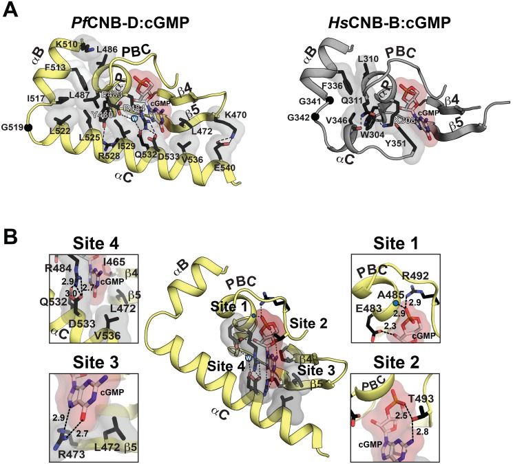
The Plasmodium falciparum cGMP-dependent protein kinase (PfPKG) is a key regulator across the malaria parasite life cycle. Little is known about PfPKG's activation mechanism. Here we report that the carboxyl cyclic nucleotide binding domain functions as a "gatekeeper" for activation by providing the highest cGMP affinity and selectivity. To understand the mechanism, we have solved its crystal structures with and without cGMP at 2.0 and 1.9 Å, respectively. These structures revealed a PfPKG-specific capping triad that forms upon cGMP binding, and disrupting the triad reduces kinase activity by 90%. Furthermore, mutating these residues in the parasite prevents blood stage merozoite egress, confirming the essential nature of the triad in the parasite. We propose a mechanism of activation where cGMP binding allosterically triggers the conformational change at the αC-helix, which bridges the regulatory and catalytic domains, causing the capping triad to form and stabilize the active conformation.


Conflict of interest statement

The authors have declared that no competing interests exist. The authors also have no affiliation or competing interest with the Eli Lilly company. Eli Lilly has no role in designing and conducting this study. This does not alter our adherence to all PLOS Pathogens policies on sharing data and materials.

Figures

Fig 1
Fig 1. Domain organization and overall structures of PfCNB-D.
(A) Domain organization of PfPKG and sequence alignment between PfCNB-D and HsCNB-B (Human PKG I). Identical residues are highlighted in yellow and the capping residues in both proteins are highlighted in red. The capping triad residues are also marked with arrows. (B) cGMP and cAMP affinities of PfCNB domains. Competition FP curves for cGMP are shown on the left and EC50 values on the right. (C) Overall structure of PfCNB-D without cGMP. The secondary structure elements are labeled. The phosphate binding cassette (PBC) is colored in yellow, the αB and αC helices in light cyan and blue, the N-terminal helices in light green and the β-barrel in gray. The N- and C-termini are labeled with their corresponding residue number seen in the final model. The sulfate ion co-crystallized with the protein is colored with its sulfur in yellow and oxygen in red. (D) Overall structure of the PfCNB-D:cGMP complex. The structure is shown with the same color scheme as above except for cGMP. The cGMP is colored by atom type (carbon, white; nitrogen, blue; oxygen, red; and phosphorus, orange). All structure images were generated using PyMOL (Delano Scientific).
Fig 2
Fig 2. Structural comparison between PfCNB-D and CNB-B and cGMP binding pocket of PfCNB-D.
(A) The cGMP pockets PfCNB-D and CNB-B from human PKG Iβ (PDB code: 4KU7) are shown. The cGMP pocket of PfCNB-D is colored in yellow (left) and the pocket of PKG Iβ CNB-B in gray (right). Key residues that stabilize the C-helix including the capping residues are shown with transparent surface in the following color theme: side chain carbon, black; oxygen, red; nitrogen. A water molecule captured between E483, R484, and Q532 is shown as a blue sphere. The C atoms of glycine residues located between at the αB and αC helices are shown as black spheres. Hydrogen bonds are shown as dotted lines. (B) Detailed interactions between PfCNB-D and cGMP. Zoomed in views for each cGMP binding site are shown on either side. The backbone amide of A485 is marked with a blue dot. The individual cGMP interacting residues are shown with the following color theme: side chain carbon, black; oxygen, red; nitrogen, blue. The residues binds cGMP with VDW contacts including the capping residues are shown with transparent surface. Hydrogen bonds are shown as dotted lines with their distances in Å units.
Fig 3
Fig 3. Role of the unique capping triad at PfCNB-D in PfPKG activation and Plasmodium parasite life cycle.
(A) Role of the capping motif forming residues in kinase activation. Individual curves with error bars denoting standard error of mean are shown on the left and corresponding Ka values for WT and capping triad mutants are shown on the right. Each data curve was normalized by designating the lowest value of the data set as 0% and the highest value as 100%. (B) The specific activities of the WT and mutants at 10 μM cGMP are shown as bar graphs with error bars denoting standard error of mean. (C) Immunoblot showing co-expression of endogenous PfPKG and ectopic PfPKG-HA in transgenic schizonts. 3D7/attB is the parental line and 3D7/PKG-HA is a previously established line where the endogenous PfPKG gene has been HA-tagged. Blots were incubated with anti-HA, anti-human PKG, and anti-PfGAPDH as a loading control. Note that the anti-human PKG antibody does not react with HA-tagged PKG (lane 2) because the free carboxyl terminus is crucial for antibody binding. This allows the differentiation between endogenous and HA-tagged PKG. (D) Growth of the transgenic lines in the presence of 1 μM compound 2 over 8 days. The established compound 2-resistant line 3D7/T618Q was included as a positive control. (E) Late trophozoites/early schizonts of the three transgenics were cultured for 12 hours in the presence of 1 μM compound 2 (lower panels) or DMSO (upper panels) and parasite development examined on Giemsa-stained blood smears. (F) Quantification of (E). >1000 cells were counted for each culture and condition and ring stage parasitaemia determined. Data represent the mean of three experiments (error bars = SD). Dark grey bars are DMSO controls, light grey bars 1 μM compound 2 treated samples. For each sample, parasitaemia was adjusted to make the DMSO control 10% to eliminate variability from differences in parasitaemia between experiments. (G) Sequence alignment of apicomplexan PKGs. Conserved residues are shaded in yellow (identical), in green (functionally similar), and in gray (identical in >66%). The capping triad residues are typed in red and marked with arrows. Toxoplasma gondii PKG, TgPKG; Theileria orientalis PKG, ToPKG; Eimeria tenella PKG, EtPKG; Babesia bovis PKG, BbPKG, and Cryptosporidium hominis, ChPKG.
Fig 4
Fig 4. Structural comparison between the apo- and cGMP bound PfCNB-D.
The apo and PfCNB-D:cGMP complex structures are aligned at the β-barrel region (not colored). The helical subdomain of the apo structure is colored in light cyan and that of the cGMP complex structure in yellow.
Fig 5
Fig 5. Conformational changes upon cGMP binding.
Conformational changes are depicted in a step-wise fashion. The hydrophobic core, hinge, and cap forming residues are shown with transparent surface. Their side chain carbons are colored in black, oxygen in red, and nitrogen in blue.. (A) Step 1: PBC assuming a closed conformation upon cGMP binding. (B) Step 2: A cogwheel-like motion between the αP- and αB-helices allowing the αB-helix to move toward the PBC. Zoomed in views highlight changes in hydrophobic interactions. (C) Step 3: The N3A motif moving away from the PBC. (D) Step 4: The αC-helix shielding the bottom of the cGMP pocket and enabling the capping triad formation.

Similar articles

Cited by

References

    1. Cowman AF, Crabb BS (2006) Invasion of red blood cells by malaria parasites. Cell 124: 755–766. - PubMed
    1. Guttery DS, Holder AA, Tewari R (2012) Sexual development in Plasmodium: lessons from functional analyses. PLoS Pathog 8: e1002404 10.1371/journal.ppat.1002404 - DOI - PMC - PubMed
    1. Doerig C, Abdi A, Bland N, Eschenlauer S, Dorin-Semblat D, et al. (2010) Malaria: targeting parasite and host cell kinomes. Biochim Biophys Acta 1804: 604–612. 10.1016/j.bbapap.2009.10.009 - DOI - PubMed
    1. Baker DA (2011) Cyclic nucleotide signalling in malaria parasites. Cell Microbiol 13: 331–339. 10.1111/j.1462-5822.2010.01561.x - DOI - PubMed
    1. Hopp CS, Bowyer PW, Baker DA (2012) The role of cGMP signalling in regulating life cycle progression of Plasmodium. Microbes Infect 14: 831–837. 10.1016/j.micinf.2012.04.011 - DOI - PMC - PubMed

Publication types

Substances

LinkOut - more resources