Mesenchymal stem cells are enriched in head neck squamous cell carcinoma, correlates with tumour size and inhibit T-cell proliferation
- PMID: 25647013
- PMCID: PMC4333504
- DOI: 10.1038/bjc.2015.15
Mesenchymal stem cells are enriched in head neck squamous cell carcinoma, correlates with tumour size and inhibit T-cell proliferation
Abstract
Background: Cancer is a multifactorial disease not only restricted to transformed epithelium, but also involving cells of the immune system and cells of mesenchymal origin, particularly mesenchymal stem cells (MSCs). Mesenchymal stem cells contribute to blood- and lymph- neoangiogenesis, generate myofibroblasts, with pro-invasive activity and may suppress anti-tumour immunity.
Methods: In this paper, we evaluated the presence and features of MSCs isolated from human head neck squamous cell carcinoma (HNSCC).
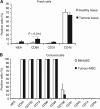
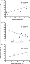
Results: Fresh specimens of HNSCC showed higher proportions of CD90+ cells compared with normal tissue; these cells co-expressed CD29, CD105, and CD73, but not CD31, CD45, CD133, and human epithelial antigen similarly to bone marrow-derived MSCs (BM-MSCs). Adherent stromal cells isolated from tumour shared also differentiation potential with BM-MSCs, thus we named them as tumour-MSCs. Interestingly, tumour-MSCs showed a clear immunosuppressive activity on in vitro stimulated T lymphocytes, mainly mediated by indoelamine 2,3 dioxygenase activity, like BM-MSCs. To evaluate their possible role in tumour growth in vivo, we correlated tumour-MSC proportions with neoplasm size. Tumour-MSCs frequency directly correlated with tumour volume and inversely with the frequency of tumour-infiltrating leukocytes.
Conclusions: These data support the concept that tumour-MSCs may favour tumour growth not only through their effect on stromal development, but also by inhibiting the anti-tumour immune response.
Figures


Similar articles
-
Adenosine metabolism of human mesenchymal stromal cells isolated from patients with head and neck squamous cell carcinoma.Immunobiology. 2017 Jan;222(1):66-74. doi: 10.1016/j.imbio.2016.01.013. Epub 2016 Jan 30. Immunobiology. 2017. PMID: 26898925
-
PDGF-AA mediates mesenchymal stromal cell chemotaxis to the head and neck squamous cell carcinoma tumor microenvironment.J Transl Med. 2016 Dec 8;14(1):337. doi: 10.1186/s12967-016-1091-6. J Transl Med. 2016. PMID: 27931212 Free PMC article.
-
A novel therapeutic approach targeting PD-L1 in HNSCC and bone marrow-derived mesenchymal stem cells hampers pro-metastatic features in vitro: perspectives for blocking tumor-stroma communication and signaling.Cell Commun Signal. 2025 Feb 10;23(1):74. doi: 10.1186/s12964-025-02073-7. Cell Commun Signal. 2025. PMID: 39930439 Free PMC article.
-
Role of Mesenchymal Stem/Stromal Cells in Head and Neck Cancer-Regulatory Mechanisms of Tumorigenic and Immune Activity, Chemotherapy Resistance, and Therapeutic Benefits of Stromal Cell-Based Pharmacological Strategies.Cells. 2024 Jul 28;13(15):1270. doi: 10.3390/cells13151270. Cells. 2024. PMID: 39120301 Free PMC article. Review.
-
Resident and bone marrow-derived mesenchymal stem cells in head and neck squamous cell carcinoma.Oral Oncol. 2010 May;46(5):336-42. doi: 10.1016/j.oraloncology.2010.01.016. Epub 2010 Mar 9. Oral Oncol. 2010. PMID: 20219413 Review.
Cited by
-
ZIF-8 as a pH-Responsive Nanoplatform for 5-Fluorouracil Delivery in the Chemotherapy of Oral Squamous Cell Carcinoma.Int J Mol Sci. 2024 Aug 27;25(17):9292. doi: 10.3390/ijms25179292. Int J Mol Sci. 2024. PMID: 39273239 Free PMC article.
-
Role of cancer-associated mesenchymal stem cells in the tumor microenvironment: A review.Tzu Chi Med J. 2022 Aug 1;35(1):24-30. doi: 10.4103/tcmj.tcmj_138_22. eCollection 2023 Jan-Mar. Tzu Chi Med J. 2022. PMID: 36866340 Free PMC article. Review.
-
Engineered sTRAIL-armed MSCs overcome STING deficiency to enhance the therapeutic efficacy of radiotherapy for immune checkpoint blockade.Cell Death Dis. 2022 Jul 14;13(7):610. doi: 10.1038/s41419-022-05069-0. Cell Death Dis. 2022. PMID: 35835756 Free PMC article.
-
Mesenchymal stem cells: From regeneration to cancer.Pharmacol Ther. 2019 Aug;200:42-54. doi: 10.1016/j.pharmthera.2019.04.005. Epub 2019 Apr 16. Pharmacol Ther. 2019. PMID: 30998940 Free PMC article. Review.
-
Cellular Barriers after Extravasation: Leukocyte Interactions with Polarized Epithelia in the Inflamed Tissue.Mediators Inflamm. 2016;2016:7650260. doi: 10.1155/2016/7650260. Epub 2016 Jan 28. Mediators Inflamm. 2016. PMID: 26941485 Free PMC article. Review.
References
-
- Annunziato F, Romagnani S. The transient nature of the Th17 phenotype. Eur J Immunol. 2010;40:3312–3316. - PubMed
-
- Baker KS, DeFor TE, Burns LJ, Ramsay NK, Neglia JP, Robison LL. New malignancies after blood or marrow stem-cell transplantation in children and adults: incidence and risk factors. J Clin Oncol. 2003;21:1352–1358. - PubMed
-
- Baron F, Lechanteur C, Willems E, Bruck F, Baudoux E, Seidel L, Vanbellinghen JF, Hafraoui K, Lejeune M, Gothot A, Fillet G, Beguin Y. Cotransplantation of mesenchymal stem cells might prevent death from graft-versus-host disease (GVHD) without abrogating graft-versus-tumor effects after HLA-mismatched allogeneic transplantation following nonmyeloablative conditioning. Biol Blood Marrow Transplant. 2010;16:838–847. - PubMed
-
- Bartholomew A, Sturgeon C, Siatskas M, Ferrer K, McIntosh K, Patil S, Hardy W, Devine S, Ucker D, Deans R, Moseley A, Hoffman R. Mesenchymal stem cells suppress lymphocyte proliferation in vitro and prolong skin graft survival in vivo. Exp Hematol. 2002;30:42–48. - PubMed
Publication types
MeSH terms
Substances
LinkOut - more resources
Full Text Sources
Other Literature Sources
Medical
Research Materials
Miscellaneous

