. 2015 Feb 3;11(2):e1004653.
doi: 10.1371/journal.pgen.1004653. eCollection 2015 Feb.

Abscission is regulated by the ESCRT-III protein shrub in Drosophila germline stem cells

Affiliations

Abscission is regulated by the ESCRT-III protein shrub in Drosophila germline stem cells

Neuza Reis Matias et al. PLoS Genet. .

Abstract

Abscission is the final event of cytokinesis that leads to the physical separation of the two daughter cells. Recent technical advances have allowed a better understanding of the cellular and molecular events leading to abscission in isolated yeast or mammalian cells. However, how abscission is regulated in different cell types or in a developing organism remains poorly understood. Here, we characterized the function of the ESCRT-III protein Shrub during cytokinesis in germ cells undergoing a series of complete and incomplete divisions. We found that Shrub is required for complete abscission, and that levels of Shrub are critical for proper timing of abscission. Loss or gain of Shrub delays abscission in germline stem cells (GSCs), and leads to the formation of stem-cysts, where daughter cells share the same cytoplasm as the mother stem cell and cannot differentiate. In addition, our results indicate a negative regulation of Shrub by the Aurora B kinase during GSC abscission. Finally, we found that Lethal giant discs (lgd), known to be required for Shrub function in the endosomal pathway, also regulates the duration of abscission in GSCs.


Conflict of interest statement

The authors have declared that no competing interests exist.

Figures
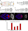
Figure 1
Figure 1. Loss of the ESCRT-III subunit Shrub in germline cells induces the formation of egg chambers with 32 cells.
(A) Schemes of a germarium (left) and germline divisions (right). At its anterior tip, the germline stem cell (GSC, green) divides asymmetrically and produces a cystoblast (CB). Cap cells (CC) maintain GSC stemness, while escort cells (EC) promote CB differentiation. The CB goes through 4 divisions forming a cyst of 16 cells, 15 nurse cells (NC) and 1 oocyte (Oo, yellow). The cyst is encapsulated by follicular cells (FC) and buds out of the germarium. The abscission of the GSC/CB is complete, while in the following 4 divisions it is incomplete. The oocyte shares the cytoplasm with the NC through 4 ring canals (RC). The spectrosome in the GSC and the fusome in its progeny (red, left scheme) are germline-specific organelles. Anterior is on the left, posterior on the right. (B and C) Schemes representing 2 possible ways of explaining the 32-cell cysts. (B) A fifth mitosis. (C) A delay in GSC abscission. (D and E) Stage 7 egg chambers from WT or UAS-shrb RNAi/+; nos-GAL4/+ stained with DAPI (DNA, blue) and phalloidin (F-actin, red). On the right, close-up on oocytes. Red arrows indicate the four ring canals in the control oocyte and the five ring canals in the mutant background. (F) Fraction of egg chambers exhibiting 32 cells on the y axis. Genotypes are on the x axis. Scale bar: 10 μm.
Figure 2
Figure 2. The loss of Shrub in germline stem cells induces the formation of 32-cell egg chambers.
(A-C) Germaria expressing UAS-GFP under the control of different promoters and stained with DAPI (DNA, blue) and α-spectrin (fusome, red). (A) nos-GAL4 is specific for germline cells; (B) bam-GAL4 is specific for early differentiated cells in the cysts; (C) nos-GAL4; bam-GAL80 is specific for germline, excluding the early cysts. Dotted lines surround GSC/CB pairs (A and B) and GSC (C). Cap cells (CC) are indicated. (D) Fraction of egg chambers exhibiting 32 cells on the y axis. Genotypes are on the x axis. Scale bar: 10 μm.
Figure 3
Figure 3. shrub loss of function leads to the formation of stem-cysts.
Germaria of WT females (A, C, and E) or females expressing UAS-shrb RNAi under the control of nos-GAL4 driver (B, D and F), stained for DAPI (DNA, blue), α-spectrin (fusome, green), and either p-Mad (A and B, red), Bag of marble (Bam, C and D, red), or Nanos (Nos, E and F, red). Dotted lines surround: GSC (A), stem-cysts (B, D and F), 4-cell cyst (C) and GSC/CB pair (E). Cap cells (CC) are indicated. (G) Fraction of germaria exhibiting at least one stem-cyst on the y axis. Genotypes are on the x axis. Scale bar: 10 μm.
Figure 4
Figure 4. Stem-cysts in shrub mutant ovaries are caused by synchronous divisions.
Germaria of WT females (A and C) or females expressing UAS-shrb RNAi under the control of nos-GAL4 driver (B and D), stained for DAPI (DNA, blue), α-spectrin (fusome, green), and either EdU (A and B, red) showing S-phase, or pH3S10 (C and D, red) to highlight mitotic cells. Dotted lines surround: GSC/CB pair (A), stem-cysts (B and D) and GSC (C). (E and F) Selected time points of live imaging experiments performed on germaria expressing H2B-RFP (chromatin, red) and G147 (tubulin, green). (E) A WT GSC (surrounded by dotted lines) undergoing mitosis alone. (F) In a female heterozygous for shrb (shrb03/+), the GSC and its daughter CB undergo mitosis synchronously (surrounded by dotted lines). Cap cells (CC) are indicated. Scale bar: 10 μm.
Figure 5
Figure 5. The cells of the stem-cyst share the same cytoplasm.
Selected time points of live imaging experiments performed on germaria expressing UAS-Par1-GFP and UAS-Tubulin-PA-GFP under the nos-GAL4 promoter. Tub-PA-GFP is photoactivated in the region defined by the red circle in the GSC, and the fluorescence diffusion to the neighboring cells is observed. (A and B) WT female GSCs in mid-G2 phase. (A) Abscission between the GSC and the CB did not yet occurred as the GFP diffuses to the CB. (B) GFP does not diffuse to the CB. Abscission has occurred and the cells no longer share their cytoplasm. (C) Stem-cyst of shrbG5/+ female. After photoactivation in the GSC, the GFP diffuses through the 2 neighboring cells. This shows that the cells of the stem-cyst do not complete abscission and stay connected, sharing their cytoplasm. Dotted lines surround: GSC/CB pair (A and B), stem-cysts of 3 cells (C). (A’, B’ and C’) Schematic representation of the Tubulin-PA-GFP diffusion from the GSC to the CB (A’ and B’) or within a stem-cyst (C’). Scale bar: 10 μm.
Figure 6
Figure 6. Drosophila Shrub localizes on the fusome and at the midbody.
Ovaries expressing UAS-GFP-shrb under the control of nos-GAL4 and stained for α-spectrin (A, red) and Pavarotti (B-E, Pav, blue) a marker of the midbody. Midbody position is indicated by a yellow arrow. (A) Before abscission, GFP-Shrb localizes to the fusome (red) and at the ring canal between the GSC and the CB (See also S3B Fig.). (B) GFP-Shrb localizes specifically at the ring canal and midbody (Pav, blue) during its constriction. (C) During abscission, GFP-Shrb is enriched at the midbody, colocalizing with Pav. The ring canal is no longer visible. (D) After abscission, the fusome retracts towards the GSC and the midbody, stained with Pav and Shrb-GFP, is segregated with it. (E) GSCs with round spectrosome (late G2, M) often show colocalization of Shrb-GFP with the midbody (Pav). Scale bar: 10 μm.
Figure 7
Figure 7. Loss of Lethal giant discs induces the formation of stem-cysts and 32-cell egg chambers.
(A) Female germline clones (GLC) of lgdd7 (hsFlp/+; lgdd7, FRT 40A/ GFP, FRT 40A) stained with DAPI (DNA, blue) and phalloidin (F-actin, red). Egg chamber with heterozygous germ cells (GFP, green) is made of 16 cells with a 4 ring canals oocyte (close up in A’). The neighboring egg chamber is a GLC, made of 32 cells with a 5 ring canals oocyte (close up in A’’). (B) Fraction of egg chambers exhibiting 32 cells on the y axis. Genotypes are on the x axis. (C) Female germline clones (GLC) of lgdd7 stained for DAPI (DNA, blue) and α-spectrin (fusome, red). A stem-cyst composed of 5 homozygous mutant cells is shown. (D) Fraction of germaria exhibiting at least one stem-cyst on the y axis. Genotypes are on the x axis. Scale bar: 10 μm.
Figure 8
Figure 8. Aurora-B and Shrub interact to regulate abscission in germline stem cells.
(A) Fraction of egg chambers exhibiting 32 cells on the y axis. Genotypes are on the x axis. (B) Fraction of germaria exhibiting at least one stem-cyst on the y axis. Genotypes are on the x axis.

Similar articles

Cited by

References

    1. Agromayor M, Martin-Serrano J (2013) Knowing when to cut and run: mechanisms that control cytokinetic abscission. Trends Cell Biol 23: 433–441. 10.1016/j.tcb.2013.04.006 - DOI - PubMed
    1. Sanger JM, Pochapin MB, Sanger JW (1985) Midbody sealing after cytokinesis in embryos of the sea urchin Arabacia punctulata. Cell Tissue Res 240: 287–292. - PubMed
    1. Pepling ME, de Cuevas M, Spradling AC (1999) Germline cysts: a conserved phase of germ cell development? Trends Cell Biol 9: 257–262. - PubMed
    1. Mullins JM, Biesele JJ (1977) Terminal phase of cytokinesis in D-98s cells. J Cell Biol 73: 672–684. - PMC - PubMed
    1. Elia N, Sougrat R, Spurlin TA, Hurley JH, Lippincott-Schwartz J (2011) Dynamics of endosomal sorting complex required for transport (ESCRT) machinery during cytokinesis and its role in abscission. Proc Natl Acad Sci U S A 108: 4846–4851. 10.1073/pnas.1102714108 - DOI - PMC - PubMed

Substances