. 2015 Mar 10;54(9):1787-806.
doi: 10.1021/bi501314t. Epub 2015 Feb 24.

Identification of destabilizing and stabilizing mutations of Ste2p, a G protein-coupled receptor in Saccharomyces cerevisiae

Affiliations

Identification of destabilizing and stabilizing mutations of Ste2p, a G protein-coupled receptor in Saccharomyces cerevisiae

Jeffrey Zuber et al. Biochemistry. .

Abstract

The isolation of mutations affecting the stabilities of transmembrane proteins is useful for enhancing the suitability of proteins for structural characterization and identification of determinants of membrane protein stability. We have pursued a strategy for the identification of stabilized variants of the yeast α-factor receptor Ste2p. Because it was not possible to screen directly for mutations providing thermal stabilization, we first isolated a battery of destabilized temperature-sensitive variants, based on loss of signaling function and decreased levels of binding of the fluorescent ligand, and then screened for intragenic second-site suppressors of these phenotypes. The initial screens recovered singly and multiply substituted mutations conferring temperature sensitivity throughout the predicted transmembrane helices of the receptor. All of the singly substituted variants exhibit decreases in cell-surface expression. We then screened randomly mutagenized libraries of clones expressing temperature-sensitive variants for second-site suppressors that restore elevated levels of binding sites for fluorescent ligand. To determine whether any of these were global suppressors, and thus likely stabilizing mutations, they were combined with different temperature-sensitive mutations. Eight of the suppressors exhibited the ability to reverse the defect in ligand binding of multiple temperature-sensitive mutations. Combining certain suppressors into a single allele resulted in levels of suppression greater than that seen with either suppressor alone. Solubilized receptors containing suppressor mutations in the absence of temperature-sensitive mutations exhibit a reduced tendency to aggregate during immobilization on an affinity matrix. Several of the suppressors also exhibit allele-specific behavior indicative of specific intramolecular interactions in the receptor.


Figures

Figure 1
Figure 1
Temperature sensitive receptors with single amino acid substitutions exhibit a temperature dependent growth phenotype. The figure shows the α-factor-dependent growth inhibition of clones that were reconstructed by transforming plasmids bearing missense mutations in either full-length STE2 or C-terminally truncated STE2 into the yeast host strain. Each panel shows the growth of four 10-fold serial dilutions of the relevant yeast strain in the presence of the indicated concentration of α-factor. The clones were scored as Functional (F), Nonfunctional (NF), Temperature Sensitive (TS), or Partially Temperature Sensitive (pTS).
Figure 2
Figure 2
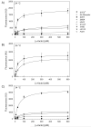
Full-length temperature-sensitive receptors isolated based on loss of signaling function exhibit reduced numbers of binding sites at the cell surface. Strains expressing the indicated full-length STE2 alleles were grown in liquid cultures at 24 °C (A) or 34 °C (B and C). The error bars represent the standard error based on assay of binding to three independent isolates of each strain. Binding was determined by flow cytometry as described in Materials and Methods. (Parameters derived from fitting are listed in Table 2.)
Figure 3
Figure 3
C-terminally truncated temperature-sensitive receptors express reduced numbers of binding sites at the cell surface. Strains expressing the indicated truncated STE2 alleles were grown in liquid cultures at 24 °C (A and B) or 34 °C (C). The error bars represent the standard error based on assay of binding to three independent isolates of each strain. Binding was determined by flow cytometry as described in Materials and Methods. (Parameters derived from fitting are listed in Table 2.)
Figure 4
Figure 4
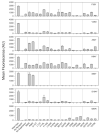
Temperature-dependence of ligand binding to cells expressing temperature sensitive receptors isolated based on the screen for loss of ligand binding. The graph shows the fluorescence of bound [K7(NBD),Nle12] α-factor determined by flow cytometry following growth at 24 °C and 34 °C and incubation with 300 nM [K7(NBD),Nle12] α-factor. The assay was performed using cells that had been re-transformed with plasmids isolated from clones derived from the original screen. The error bars represent the standard error based on assay of binding to three independent isolates of each strain.
Figure 5
Figure 5
Pattern of additivity of individual temperature-sensitive mutations that did not yield temperature sensitive growth arrest as single substitutions. On the axes, the color of the circles indicates the effect of the mutation alone on the number of cell surface ligand binding sites. The fill pattern of the squares at the intersection of two mutations indicates the effect of the combination of the two mutations, as measured by growth arrest assay.
Figure 6
Figure 6
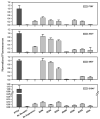
Ligand binding assays performed on yeast strains in the double mutant array. Strains were cultured overnight at 30 °C (34 °C for F38Y and A52T mutants) and assayed for ligand binding with 300 nM fluorescent α-factor. The error bars represent the standard error based on assay of binding to three independent isolates of each strain.
Figure 7
Figure 7
Summary of the effects of combinations of suppressor and temperatures sensitive mutations on numbers of ligand binding sites at the cell surface. The temperature-sensitive mutations are listed across the top of the tables. Suppressor mutations are listed along the sides. A) Each mutation pair was scored using the metric R, defined as the difference between the fluorescence of the double mutant and the temperature-sensitive single mutant divided by the difference between wild-type and the temperature-sensitive single mutant, as a measure of recovery of ligand binding levels due to the suppressor. Magenta boxes indicate positive effect of the suppressor mutation on ligand binding levels, while cyan indicates that the suppressor mutation had a negative effect on ligand binding levels. Note that the scale is clipped to better highlight the intermediate effects. B) The indicated p-values refer to the comparison of the ligand binding of the double mutant to that of the temperature sensitive mutant alone. Only p-values for double mutants with an R-value > 5 or < −5 were mapped. Magenta boxes indicate the significance of the p values for alleles showing enhanced ligand binding resulting from suppression. Blue boxes indicate the significance of the p-values for alleles showing decreased ligand binding resulting from combining the two mutations.
Figure 8
Figure 8
Suppression of different temperature sensitive mutations by site-direct substitutions at position R58. Binding of [K7(NBD),Nle12] α-factor to cells was conducted as described in Materials and Methods at a concentration of 300 nM ligand. For the clones containing the S95Y and S104Y mutations, ligand binding was measured after growth at 30 °C. For the clones containing the A52T and F38Y mutations, ligand binding was measured after growth at 34 °C. The error bars represent the standard error based on assay of binding to three independent isolates of each strain.
Figure 9
Figure 9
Global suppressors identified from the double mutant array exhibit additive effects on ligand binding. Combinations of the global suppressor mutations R58S, F119L, and M218T with the temperature sensitive mutations A52T (A) and F38Y (B) were constructed by site-directed mutagenesis. Assays of binding of [K7(NBD),Nle12] α-factor were performed using 300 nM ligand on cells cultured at 34 °C. The error bars represent the standard error based on assay of binding to three independent isolates of each strain.
Figure 10
Figure 10
Effects of the presence of α-factor and suppressor mutations on the yield of immobilization of Ste2p. The indicated C-terminally truncated Ste2p variants were solubilized using dodecylmaltoside, either in the presence or absence of 300nM α-factor then incubated for 2 hours at 24°C with beads coated with anti-c-myc antibodies. To determine the amount bound, the Ste2p was then stripped from the beads using SDS/urea buffer and subjected to immunoblotting, probed with anti-c-myc antibodies.
Figure 11
Figure 11
Distribution of temperature-sensitive mutation in Ste2p. The figure shows the predicted topology of Ste2p . Residues in circles with black fills are sites at which single substitutions cause temperature-sensitive phenotypes. The other highlighted residues have only been found to confer temperature sensitivity when they are present in alleles with multiple amino acid substitutions. The residues in enlarged circles had substitutions that caused large decreases in ligand binding. The residues in hexagons had substitutions that caused moderate decreases in ligand binding, while the residues in diamonds had substitutions that caused little decreases in ligand binding.
Figure 12
Figure 12
Distribution of suppressor mutations in Ste2p mapped on to the predicted transmembrane topology of Ste2p. The residues highlighted in black are the locations of the temperature sensitive mutations. The other highlighted residues indicate the positions of suppressors. Large grey circles show the locations of global suppressors, defined as mutations that restore at least 10% of ligand binding to a 99% confidence level for more than one temperature-sensitive mutation. Residues depicted as smaller dark grey circles are allele-specific suppressors, defined as being able to restore at least 10% of ligand binding to a 99% confidence level for no more than one of the tested temperature-sensitive mutations.

Similar articles

Cited by

References

    1. Wallin E, Heijne GV. Genome-wide analysis of integral membrane proteins from eubacterial, archaean, and eukaryotic organisms. Protein Science. 1998;7:1029–1038. - PMC - PubMed
    1. Overington JP, Al-Lazikani B, Hopkins AL. How many drug targets are there? Nature Reviews Drug Discovery. 2006;5:993–996. - PubMed
    1. White SH. Biophysical dissection of membrane proteins. Nature. 2009;459:344–346. - PubMed
    1. Lundstrom K. Structural biology of G protein-coupled receptors. Bioorganic & Medicinal Chemistry Letters. 2005;15:3654–3657. - PubMed
    1. Miller JL, Tate CG. Engineering an Ultra-Thermostable β1-Adrenoceptor. Journal of Molecular Biology. 2011;413:628–638. - PMC - PubMed

Publication types

MeSH terms

Substances