. 2015 Feb 3;10(2):e0117450.
doi: 10.1371/journal.pone.0117450. eCollection 2015.

Dopamine increases CD14+CD16+ monocyte migration and adhesion in the context of substance abuse and HIV neuropathogenesis

Affiliations

Dopamine increases CD14+CD16+ monocyte migration and adhesion in the context of substance abuse and HIV neuropathogenesis

Jacqueline S Coley et al. PLoS One. .

Abstract

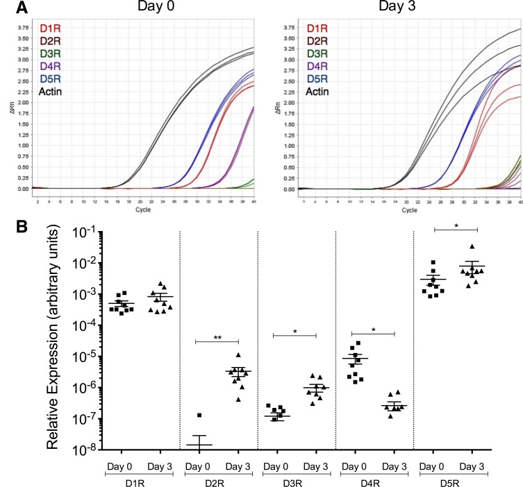
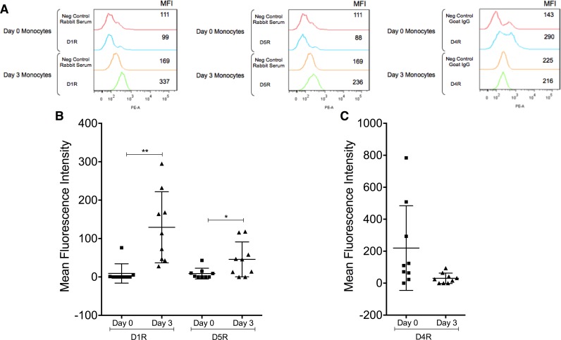
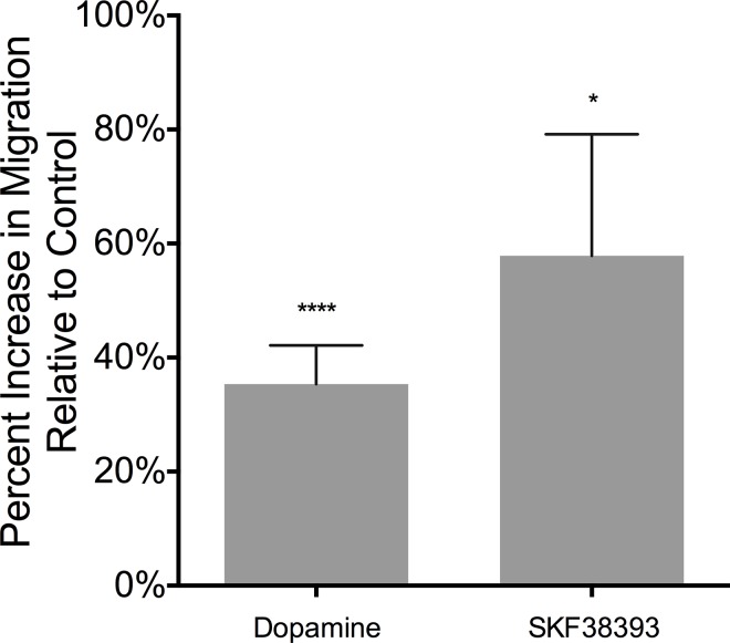
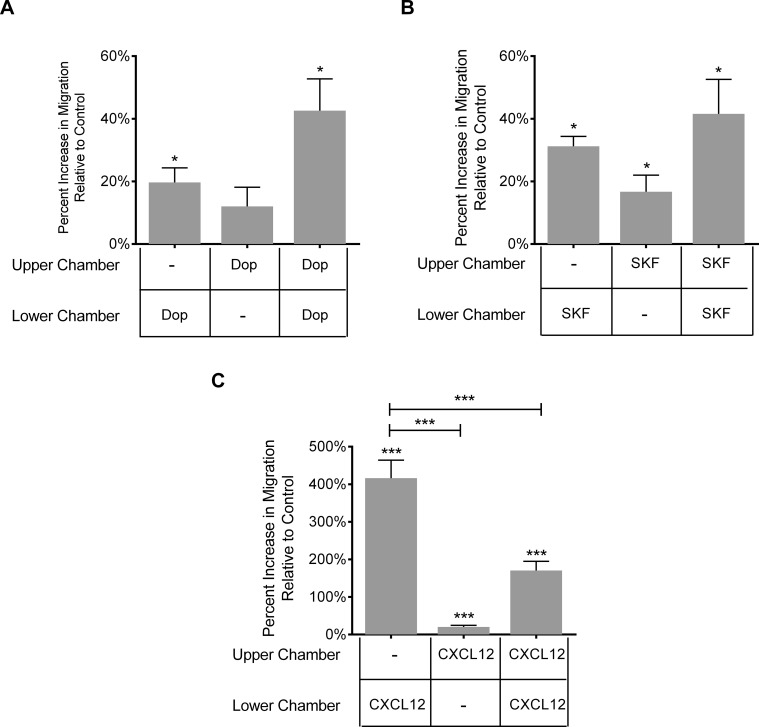
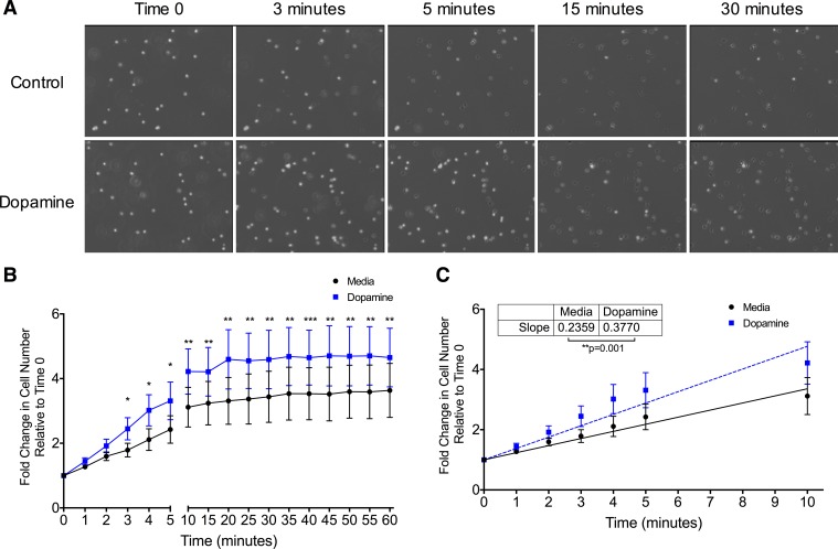
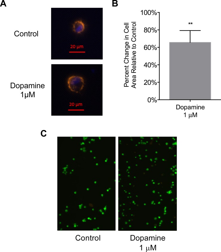
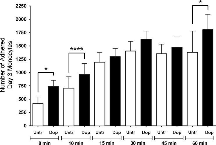
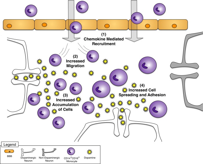
Drug abuse is a major comorbidity of HIV infection and cognitive disorders are often more severe in the drug abusing HIV infected population. CD14+CD16+ monocytes, a mature subpopulation of peripheral blood monocytes, are key mediators of HIV neuropathogenesis. Infected CD14+CD16+ monocyte transmigration across the blood brain barrier mediates HIV entry into the brain and establishes a viral reservoir within the CNS. Despite successful antiretroviral therapy, continued influx of CD14+CD16+ monocytes, both infected and uninfected, contributes to chronic neuroinflammation and the development of HIV associated neurocognitive disorders (HAND). Drug abuse increases extracellular dopamine in the CNS. Once in the brain, CD14+CD16+ monocytes can be exposed to extracellular dopamine due to drug abuse. The direct effects of dopamine on CD14+CD16+ monocytes and their contribution to HIV neuropathogenesis are not known. In this study, we showed that CD14+CD16+ monocytes express mRNA for all five dopamine receptors by qRT-PCR and D1R, D5R and D4R surface protein by flow cytometry. Dopamine and the D1-like dopamine receptor agonist, SKF38393, increased CD14+CD16+ monocyte migration that was characterized as chemokinesis. To determine whether dopamine affected cell motility and adhesion, live cell imaging was used to monitor the accumulation of CD14+CD16+ monocytes on the surface of a tissue culture dish. Dopamine increased the number and the rate at which CD14+CD16+ monocytes in suspension settled to the dish surface. In a spreading assay, dopamine increased the area of CD14+CD16+ monocytes during the early stages of cell adhesion. In addition, adhesion assays showed that the overall total number of adherent CD14+CD16+ monocytes increased in the presence of dopamine. These data suggest that elevated extracellular dopamine in the CNS of HIV infected drug abusers contributes to HIV neuropathogenesis by increasing the accumulation of CD14+CD16+ monocytes in dopamine rich brain regions.


Conflict of interest statement

Competing Interests: The co-author Eliseo A. Eugenin is a PLOS ONE Editorial Board member. This does not alter the authors' adherence to PLOS ONE Editorial policies and criteria.

Figures

Fig 1
Fig 1. Dopamine receptor mRNA expression changes with monocyte maturation.
Day 0 and Day 3 monocytes were analyzed by qRT-PCR for dopamine receptor mRNA expression. (A) Amplification plots from a representative donor showing mRNA expression of D1R, D2R, D3R, D4R, D5R and actin in Day 0 and Day 3 monocytes. (B) Relative expression of dopamine receptors normalized to actin for all donors tested. Data points for D1R, D3R and D4R distributed normally and were analyzed by two-tailed paired Student’s t-test for significance. Data points for D2R and D5R did not have a normal distribution and were analyzed by two-tailed Wilcoxon matched-pairs signed rank test for significance (*p<0.05, **p<0.01, N = 9).
Fig 2
Fig 2. Dopamine receptor surface protein expression changes with monocyte maturation.
Day 0 and Day 3 monocytes were analyzed by flow cytometry for surface expression of D1R, D5R, and D4R. (A) Representative histograms from a single donor showing increased surface expression of D1R and D5R, and decreased expression of D4R with maturation. Analysis of cell cultures from all donors shows surface expression of (B) D1-like dopamine receptors, D1R and D5R, increasing with maturation (*p<0.05, **p<0.01, N = 9) and (C) D4R surface expression trending towards a decrease with maturation (p = 0.07, N = 9) (two-tailed Wilcoxon matched-pairs signed rank test).
Fig 3
Fig 3. Dopamine receptor activation increases Erk2 phosphorylation.
Day 3 monocytes were treated with dopamine (100 nM, 500 nM, 1 μM) for 5 or 15 minutes, and protein lysates were analyzed by Western blot. (A) Representative blot showing changes in Erk1/2 phosphorylation in response to dopamine treatment. Total Erk1/2 was used as a loading control. (B) Quantification of changes in Erk2 phosphorylation normalized to total Erk2, shown as maximal percent change at 5 or 15 minutes relative to untreated cells, which is set to 0% (*p<0.05, N = 6) (two-tailed Wilcoxon matched-pairs signed rank test).
Fig 4
Fig 4. Dopamine and D1-like dopamine receptor activation increase migration of Day 3 monocytes.
Day 3 monocytes were added to the top wells of a microchemotaxis chamber. Dopamine (100 nM, 500 nM, 1 μM) (N = 19) or D1-like receptor agonist SKF38393 (1 nM, 10 nM, 100 nM) (N = 6) was added to the bottom chamber. Cells were allowed to migrate for 1 hour at 37°C through a polycarbonate membrane. Cells that migrated and adhered to the underside of the membrane were fixed, stained, and quantified by densitometry. Migration is shown as the maximal percent increase with 100 nM, 500 nM or 1 μM dopamine, relative to baseline migration, which is set to 0% (*p<0.05, ****p<0.0001) (two-tailed Wilcoxon matched-pairs signed rank test).
Fig 5
Fig 5. Dopamine induced migration is not gradient dependent.
A checkerboard assay was performed by generating positive, negative, and null gradients using a microchemotaxis chamber. (A) Dopamine (100 nM, 500 nM or 1 μM) and (B) D1-like dopamine receptor agonist SKF38393 (1, 10 or 100 nM) increased migration of Day 3 monocytes in a gradient independent manner, as evidenced by increased transmigration when added to the top chamber as well as to both the top and the bottom chamber (N = 6). (C) CXCL12 (1 ng/ml), used as a positive control for chemotaxis, induced maximal migration when present only in the bottom chamber, indicative of gradient dependent transmigration. Migration is shown as maximal percent increase relative to baseline migration, which is set to 0% (*p<0.05, ***p<0.001, N = 12) (two-tailed Wilcoxon matched-pairs signed rank test).
Fig 6
Fig 6. Dopamine increases the accumulation of Day 3 monocytes and their rate of settling.
Day 3 monocytes were added to tissue culture dishes in the presence of dopamine (1 μM) or media, and the surface of each dish was imaged for 1 hour at 15 second intervals to record cells as they settled. Brightness was increased equally for all images. (A) Images from a representative experiment showing more monocytes settling to the surface of tissue culture dishes in the presence of dopamine at multiple time points when compared to cells without dopamine (control). (B) The addition of dopamine resulted in increased numbers of settled cells, as shown by fold change relative to Time 0, which is set to 1 (*p<0.05, **p<0.01, ***p<0.001 relative to media, N = 6) (two-tailed paired Student’s t-test). (C) Dopamine significantly increased the rate at which Day 3 monocytes settled to the surface of tissue culture dishes (**p = 0.001, N = 6) (two-tailed paired Student’s t-test).
Fig 7
Fig 7. Dopamine increases the area of Day 3 monocytes during the early stages of adhesion.
(A) Representative images from a single donor showing increased area of a Day 3 monocyte in the presence of 1 μM dopamine after 10 minutes of adhesion as compared to a non-dopamine treated control cell. (B) Quantification of percent change in cell area as compared to control, which is set to 0%. Data are shown as the maximal increase in area per cell, relative to control. The time of maximal increase varied between 8, 10, and 15 minutes among independent donors due to inherent variability in primary cells (**p<0.01, N = 5) (two-tailed paired Student’s t-test). (C) Viability assay of Day 3 monocytes after incubation in media (control) or 1 μM dopamine for 30 min. Live cells exhibit green fluorescence and dead cells exhibit red fluorescence.
Fig 8
Fig 8. Dopamine increases the adhesion of Day 3 monocytes.
(A) Dopamine increases the adhesion of Day 3 monocytes after 8, 10 and 60 minutes. Data are shown as the number of adherent Day 3 monocytes in the presence of 1 μM dopamine or diluent (untreated) after multiple time points (*p<0.05, ***p<0.001, N = 5 for 8 and 10 min and N = 6 for 15, 30, 45 and 60 min) (two-tailed paired Student’s t-test).
Fig 9
Fig 9. Dopamine increases CD14+CD16+ monocyte migration and adhesion.
(1) CD14+CD16+ monocytes enter the CNS in response to increased chemokines and then encounter increased extracellular dopamine due to drug abuse. (2) CD14+CD16+ monocytes migrate more in response to dopamine. Dopamine increases the (3) accumulation and (4) spreading and adhesion of CD14+CD16+ monocytes. This may alter the distribution of CD14+CD16+ monocytes in the CNS, leading to accumulation of these cells in dopaminergic brain regions. Increased CD14+CD16+ monocyte accumulation may contribute to neuroinflammation and neuronal damage.

Similar articles

Cited by

References

    1. Davis LE, Hjelle BL, Miller VE, Palmer DL, Llewellyn AL, et al. (1992) Early viral brain invasion in iatrogenic human immunodeficiency virus infection. Neurology 42: 1736–1739. - PubMed
    1. Valcour V, Chalermchai T, Sailasuta N, Marovich M, Lerdlum S, et al. (2012) Central Nervous System Viral Invasion and Inflammation During Acute HIV Infection. J Infect Dis 206: 275–282. 10.1093/infdis/jis326 - DOI - PMC - PubMed
    1. Heaton RK, Clifford DB, Franklin DR, Woods SP, Ake C, et al. (2010) HIV-associated neurocognitive disorders persist in the era of potent antiretroviral therapy. Neurology 75: 2087–2096. 10.1212/WNL.0b013e318200d727 - DOI - PMC - PubMed
    1. Heaton RK, Franklin DR, Ellis RJ, McCutchan JA, Letendre SL, et al. (2011) HIV-associated neurocognitive disorders before and during the era of combination antiretroviral therapy: differences in rates, nature, and predictors. J Neurovirol 17: 3–16. 10.1007/s13365-010-0006-1 - DOI - PMC - PubMed
    1. Simioni S, Cavassini M, Annoni JM, Rimbault Abraham A, Bourquin I, et al. (2010) Cognitive dysfunction in HIV patients despite long-standing suppression of viremia. AIDS 24: 1243–1250 1210.1097/QAD.1240b1013e3283354a3283357b. 10.1097/QAD.0b013e3283354a7b - DOI - PubMed

Publication types