. 2015 Feb 3;11(2):e1004965.
doi: 10.1371/journal.pgen.1004965. eCollection 2015.

Identification and functional analysis of healing regulators in Drosophila

Affiliations

Identification and functional analysis of healing regulators in Drosophila

Carmen Álvarez-Fernández et al. PLoS Genet. .

Abstract

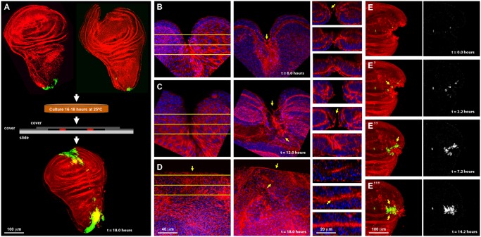
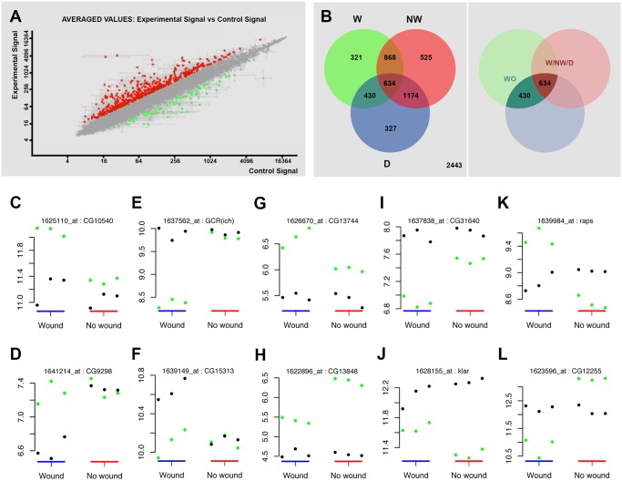
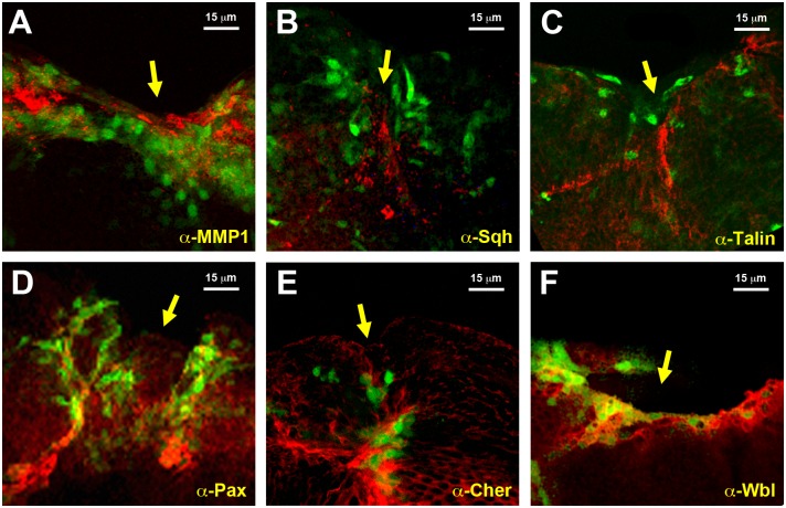
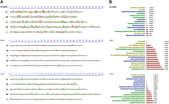
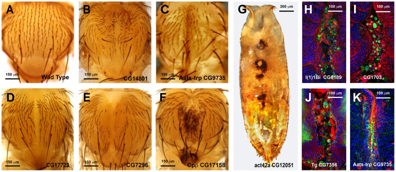
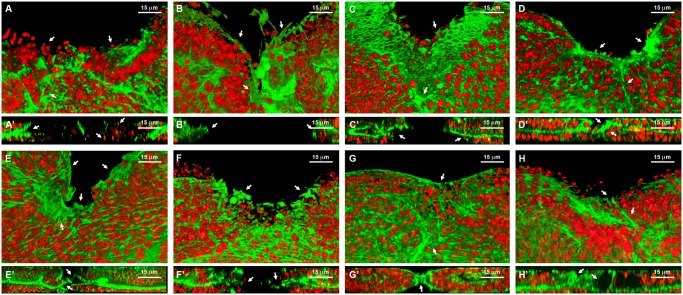
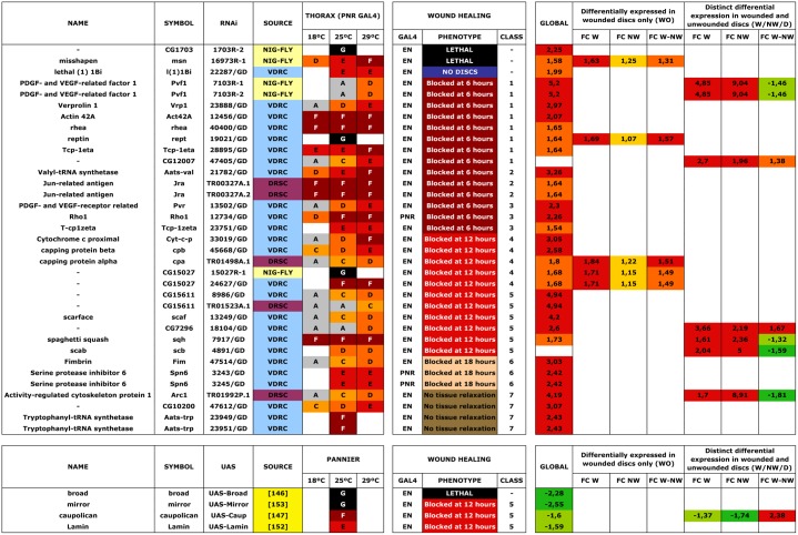
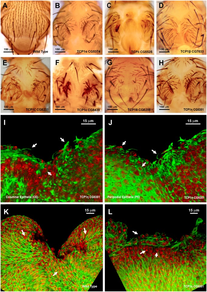
Wound healing is an essential homeostatic mechanism that maintains the epithelial barrier integrity after tissue damage. Although we know the overall steps in wound healing, many of the underlying molecular mechanisms remain unclear. Genetically amenable systems, such as wound healing in Drosophila imaginal discs, do not model all aspects of the repair process. However, they do allow the less understood aspects of the healing response to be explored, e.g., which signal(s) are responsible for initiating tissue remodeling? How is sealing of the epithelia achieved? Or, what inhibitory cues cancel the healing machinery upon completion? Answering these and other questions first requires the identification and functional analysis of wound specific genes. A variety of different microarray analyses of murine and humans have identified characteristic profiles of gene expression at the wound site, however, very few functional studies in healing regulation have been carried out. We developed an experimentally controlled method that is healing-permissive and that allows live imaging and biochemical analysis of cultured imaginal discs. We performed comparative genome-wide profiling between Drosophila imaginal cells actively involved in healing versus their non-engaged siblings. Sets of potential wound-specific genes were subsequently identified. Importantly, besides identifying and categorizing new genes, we functionally tested many of their gene products by genetic interference and overexpression in healing assays. This non-saturated analysis defines a relevant set of genes whose changes in expression level are functionally significant for proper tissue repair. Amongst these we identified the TCP1 chaperonin complex as a key regulator of the actin cytoskeleton essential for the wound healing response. There is promise that our newly identified wound-healing genes will guide future work in the more complex mammalian wound healing response.


Conflict of interest statement

The authors have declared that no competing interests exist.

Figures

Fig 1
Fig 1. Imaginal discs wound healing in vitro.
A) On the top row are shown dissected wing imaginal discs before injury (left) and just after injury (right) displaying puc expression at the stalk and some PE cells. Imaginal discs were cultured in a modified MM3 medium up to 24 hours on chambered slides as shown (center), which prevents discs folding and allows their imaging in vivo. At the bottom, a healed imaginal disc after 18 hours of culture displays strong ectopic puc expression at the wound edge and surrounding areas. Double staining for puc (green; pucE69-A—Gal4; UAS-GFP) and Actin (Phalloidin—red). B) Injured disc after 6 hours of in vitro culture. PE view (left) displaying a wide wound gap (yellow lines indicate the positions chosen for Z reconstruction). CE view (middle) of the same disc, showing elongated cells at the leading edge, filopodia and the initial zippering (arrow) of the epithelia. Orthogonal sections at three different levels (right) with the CE facing upwards and the PE downwards. The CE becomes partially disorganized establishing strong heterotypic contacts with the PE (arrow). C) Injured disc after 12 hours of in vitro culture. PE, CE and orthogonal views are shown as in B. There is a remarkable reduction in wound size and a notable actin accumulation (arrows). D) Injured imaginal disc after 18 hours of in vitro culture. Complete wound closure is observed for both, the PE (left), and CE (middle). Orthogonal sections show the basolateral zippering of the columnar epithelia (right). For B to D, phalloidin (actin) is shown in red; DAPI (nuclei) in blue. E, E', E'' and E''') Time-lapse snapshots from S1 Movie of the healing process of a wounded imaginal disc cultured in modified MM3 medium. As culture progresses, puc expression (arrows) builds up at the edges on those cells actively engaged in healing. Cell membranes are shown in red (FM4–64) and puc expression in green (pucE69-A—Gal4; UAS-GFP) (left). The green channel is shown on the right. Scale bars are indicated for each panel.
Fig 2
Fig 2. Transcriptomic healing response in imaginal discs.
A) log-log scatter plot of the averaged microarray data for the global comparison between JNK signaling positive healing-engaged cells and their siblings in wounded imaginal discs (733 upregulated (red) and 748 downregulated (green) genes—FC 1.5, p-value < 0.05). B) A Venn diagram was used to represent the interactions of the different identified populations. The W subpopulation corresponds to those 2253 genes whose expression is modified in wounded discs. The NW subpopulation corresponds to those 3201 genes whose expression is modified in non-wounded discs. The D subpopulation corresponds to those 2565 genes in wounded or non-wounded discs that appear differentially modified between JNK-positive and negative cells. The WO and W/NW/D intersections highlighted on the right covers the 430 genes differentially expressed in wounded discs only and the 634 genes differentially expressed in both, wounded and in non-wounded discs but distinctly in both conditions. C to L) Representative genes for different subpopulations covering the WO and W/NW/D sets. Green spots show the level of expression for each replica in JNK-positive cells. Black spots show the levels of expression for each replica in JNK-negative cells. C) Genes upregulated in JNK-positive cells in wounded discs only (CG10540). D) Genes downregulated in JNK-negative cells in wounded discs only (CG9298). E) Genes downregulated in JNK-positive cells in wounded discs only [GCR(ich)—CG5812]. F) Genes upregulated in JNK-negative cells in wounded discs only (CG15313). G) Genes more upregulated in JNK-positive cells in wounded than in non-wounded discs (CG13744). H) Genes less upregulated in JNK-positive cells in wounded than in non-wounded discs (CG13848). I) Genes more downregulated in JNK-positive cells in wounded than in non-wounded discs (CG31640). J) Genes less downregulated in JNK-positive cells in wounded than in non-wounded discs (klar—CG17046). K) Genes upregulated in JNK-positive cells in wounded and downregulated in non-wounded discs (raps—CG5692). L) Genes downregulated in JNK-positive cells in wounded and upregulated in non-wounded discs (CG12255).
Fig 3
Fig 3. Validation of the genomic expression analysis.
A) Imaginal disc, 12 hours after incisive wounding, showing enhanced expression of MMP1, a secreted matrix metalloproteinase upregulated in the healing transcriptome, in those cells engaged in healing (red). Similar results were observed for B) Spaghetti squash, the homologue of the regulatory light chain of non-muscle myosin II; C) Talin, a FERM domain protein linking the cytoplasmic tail of integrins to the actin cytoskeleton; D) Paxillin, a cytoskeleton scaffolding protein present at focal adhesions; E) Cheerio coding for filamin, an actin binding protein; and F) Windbeutel, a resident protein of the endoplasmic reticulum. All these genes were found to be upregulated in the healing transcriptome. GFP expression highlighting puc upregulation in all cases is shown in green. Scale bars are indicated for each panel.
Fig 4
Fig 4. Chromosomal clustering and GO enrichment analysis.
A) Graphical representation of the chromosomal clustering of the global comparison (JNK-positive vs negative cells in wounded discs) (upper panel), WO (middle panel) and W/NW/D (lower panel) contrasts. Upregulated (red bars) and downregulated (green bars) genes are positioned in the different Drosophila chromosome arms (X, 2L, 2R, 3L and 3R). Identified clusters are represented by inverted triangles. B) GO term enrichment analysis. The highest enriched GO terms (15) with lowest p-values (horizontal histograms) are displayed. These terms are distributed between the different terms categories: Cellular Component (CCs—yellow), Biological Process (BPs—blue) and Molecular Function (MFs—green) and represented for the global (upper panel), WO (middle panel) and W/NW/D (lower panel) contrasts.
Fig 5
Fig 5. Thorax fusion phenotypes.
A to G) Thorax adult phenotypes grouped in different categories resulting from RNAi interference in the expression of selected genes. UAS-RNAi transgenes were expressed in the presumptive central area of the thorax with a Pnr-Gal4 driver. A) Wild Type adult. It shows a canonical midline fusion of the thoracic hemisegments and perfect patterning of sensory bristles B) Interference with CG14801 expression (Class 1TC phenotype) results in mirrored bristle polarity defects in the adult thorax. C) Interference with Aats-trp (CG9735) expression (Class 2TC phenotype) results in an excess of fusion with the hemithoraces collapsing at the midline. D) Interference with CG17723 expression (Class 3TC phenotype) results in a weak thorax cleft. E) Interference with CG7296 expression (Class 4TC phenotype) results in an intermediate thorax cleft. Some bristle defects are also observed. F) Interference with cpβ expression (Class 5TC phenotype) results in a big thorax cleft and scutellum abnormalities. G) Interference with Act42a (CG12051) expression (Class 6TC phenotype) results in pupal lethality and failure of discs eversion and/or fusion. Necrotic areas are found along the dorsal midline of an uneverted pupae. H to K) Expansion and fusion of imaginal discs are driven by leading edge cells, which extend long filopodia and accumulate an actin cable at the front edge. The JNK signaling pathway is activated in the leading edge cells during the fusion process. 7–7.5 h APF thoraces stained with Phalloidin (actin—red), DAPI (nuclei—blue) and Anti-LacZ (puc expression—Green). are shown. Anterior is up. H) Dorsal view of the thoracic area of a pupa which has failed to properly close upon downregulation of l(1)1Bi (CG6189) expression (Class 5TC). Phalloidin staining shows loss of actin accumulation or filopodia extensions in the leading edge of the discs, while puc-expressing cells are present along the full edge of both hemithoraces. I) Dorsal view of the thoracic area of a pupa showing irregular closure at the midline upon downregulation of CG1703 expression (Class 5TC). Phalloidin staining shows fusion defects, enlarged leading edge cells that seem to detach, which sustain strong puc expression. Picnotic nuclei are present in the scutellum area. J) Dorsal view of the thoracic area of a pupa which has failed to fully close upon downregulation of tg (CG7356) (Class 4TC). Phalloidin staining points to a clumsy accumulation of actin, most evident anteriorly. Several cells at the leading edge detach and round up and puc expression is sustained at the thorax midline. K) Aats-trp downregulation during thorax closure results in an excess of fusion (Class 2TC) that does not seem to affect actin accumulation or puc expression (dorsal thoracic area). Scale bars are indicated for each panel.
Fig 6
Fig 6. Healing phenotypes categories.
3D reconstruction of the peripodial and columnar epithelia at 18 hours after wounding. Phalloidin (actin) is shown in green; DAPI (nuclei) in red. A) Interference with Act42a expression (Class 1 phenotype) results in a large open wound and extensive cell death (arrows). A’) Orthogonal section. No heterotypic contacts, absence of actin cytoskeleton and occasional picnotic nuclei are depicted with arrows. B) Interference with Aats-Val expression (Class 2 phenotype) results in an extensive open wound with actin and active filopodia present at the leading edge (arrows). B’) Orthogonal section. Early approximation of the leading edges but failure on fusion. C) Interference with pvr expression (Class 3 phenotype) results in a large open wound. The CE has initiated healing but the PE fails to heal (arrows). C’) Orthogonal section. Arrows point to the delay on healing of the peripodial epithelia. D) Interference with cpβ expression (Class 4 phenotype) shows a large open wound and abnormal accumulation of actin (arrows). D’) Orthogonal section. Actin accumulation impedes the normal fusion of the epithelia (arrows). E) Interference with scab expression (Class 5 phenotype) shows a large open wound and a gap between the peripodial and the columnar epithelia (arrows). E’) Orthogonal section. The gaps at the edge between layers are more evident. The columnar epithelium initiates sealing apically (arrows). F) Overexpression of mirror (Class 5 phenotype) shows a partial closure and an abnormal distribution of actin (arrows). F’) Orthogonal section. Apical sealing precedes basal attachments that seem disorganized (arrows). G) Interference with fimbrin expression (Class 6 phenotype) displays advanced closure but disorganized actin cytoskeleton in the peripodial epithelia (arrows). G’) Orthogonal section. Actin accumulation at the junction is highlighted. The peripodial epithelia remains disorganized at the fusion point (arrows). H) Interference with arc1 expression (Class 7 phenotype) displays a full closure but shape abnormalities at the junction and adjacent territories (arrows). H’) Orthogonal section. Apical gaps are observed at the fusion area and surrounding. Tissue layers are shifted (arrows). Scale bars are indicated for each panel.
Fig 7
Fig 7. Wound healing phenotypes.
53 RNAi and 4 UAS lines were employed for interference or overexpression in the Pnr- or En-Gal4 domains. 35 and 4 lines respectively yielded distinguishable phenotypes. The thorax phenotypes (PANNIER GAL4) at different temperatures (18°C, 25°C and 29°C) are indicated (A -Wild Type; B—Bristle Defects; C—Weak Thorax Cleft; D—Thorax Cleft; E—Strong Thorax Cleft; F—Pupal Lethal and G—Embryonic lethal). Healing was assayed at 25°C with different GAL4 (EN or PNR). Healing phenotype is indicated and phenotypic classes are coded as in the text (1 -Early (6 hours) defects—No Actin; 2—Early (6 hours) defects—Unstructured Actin; 3—Early (6 hours) defects—Actin present; 4—Intermediate (12 hours) defects—CE zippering fails; 5—Intermediate (12 hours) defects—Gaps between the epithelia; 6—Late (18 hours) defects—Incomplete closure and 7—No tissue Relaxation—Tissue folds) (see Functional analysis of “healing” genes section). Transcriptomic results are presented as described in S1, S2 and S3 Tables in progressive color series by fold change, p-value < 0.05. GLOBAL—global comparison of healing-competent JNK-positive cells vs their non-engaged siblings in wounded discs; WO—Genes differentially expressed in wounded discs only [FC W—comparison of JNK-positive and negative cells in wounded discs (W)—FC NW—comparison of JNK-positive and negative cells in non-wounded discs (NW)—FC W-NW—ratio of expression differences for JNK-positive vs negative cells between wounded and non-wounded discs (D)]; W/NW/D—Distinct differential expression in wounded and unwounded discs [FC W—comparison of JNK-positive and negative cells in wounded discs (W)—FC NW—comparison of JNK-positive and negative cells in non-wounded discs (NW)—FC W-NW—ratio of expression differences for JNK-positive vs negative cells between wounded and non-wounded discs (D)].
Fig 8
Fig 8. Thorax fusion and impaired healing phenotypes of TCP1 subunits.
A) Wild type notum of an adult Drosophila. B to H) Thorax malformations observed after interference (RNAi) with the expression of different TCP1 subunits as labeled (tested with a Pnr-GAL4 driver). I and J) 3D reconstruction of the peripodial and columnar epithelia at 18 hours after wounding. (I) PE and (J) CE views. TCP1η RNAi knockdown results in impaired healing after 20–24 hours of culture in vitro (tested with a En-GAL4 driver). Arrows point to abnormal actin-rich structures and arrowheads to dead cells. Phalloidin (actin) is shown in green; DAPI (nuclei) in red. K) Injured wild type disc after 12 hours of in vitro culture. CE view showing the perpendicular alignment of microtubules to the sealing front (arrows) and elongated cells at the leading edge. A remarkable reduction in wound size is observed. L) Injured TCP1η RNAi knockdown disc after 12 hours of culture in vitro. CE view showing conspicuous picnotic nuclei (arrowheads) and an abnormal transverse alignment of microtubules (arrows) at the leading healing front. Wound closure fails to proceed. Tubulin is shown in green; DAPI (nuclei) in red. Scale bars are indicated for each panel.

Similar articles

Cited by

References

    1. Martin P, Parkhurst SM (2004) Parallels between tissue repair and embryo morphogenesis. Development 131: 3021–3034. 10.1242/dev.01253 - DOI - PubMed
    1. Harden N (2002) Signaling pathways directing the movement and fusion of epithelial sheets: lessons from dorsal closure in Drosophila. Differentiation 70: 181–203. 10.1046/j.1432-0436.2002.700408.x - DOI - PubMed
    1. Wood W, Jacinto A, Grose R, Woolner S, Gale J, et al. (2002) Wound healing recapitulates morphogenesis in Drosophila embryos. Nature Cell Biology 4: 907–912. 10.1038/ncb875 - DOI - PubMed
    1. Martin P (1997) Wound healing--aiming for perfect skin regeneration. Science 276: 75–81. 10.1126/science.276.5309.75 - DOI - PubMed
    1. McCluskey J, Martin P (1995) Analysis of the tissue movements of embryonic wound healing--DiI studies in the limb bud stage mouse embryo. Developmental Biology 170: 102–114. 10.1006/dbio.1995.1199 - DOI - PubMed

Publication types

MeSH terms

Substances