. 2015 May;20(3):507-16.
doi: 10.1007/s12192-015-0575-2. Epub 2015 Feb 4.

Hypoxia-inducible factor-1α regulates the expression of L-type voltage-dependent Ca(2+) channels in PC12 cells under hypoxia

Affiliations

Hypoxia-inducible factor-1α regulates the expression of L-type voltage-dependent Ca(2+) channels in PC12 cells under hypoxia

Ran Li et al. Cell Stress Chaperones. 2015 May.

Abstract

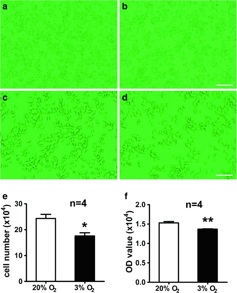
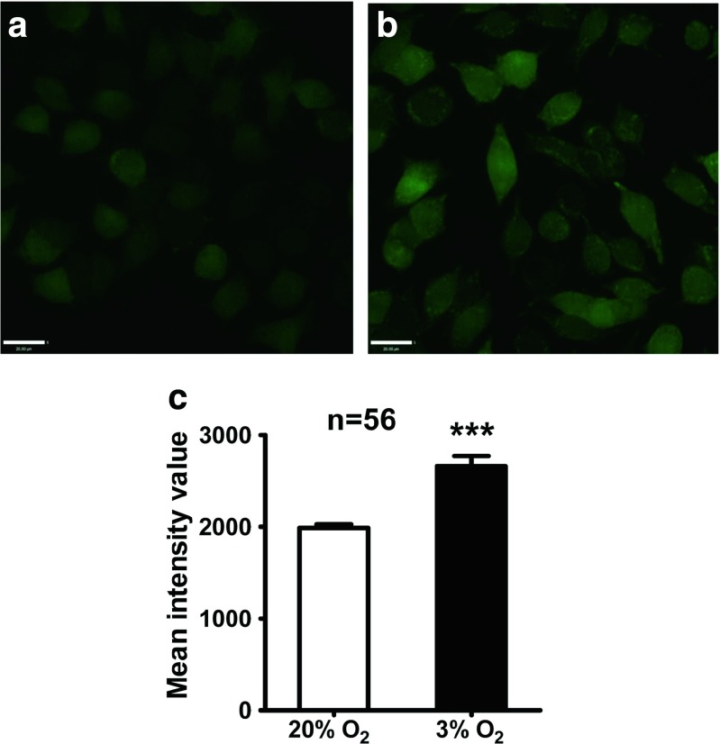
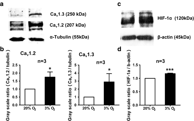
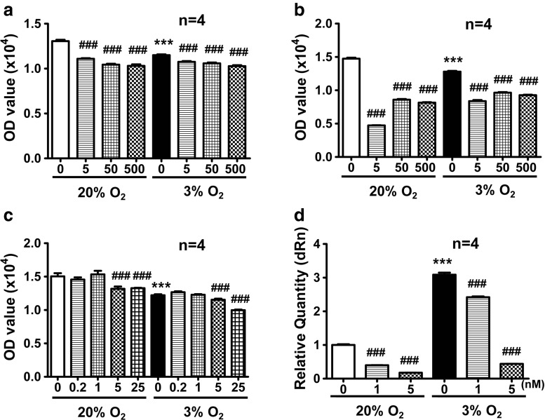
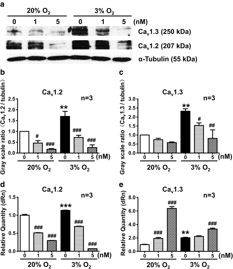
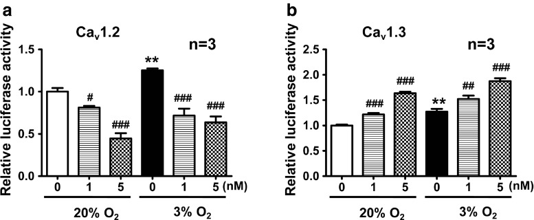
Hypoxia is an important factor in regulation of cell behavior both under physiological and pathological conditions. The mechanisms of hypoxia-induced cell death have not been completely elucidated yet. It is well known that Ca(2+) is critically related to cell survival. Hypoxia-inducible factor-1α (HIF-1α) is a core regulatory factor during hypoxia, and L-type voltage-dependent Ca(2+) channels (L-VDCCs) have been reported to play a critical role in cell survival. This study was conducted to explore the relationship between L-VDCC expression and HIF-1α regulation in PC12 cells under hypoxia. PC12 cells were treated at 20 or 3 % O2 to observe its proliferation and the intracellular calcium concentration. Then, we detected the protein expression of HIF-1α and L-VDCCs subtypes, Cav1.2 and Cav1.3. At last, to verify the relationship between HIF-1α and Cav1.2 and Cav1.3, we got the expression of Cav1.2 and Cav1.3 with Western blot and luciferase report gene assays after PC12 cells were treated by echinomycin, which is an HIF-1α inhibitor. Compared with 20 % O2 (normoxia), 3 % O2 (hypoxia) inhibited cell proliferation, increased the intracellular calcium concentration, and induced protein expression of HIF-1α. The protein expression of two L-VDCCs subtypes expressed in the nervous system, Cav1.2 and Cav1.3, was upregulated by hypoxia and reduced dose dependently by treatment with echinomycin, a HIF-1α inhibitor. Luciferase report gene assays showed that the expression of Cav1.2 and Cav1.3 genes was augmented under 3 % O2. However, echinomycin only slightly and dose dependently decreased expression of the Cav1.2 gene, but not that of the Cav1.3 gene. These data indicated that Cav1.2 might be regulated by HIF-1α as one of its downstream target genes and involved in regulation of PC12 cells death under hypoxia.


Figures

Fig. 1
Fig. 1
Hypoxia (3 % O2) inhibits the proliferation of PC12 cells. PC12 cells were cultured under hypoxia (3 % O2) (b, d) and normoxia (20 % O 2) (a, c) for 24 h. Representative photos under hypoxia (b, d) and normoxia (a, c). Statistic analysis of cell numbers by cell counting (e). Statistic analysis of cell viability by MTS assay (f). Scale bars = 25 μm (a, b) and 100 μm (c, d). *P < 0.05, **P < 0.01; “n” stands for the number of parallel wells
Fig. 2
Fig. 2
[Ca2+]i of PC12 cells in 20 and 3 % O2 as detected by calcium imaging. Hypoxic culture in 3 % O2 resulted in elevated [Ca2+]i in most PC12 cells (b), whereas calcium signals were relatively lower in PC12 cells cultured in 20 % O2 (a). The mean intensity value of basal [Ca2+]i in PC12 cells was higher in 3 % O2 compared with that in 20 % O2 (c). Scale bar = 20 μm; ***P < 0.001; “n” indicates the number of analyzed cells
Fig. 3
Fig. 3
Hypoxia induces protein expression of Cav1.2, Cav1.3 and HIF-1α. Expression of Cav1.2, Cav1.3, and HIF-1α protein in PC12 cells under 20 and 3 % O2 were examined by Western blot. Both Cav1.2, Cav1.3 protein (a, b), and HIF-1α protein (c, d) expression in PC12 cells was higher under 3 % O2 than that under 20 % O2. *P < 0.05; “n” stands for the number of repeated experiments
Fig. 4
Fig. 4
Screening for appropriate concentrations of echinomycin by MTS assays. The viability of PC12 cells was decreased by culturing in 3 % O2 for 24 h. Echinomycin at 5, 50, and 500 nM was toxic to PC12 cells at 24 and 48 h (a, b). Echinomycin at 0.2 and 1 nM had little effect on PC12 cell viability, whereas 5 and 25 nM echinomycin slightly decreased the viability of PC12 cells (c). VEGF mRNA expression was increased in 3 % O2 and echinomycin treatment inhibited VEGF mRNA expression dose dependently (d). ***P < 0.001 vs 20 % O2; ### P < 0.001 vs 20 or 3 % O2; “n” indicates the number of parallel wells
Fig. 5
Fig. 5
The protein and mRNA expression of Cav1.2 and Cav1.3 during 20 and 3 % O2 was detected by Western blot and real-time PCR. Cav1.2 and Cav1.3 protein expression in PC12 cells was increased in 3 % O2 compared with that in 20 % O2, and decreased in both 3 and 20 % O2 after treatment with echinomycin for 24 h. The mRNA expression of Cav1.2 and Cav1.3 was increased in 3 % O2 compared with 20 % O2. However, when PC12 cells were treated by echinomycin, the mRNA expression of Cav1.2 decreased while the mRNA expression of Cav1.3 increased dose dependently. **P < 0.01 vs 20 % O2; ***P < 0.001 vs 20 % O2; # P < 0.05 vs 20 or 3 % O2; ## P < 0.01 vs 3 % O2; ### P < 0.001 vs 20 or 3 % O2; “n” indicates the number of independent experiments
Fig. 6
Fig. 6
Inhibition of HIF-1α regulates gene transcription of Cav1.2 and Cav1.3 detected by luciferase reporter assays. The binding of HIF-1α to Cav1.2 and Cav1.3 gene promoters was augmented in 3 % O2 compared with that in 20 % O2. After echinomycin treatment, binding of HIF-1α to the Cav1.2 gene promoter (a) was attenuated, while there was an enhancement of the binding of HIF-1α to the Cav1.3gene promoter (b). **P < 0.01 vs 20 % O2; # P < 0.05 vs 20 or 3 % O2; ## P < 0.01 vs 20 or 3 % O2; ### P < 0.001 vs 20 or 3 % O2; “n” stands for the number of independent experiments

Similar articles

Cited by

References

    1. Alberdi E, Sánchez-Gómez MV, Cavaliere F, Pérez-Samartín A, Zugaza JL, Trullas R, Domercq M, Matute C. Amyloid β oligomers induce Ca2+ dysregulation and neuronal death through activation of ionotropic glutamate receptors. Cell Calcium. 2010;47(3):264–272. doi: 10.1016/j.ceca.2009.12.010. - DOI - PubMed
    1. Baranova O, Miranda LF, Pichiule P, Dragatsis I, Johnson RS, Chavez JC. Neuron-specific inactivation of the hypoxia inducible factor 1α increases brain injury in a mouse model of transient focal cerebral ischemia. J Neurosci. 2007;27(23):6320–6332. doi: 10.1523/JNEUROSCI.0449-07.2007. - DOI - PMC - PubMed
    1. Barsukova AG, Bourdette D, Forte M. Mitochondrial calcium and its regulation in neurodegeneration induced by oxidative stress. Eur J Neurosci. 2011;34(3):437–447. doi: 10.1111/j.1460-9568.2011.07760.x. - DOI - PMC - PubMed
    1. Bickler PE, Fahlman CS, Ferriero DM. Hypoxia increases calcium flux through cortical neuron glutamate receptors via protein kinase C. J Neurochem. 2004;88(4):878–884. doi: 10.1046/j.1471-4159.2003.02203.x. - DOI - PubMed
    1. Brown RC, Davis TP. Calcium modulation of adherens and tight junction function: a potential mechanism for blood–brain barrier disruption after stroke. Stroke. 2002;33(6):1706–1711. doi: 10.1161/01.STR.0000016405.06729.83. - DOI - PubMed

Publication types

Substances

LinkOut - more resources