. 2015 Feb 4:16:5.
doi: 10.1186/s12858-015-0034-9.

Identification of fragments from Autographa californica polyhedrin protein essential for self-aggregation and exogenous protein incorporation

Affiliations

Identification of fragments from Autographa californica polyhedrin protein essential for self-aggregation and exogenous protein incorporation

Alicia Sampieri et al. BMC Biochem. .

Abstract

Background: Baculoviruses are widely used for the production of recombinant proteins, biopesticides and as gene delivery systems. One of the viral forms called polyhedra has been recently exploited as a scaffold system to incorporate or encapsulate foreign proteins or peptide fragments. However, an efficient strategy for foreign protein incorporation has not been thoroughly studied.

Results: Based on the crystal structure of polyhedrin, we conducted an in silico analysis of the baculovirus Autographa californica nucleopolyhedrovirus (AcMNPV) polyhedrin protein to select the minimum fragments of polyhedrin that could be incorporated into polyhedra. Using confocal and transmission electron microscopy we analyzed the expression and cellular localization of the different polyhedrin fragments fused to the green fluorescent protein (EGFP) used as reporter. The amino fragment 1-110 contains two repeats formed each of two β sheets followed by a α helix (amino acids 1-58 and 58-110) that are important for the formation and stability of polyhedra. These fragments 1-58, 58-110 and 1-110 could be incorporated into polyhedra. However, only fragments 1-110 and 58-110 can self-aggregate.

Conclusions: These results demonstrate that 58-110 is the minimum fragment that contributes to the assembly of the recombinant polyhedra via self-aggregation. This is the minimum sequence that can be used to efficiently incorporate foreign proteins into polyhedra.


Figures

Figure 1
Figure 1
Identification of different motifs for nuclear localization, self-aggregation and incorporation into polyhedra in polyhedrin. A, diagram illustrating the different fragments from the polyhedrin protein tested in this study. In red as depicted the fragments that are not incorporated into the polyhedra crystal (when co-infected with a virus carrying a copy of WT polyhedrin). Inside the rectangles are indicated the amino acid numbers for the different fragments. B, analysis of the secondary structure of wild type polyhedrin, obtained from the crystallographic structure (2WUY.pdb, http://www.rcsb.org/pdb/explore.do?structureId=2wuy)). Red barrel depict α helices and green cubes β sheets, while coils are depicted as straight lines. C, identification of the self-aggregating fragments from polyhedrin and its cellular localization. Red indicates only nuclear and yellow nuclear and cytosolic. D, diagram indicating the self-aggregating domain and nuclear localization domain in the N-terminal region from polyhedrin. Domains are shaded in color for easier identification.
Figure 2
Figure 2
Different efficiencies of incorporation into the polyhedra crystals obtained with the different fragments studied . A, flow cytometry studies of purified polyhedra crystals obtained with the different fragments indicated in the figure. In all cases a MOI of 1 was used for each EGFP containing polyhedrin fragment and a MOI of 3 for wild type polyhedrin. Crystals purified from Sf9 cells subjected to sonication (Methods). Fluorescence intensity collected in the 525 nm emission channel (Methods). In all cases 10,000 events were collected for each polyhedra. Autofluorescence (fluorescence background) was determined using wild type polyhedra (without EGFP), as indicated in the first panel at the top. Using this background level we identified the EGFP positive fluorescence (EGFP+, indicated by the gray rectangle). B, percentage of EGFP+ events (individual polyhedra crystals) obtained from the histograms shown in A. Notice polyhedra crystals produced with fragment PH(58–110)­EGFP produced the highest EGFP intensity values, followed by PH(1–58)­EGFP and PH(1–110)­EGFP. Notice that PH(1–25)­EGFP, PH(25–58)­EGFP and PH(110–245)­EGFP did not produce fluorescent polyhedra. Flow cytometry data is in agreement with the results obtained with confocal microscopy (Figure 7).
Figure 3
Figure 3
Polyhedrin 58–110 and 1–110 produce electron dense nanoparticles. A, Representative transmission electron microscopy (TEM) of the segment of the cell nucleus from a Sf9 cell infected with a recombinant baculovirus expressing PH(1–110)­EGFP. The bar scale indicates 1 micron and applies to all panels in the figure. B, representative scanning electron microscopy of purified nanoparticles produced by the expression of PH(1–110)­EGFP. C, confocal microscopy of purified nanoparticles produced by the expression of PH(1–110)­EGFP. D, identification of main nanoparticle sizes in solution using Nanoparticle Tracking Analysis (NTA). Notice that all nanoparticles identified are multiples of the smallest size identified with NTA of approximately 100 nm. Numbers next to each peak identify the mean peak nanoparticle size value in nanometers (nm). All nanoparticles were purified from Sf9 lysates and isolated by low speed centrifugation, as indicated in material and methods. Identical nanoparticles were observed when using PH(58–110)­EGFP (data not shown).
Figure 4
Figure 4
Polyhedrin fused to EGFP forms cytosolic aggregates. A, confocal microscopy visualization of the cellular localization of full-length polyhedrin fused to EGFP (PH(1–245)-EGFP). Panel on the left side shows EGFP fluorescence, and panel on the right DAPI staining to illustrate the localization of the nucleus. Lower panel shows the merge (EGFP + DAPI) and to the right the merge + differential interference contrast (DIC). B, confocal tridimensional projection of a cell expressing PH(1–245)-EGFP. Notice that DAPI labeling is covered by the PH(1–245)-EGFP fluorescence, because PH(1–245)-EGFP is expressed in the cytosol. Notice the formation of aggregates by PH(1–245)-EGFP.
Figure 5
Figure 5
Titration of the amount of WT polyhedrin required to integrate PH (1–245) -EGFP into the polyhedra crystal. A, upper panels show the fluorescence of PH(1–245)-EGFP and DAPI, while lower panels show differential interference contrast (DIC) in cells co-infected with baculoviruses carrying PH(1–245)-EGFP (multiplicity of infection , MOI =1) and a second baculovirus carrying a wild type copy of polyhedrin. The second baculovirus was utilized at increasing MOIs of 0.5, 1, 2, 3, 4 and 5 (only 4 MOIs shown for illustration purposes). Notice that at a MOI of 3 (and above), all PH(1–245)-EGFP was contained inside the polyhedra crystals. B, percentage of pixel co-localization between EGFP and DAPI, to determine the amount of PH(1–245)-EGFP present inside the nucleus. Notice that with a MOI of 3, all PH(1–245)-EGFP localizes in the nucleus (no differences were observed between MOIs of 3 and 5). C, polyhedra containing PH(1–245)-EGFP were purified by centrifugation from Sf9 insect cells subjected to sonication. Notice that all polyhedra were fluorescent, indicating that PH(1–245)-EGFP was present.
Figure 6
Figure 6
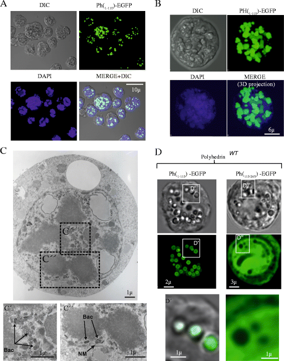
A wild type copy of polyhedrin is required to incorporate polyhedrin fragments into the crystal . A, confocal microscopy images illustrating that the polyhedrin fragment PH(1–110)-EGFP forms aggregates inside the nuclei of infected cells (when expressed in the absence of WT polyhedrin). B, Tridimensional confocal reconstruction of a cell expressing PH(1–110)-EGFP. Notice the formation of aggregates inside the nucleus. C, The PH(1–110)-EGFP aggregates are visible in TEM as dense amorphous particles, and they do not contain baculoviruses inside. Notice that in fact viruses are excluded from the aggregates (C’ and C”, indicates as Bac and arrows, NM = nuclear membrane). D, Only the amino terminal fragment from polyhedrin can be incorporated into polyhedra crystals (when co-expressed with wild type polyhedrin). Notice that only the fragment PH(1–110)-EGFP form polyhedra (D’). The carboxyl terminus fragment PH(1–110)-EGFP is not incorporated into the polyhedra crystals, in fact it is excluded from the crystal and observed as a soluble protein in the cell cytosol (D”).
Figure 7
Figure 7
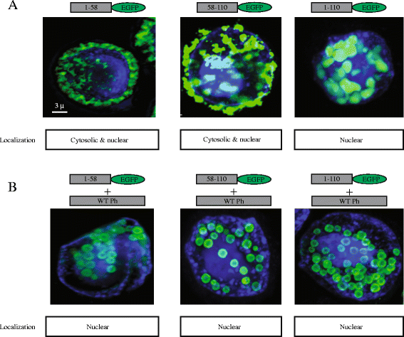
Identification of the minimum fragment from polyhedrin that retains the self-aggregation property. A, confocal microscopy studies using the fragments PH(1–58)­EGFP, PH(58–110)­EGFP and PH(1–110)­EGFP alone, or in co-expression with WT polyhedrin (B). Boxes below each panel indicate cellular localization based on the degree of co-localization with the nuclear marker DAPI. Notice that fragment PH(1–58)­EGFP is soluble and found in both the nucleus and the cytosol. Fragments PH(58–110)­EGFP and PH(1–110)­EGFP can form self-aggregates when expressed alone or be incorporated into polyhedra, when co-expressed with WT polyhedrin. Thus the minimum self-aggregating fragment identified in this study was PH(58–110)­EGFP.

Similar articles

Cited by

References

    1. Elias CB, Jardin B, Kamen A. Recombinant protein production in large-scale agitated bioreactors using the baculovirus expression vector system. Methods Mol Biol. 2007;388:225–46. doi: 10.1007/978-1-59745-457-5_11. - DOI - PubMed
    1. Trowitzsch S, Bieniossek C, Nie Y, Garzoni F, Berger I. New baculovirus expression tools for recombinant protein complex production. J Struct Biol. 2010;172:45–54. doi: 10.1016/j.jsb.2010.02.010. - DOI - PubMed
    1. Patterson RM, Selkirk JK, Merrick BA. Baculovirus and insect cell gene expression: review of baculovirus biotechnology. Env Heal Perspect. 1995;103:756–9. doi: 10.1289/ehp.95103756. - DOI - PMC - PubMed
    1. Carstens EB, Krebs A, Gallerneault CE. Identification of an amino acid essential to the normal assembly of Autographa californica nuclear polyhedrosis virus polyhedra. J Virol. 1986;58:684–8. - PMC - PubMed
    1. Ramoska WA, Stairs GR, Hink WF. Ultraviolet light activation of insect nuclear polyhedrosis virus. Nature. 1975;253:628–9. doi: 10.1038/253628a0. - DOI - PubMed

Publication types

MeSH terms

Substances

LinkOut - more resources