. 2015 Feb 4;15(1):17.
doi: 10.1186/s12866-015-0354-3.

Tn5/7-lux: a versatile tool for the identification and capture of promoters in gram-negative bacteria

Affiliations

Tn5/7-lux: a versatile tool for the identification and capture of promoters in gram-negative bacteria

Steven T Bruckbauer et al. BMC Microbiol. .

Abstract

Background: The combination of imaging technologies and luciferase-based bioluminescent bacterial reporter strains provide a sensitive and simple non-invasive detection method (photonic bioimaging) for the study of diverse biological processes, as well as efficacy of therapeutic interventions, in live animal models of disease. The engineering of bioluminescent bacteria required for photonic bioimaging is frequently hampered by lack of promoters suitable for strong, yet stable luciferase gene expression.

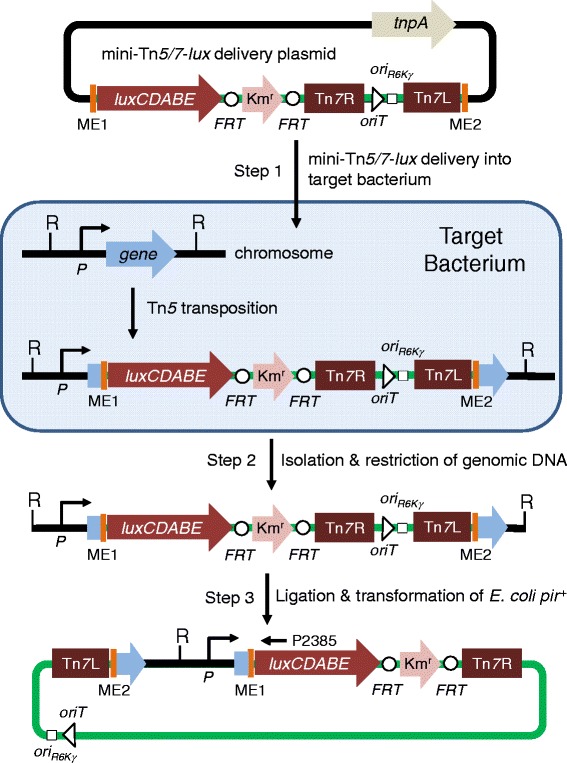
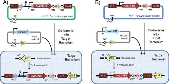
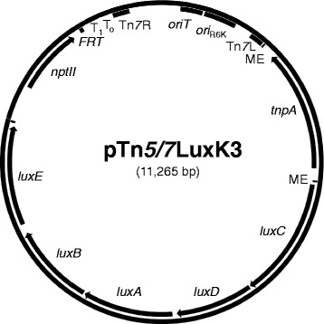
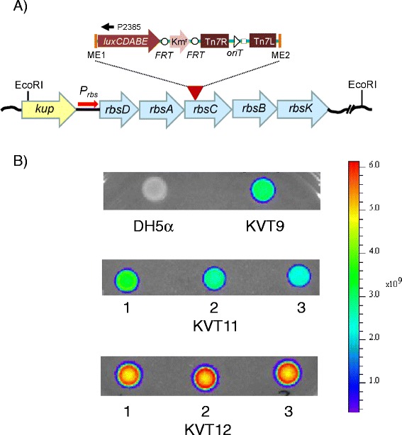
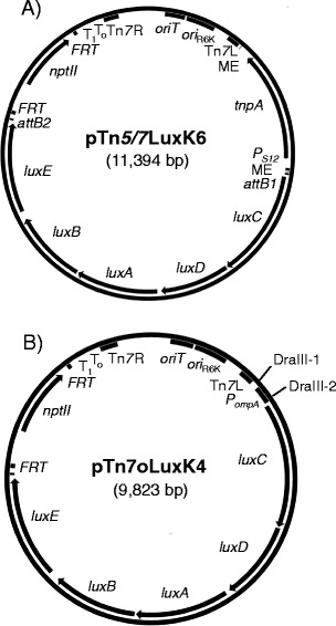
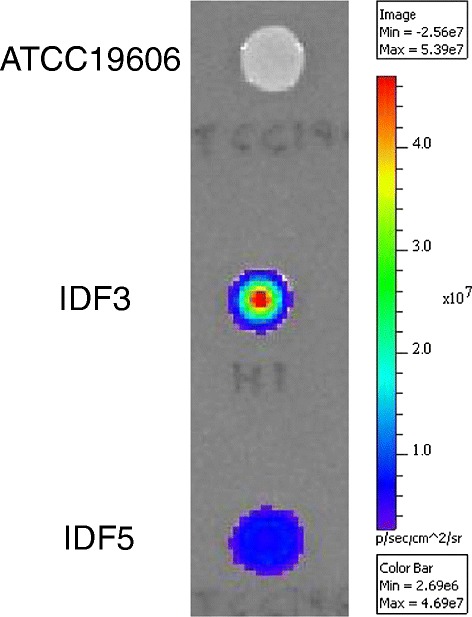
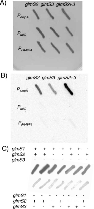
Results: We devised a novel method for identification of constitutive native promoters in Gram-negative bacteria. The method is based on a Tn5/7 transposon that exploits the unique features of Tn5 (random transposition) and Tn7 (site-specific transposition). The transposons are designed such that Tn5 transposition will allow insertion of a promoter-less bacterial luxCDABE operon downstream of a bacterial gene promoter. Cloning of DNA fragments from luminescent isolates results in a plasmid that replicates in pir (+) hosts. Sequencing of the lux-chromosomal DNA junctions on the plasmid reveals transposon insertion sites within genes or operons. The plasmid is also a mini-Tn7-lux delivery vector that can be used to introduce the promoter-lux operon fusion into other derivatives of the bacterium of interest in an isogenic fashion. Alternatively, promoter-containing sequences can be PCR-amplified from plasmid or chromosomal DNA and cloned into a series of accompanying mini-Tn7-lux vectors. The mini-Tn5/7-lux and mini-Tn7-lux vectors are equipped with diverse selection markers and thus applicable in numerous Gram-negative bacteria. Various mini-Tn5/7-lux vectors were successfully tested for transposition and promoter identification by imaging in Acinetobacter baumannii, Escherichia coli, and Burkholderia pseudomallei. Strong promoters were captured for lux expression in E. coli and A. baumannii. Some mini-Tn7-lux vectors are also equipped with attB sites for swapping of the lux operon with other reporter genes using Gateway technology.

Conclusions: Although mini-Tn5-lux and mini-Tn7-lux elements have previously been developed and used for bacterial promoter identification and chromosomal insertion of promoter-lux gene fusions, respectively, the newly developed mini-Tn5/7-lux and accompanying accessory plasmids streamline and accelerate the promoter discovery and bioluminescent strain engineering processes. Availability of vectors with diverse selection markers greatly extend the host-range of promoter probe and lux gene fusion vectors.


Figures

Figure 1
Figure 1
Promoter identification and capture using mini-Tn 5/7-lux elements. Step 1 involves random Tn5 transposition into the chromosome of the bacterial host of interest (target bacterium) after conjugal transfer of the mini-Tn5/7-lux delivery plasmid. This plasmid also contains the Tn5 transposase encoding tnpA gene. TnpA acts on the Tn5 mosaic ends (ME), which results in random mini-Tn5/7-lux transposition into the bacterial chromosome, including insertion in a gene where the Photorhabdus luminescens luxCDABE operon is transcribed from the gene’s promoter (P). The mini-Tn7 element, i.e. sequences flanked by the Tn7 left (Tn7L) and right (Tn7R) ends, located on the mini-Tn5/7-lux delivery plasmid does not transpose in this step because the delivery plasmid does not encode Tn7 transposase. Mini-Tn5/7-lux chromosomal insertion is stable because the chromosomally-integrated elements do neither encode Tn5 nor Tn7 transposase. In step 2, chromosomal DNA of in this example kanamycin resistance (Kmr) and light-emitting transformants is isolated and digested with a restriction enzyme (R) that does not cleave within the transposed element. In step 3, chromosomal DNA fragments are religated and a plasmid containing the R6K origin of replication (ori R6K) and origin of conjugal transfer (oriT) is recovered by transformation of a pir + E. coli host and selecting Kmr transformants. Sequencing of plasmid DNA with a luxC-specific primer (P2385) will reveal the transposon insertion site and putative promoter sequences. The Kmr selection marker contained on the chromosomally integrated mini-Tn7-lux elements is flanked by Flp recombinase target (FRT) sites for optional marker excision with Saccharomyces cerevisiae Flp recombinase.
Figure 2
Figure 2
Methods for site-specific insertion of promoter- lux fusions using mini-Tn 7-lux elements. Chromosomal integration of promoter-lux fusions can be achieved in two ways. (A ) The plasmid recovered in step 3 of the promoter identification and capture procedure illustrated in Figure 1 is a functional mini-Tn7 delivery plasmid which in some instances (e.g. when the plasmid contains short regions of promoter-containing chromosomal DNA or when RecA-deficient target strains are available) may be used to directly transpose site-specifically into the glmS gene-associated Tn7 attachment site (attTn7) in the chromosome of the bacterium under study to obtain luminescent derivatives. Site-specific mini-Tn7-lux insertion is achieved by co-transfer of the mini-Tn7-lux delivery plasmid and a helper plasmid that encodes the site-specific TnsABCD transposition pathway, which acts on the Tn7 left (Tn7L) and right (Tn7R) ends. Both plasmids contain the origin of transfer (oriT) for conjugal transfer into the target bacterium and the conditional R6K origin of replication (ori R6K), which limits their replication to E. coli hosts expressing the plasmid R6K π protein. The mini-Tn5 element (sequences flanked my the mosaic ends, ME) contained on the mini-Tn7-lux delivery plasmid does not transpose because the delivery plasmid does not encode Tn5 transposase. (B ) Alternatively, the promoter identified by sequencing the mini-Tn5/7-lux-chromosomal junction sequences located on the plasmid rescued in step 3 of the procedure illustrated in Figure 1 can be cloned into other mini-Tn7-lux elements. These are then transposed into the target bacterium for obtaining bioluminescent bacteria by site-specific mini-Tn7-lux transposition as described above. In both scenarios, A and B, the Kmr selection marker contained on the chromosomally integrated mini-Tn7-lux elements is flanked by Flp recombinase target (FRT) sites for optional marker excision with Saccharomyces cerevisiae Flp recombinase. The ampicillin resistance (Apr) marker is used for selection and maintenance of the helper plasmid in E. coli.
Figure 3
Figure 3
Map of mini-Tn 5/7 - lux delivery plasmid pTn 5/7 LuxK3. The mini-Tn5/7-lux element flanked by the Tn5 mosaic ends (ME) carries a promoter-less P. luminescens luxCDABE operon, the kanamycin resistance encoding nptII gene, the Tn7 left (Tn7L) and right (Tn7R) ends, the R6K origin of replication (ori R6K), and an origin of conjugative transfer (oriT). The Tn5 transposase-encoding tnpA gene is located outside of the transposable element. Other abbreviations: FRT, single Flp recombinase target site; T0T1, transcriptional terminators T0 and T1 from bacteriophage λ and E. coli rrnB operon, respectively.
Figure 4
Figure 4
Mini-Tn 5/7 - lux aided promoter identification and capture in E. coli . A) The promoter-less mini-Tn5/7-lux element (indicated by the red arrowhead and expanded inset above it) from pTn5/7LuxK3 was transposed into the DH5α chromosome. Chromosomal DNA from a Kmr and luminescent exconjugant (KVT9; panel B) was isolated, digested with EcoRI, religated and transformed into CC118(λpir +). The lux operon-chromosomal DNA junction on the plasmid was sequenced with the luxC-specific primer P2385. The mini-Tn5/7-lux transposon was inserted in rbsC, the third gene of the rbsDACBK operon required for the transport and initial metabolic steps of ribose. The kup gene located upstream of the rbsDACBK operon encodes a potassium transporter. B) The recovered plasmid was used to transpose the mini-Tn7-P rbs-lux element residing on it to the attTn7 site on the DH5α chromosome resulting in strain KVT11. In a parallel effort, The rbs operon promoter (P rbs) located in the 167 bp kup-rbsD intergenic region was PCR-amplified and cloned on a 173 bp StuI-DraIII fragment into pTn5/7LuxK4 where it replaced the tnpA gene and flanking MEs to drive transcription of the lux operon. The resulting mini-Tn7-P rbs-lux element was transposed into the attTn7 site on the DH5α chromosome resulting in strain KVT12. Ten μl samples of an overnight culture of the indicated strains were spotted on an LB plate, grown overnight at 37°C and light emission was measured using a Xenogen IVIS imager.
Figure 5
Figure 5
Maps of next generation mini-Tn 5/7 - lux and mini-Tn 7 - lux delivery vectors. A) pTn5/7LuxK6. As in pTn5/7LuxK3 (Figure 2) the mini-Tn5/7-lux element is flanked by the Tn5 mosaic ends (ME) and carries a promoter-less P. luminescens luxCDABE operon, in this example the kanamycin resistance encoding nptII gene, the Tn7 left (Tn7L) and right (Tn7R) ends, the R6K origin of replication (ori R6K), and an origin of conjugative transfer (oriT). The new generation of vectors is further functionalized by 1) transcription of the Tn5 transposase-encoding tnpA gene located outside of the transposable element by the constitutive S12 gene promoter (P S12) from Burkholderia thailandensis; 2) flanking of the antibiotic resistance marker by functional Flp recombinase target (FRT) sites; and 3) flanking of the lux operon by attB sites for Gateway recombineering. The pTn5/7LuxG6, pTn5/7LuxT6 and pTn5/7LuxTc6 contain gentamicin, trimethoprim and tetracycline resistance markers, respectively. Other abbreviations are defined in the Figure 2 legend. B) pTn7oLuxK4. This vector contains many features of the pTn5/7Lux series but does not contain the Tn5 transposase gene or mosaic ends. Its unique features include two DraIII sites that because of the 3-nucleotide ambiguity (indicated by bold letters) in the DraIII recognition sites (DraIII-1 5′-CACTATGTG and DraIII-2 5′- CACCGCGTG) can be used for directional cloning of promoter-containing DNA fragments for transcription of the lux operon genes. In the illustrated example the lux operon is transcribed from the B. pseudomallei ompA promoter (P ompA). This is also indicated by the “o” in the plasmid name. The pTn7oLuxG4 and pTn7oLuxT4 contain P ompA and the gentamicin (aacC1) and trimethoprim (dhfRII) resistance markers, respectively. Other abbreviations are as in A).
Figure 6
Figure 6
Mini-Tn 5/7 - lux aided promoter identification and capture in A. baumannii . The promoter-less mini-Tn5/7-lux element from pTn5/7LuxK5 was transposed into the A. baumannii strain ATCC19606 chromosome. Chromosomal DNA from a Kmr and luminescent exconjugant (IFD3) was isolated, digested with Acc65I, religated and transformed into E. coli pir-116 + strain MaH1. DNA from a Kmr transformants was sequenced with the luxC-specific primer P2385. The mini-Tn5/7-lux transposon was inserted in a gene annotated as A1S_0947 in strain ATCC17978 and the upstream A1S_0944-A1S_0945 intergenic region containing several possible promoters was PCR-amplified and cloned on a 329-bp StuI-DraIII fragment into pTn5/7LuxK5. The mini-Tn7-P H1A-lux element was transposed into the attTn7 site on the ATCC19606 chromosome resulting in strain IFD5. Ten μl samples of an overnight culture of the indicated strains were spotted on an LB plate, grown overnight at 37°C and light emission was measured using a Xenogen IVIS imager.
Figure 7
Figure 7
Promoter strength and insertion site location determine bioluminescence signal strength in a bacterium with multiple Tn 7 insertion sites. Mini-Tn7 elements in which lux operon expression is directed from the indicated promoters were transposed into the genome of B. pseudomallei strain Bp82.27 and insertion at either glmS1-, glmS2- or glmS3-associated attTn7 sites was determined by PCR. Colonies were patched on LB agar plates with 35 μg/ml kanamycin and incubated at 37°C. Patches were imaged using a Bio-Rad Universal Hood II ChemiDocXRS using high sensitivity chemiluminescence settings. A) and B) Bp82.27 with mini-Tn7-lux insertions derived from transposition from pTn7oLuxK3, pTn7tLuxK3 and pTn7xLuxK3. The promoters directing lux operon expression in these constructs are B. pseudomallei P ompA, B. pseudomallei P tolC and P. aeruginosa P PA4974, respectively. Patches were grown overnight at 37°C and either imaged using white light and a 0.007 s exposure time (A) or imaged for luminescence using a 30 s exposure time (B). C) Insertion site dependence of bioluminescence intensity. The mini-Tn7-P ompA -lux element from pTn7oLuxK4 was transposed into the Bp82.27 genome. Insertion sites were determined by PCR. Patches were grown overnight at 37°C and imaged for luminescence using a 30 s exposure time.

Similar articles

Cited by

References

    1. Contag CH, Contag PR, Mullins JI, Spilman SD, Stevenson DK, Benaron DA. Photonic detection of bacterial pathogens in living hosts. Mol Microbiol. 1995;18:593–603. doi: 10.1111/j.1365-2958.1995.mmi_18040593.x. - DOI - PubMed
    1. Contag CH, Bachmann MH. Advances in in vivo bioluminescence imaging of gene expression. Annu Rev Biomed Eng. 2002;4:235–60. doi: 10.1146/annurev.bioeng.4.111901.093336. - DOI - PubMed
    1. Piwnica-Worms D, Schuster DP, Garbow JR. Molecular imaging of host-pathogen interactions in intact small animals. Cell Microbiol. 2004;6:319–31. doi: 10.1111/j.1462-5822.2004.00379.x. - DOI - PubMed
    1. Kadurugamuwa JL, Francis KP. Bioluminescent imaging of bacterial biofilm infections in vivo. Methods Mol Biol. 2008;431:225–39. - PubMed
    1. Kong Y, Shi Y, Chang M, Akin AR, Francis KP, Zhang N, et al. Whole-body imaging of infection using bioluminescence. Curr Prot Microbiol. 2011;Chapter 2:Unit 2C 4. - PMC - PubMed

Publication types

MeSH terms

LinkOut - more resources