

Histochemical and immunohistochemical characterization of chordoma in ferrets
- PMID: 25648567
- PMCID: PMC4427749
- DOI: 10.1292/jvms.14-0488
Histochemical and immunohistochemical characterization of chordoma in ferrets
Abstract
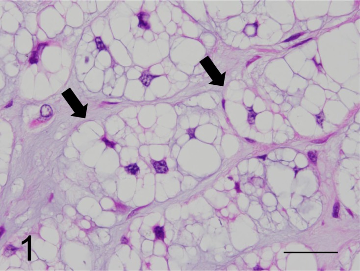
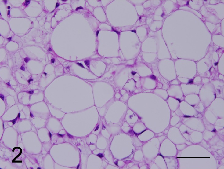
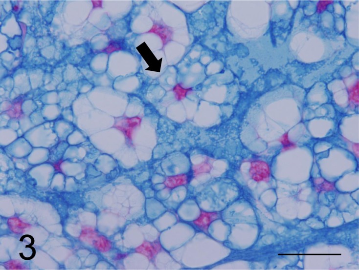
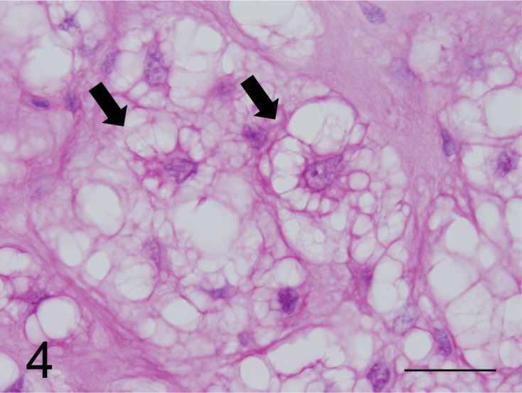
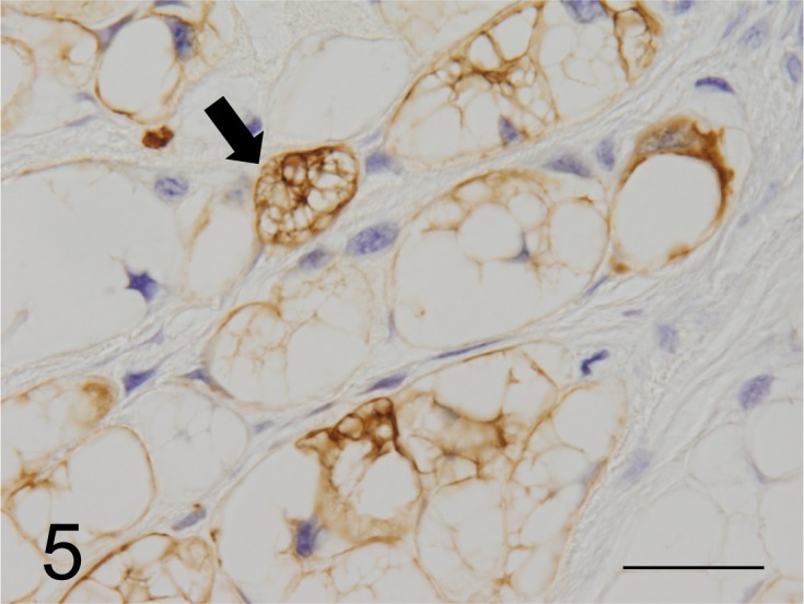
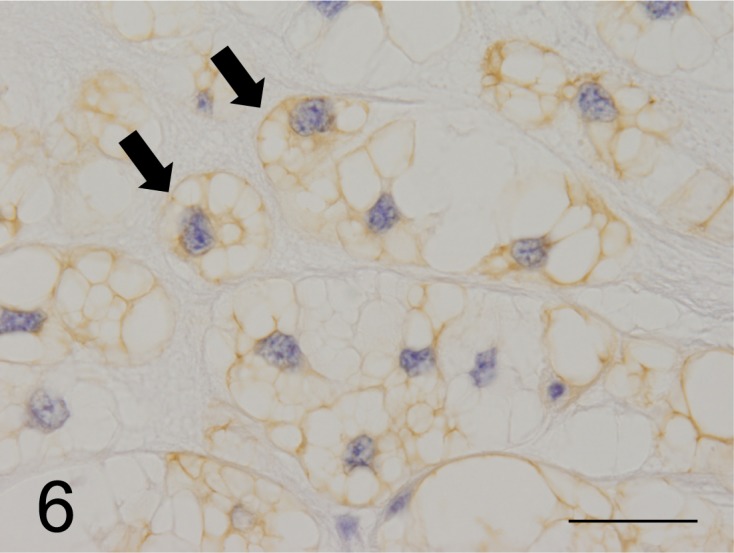
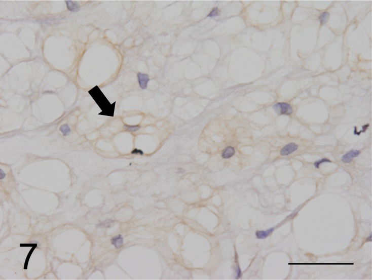
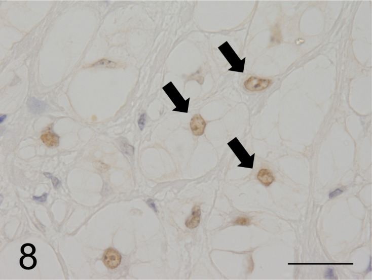
Chordomas of the tip of the tail in 6 ferrets were examined using histopathological, histochemical and immunohistochemical procedures. Histopathologically, round neoplastic cells containing numerous cytoplasmic vacuoles of varying sizes, categorized as "physaliphorous cells", were observed in the amorphous eosinophilic or pale basophilic myxoid stroma. Physaliphorous cells were arranged in lobules and in a "chordoid" or "cobblestone" manner. The neoplasms were diagnosed as benign chordoma without local invasion and metastasis. Histochemically, the cytoplasm of small neoplastic cells was positive for periodic acid-Schiff stain and alcian blue (AB) pH 2.5 and pH 1.0 stains, but negative for hyaluronidase digestion-AB pH 2.5 stain. All neoplastic cells were strongly stained with colloidal ion, negative for high iron diamine AB pH 2.5 and toluidine blue pH 2.5 stains, and positive for Mayer's mucicarmine stain. Immunohistochemistry using antibodies directed against low-molecular-weight cytokeratins (CK18, CK19 and CK20), vimentin and mucin core protein (MUC5AC) revealed that neoplastic cells had both epithelial and mesenchymal elements. The expression of low-molecular-weight cytokeratins suggests that neoplastic cells acquired the properties of glandular epithelial cells and produced epithelial mucus. Furthermore, the expression of cytokeratins, vimentin, S100 protein, brachyury and epithelial membrane antigen indicates that the neoplasms were equivalent to the classic type of human chordoma. Therefore, immunohistochemistry using these antibodies can be useful for the characterization of ferret chordoma.
Figures
References
-
- Flanagan A. M., Yamabuchi T.2013. Chordoma. pp. 328–329. In: WHO Classification of Tumours of Soft Tissue and Bone, 4th ed. (Christopher, D. M., Fletcher, Julia A., Bridge, Pancras C. W. Hogendoorn. and Fredrik, M. eds.), International Agency for Research on Cancer (IARC), Lyon.
MeSH terms
LinkOut - more resources
Full Text Sources
Other Literature Sources
Medical