

Tract Profiles of the Cerebellar White Matter Pathways in Children and Adolescents
- PMID: 25648754
- PMCID: PMC4524802
- DOI: 10.1007/s12311-015-0652-1
Tract Profiles of the Cerebellar White Matter Pathways in Children and Adolescents
Abstract
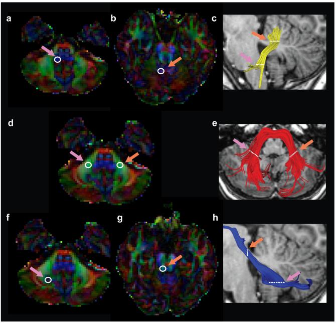
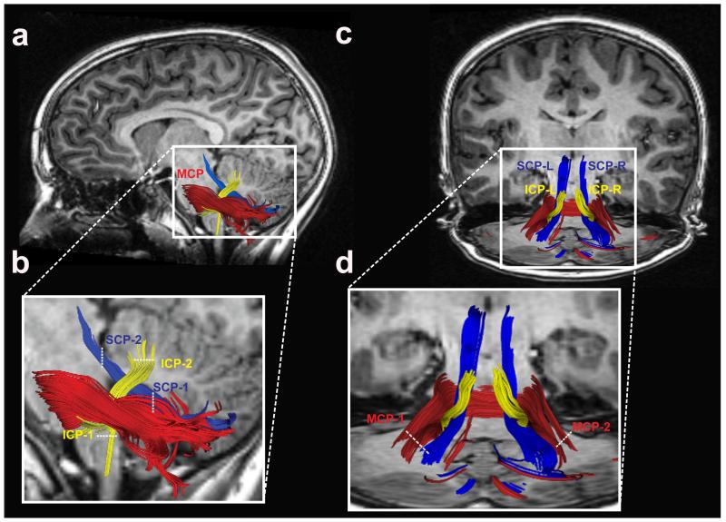
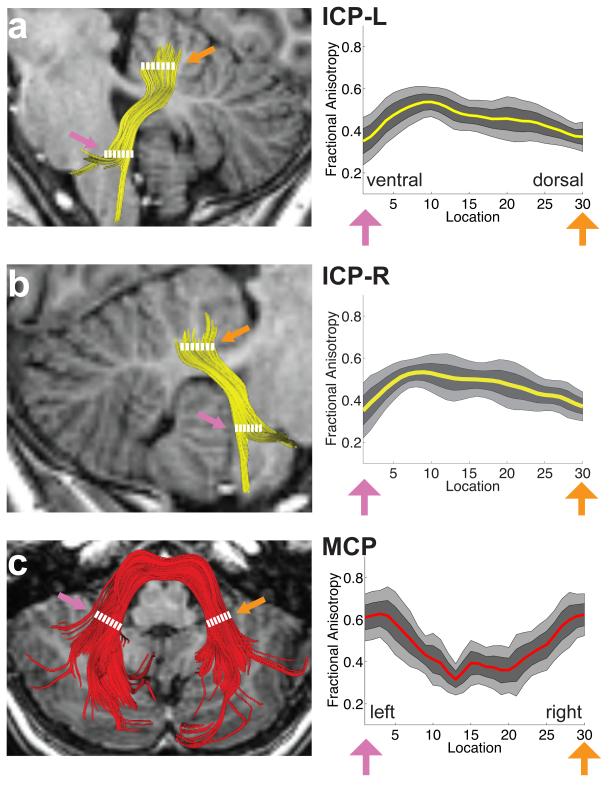
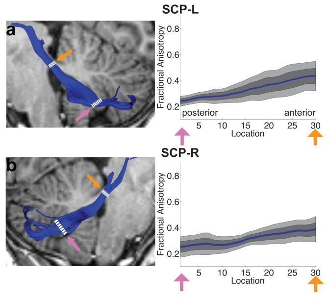
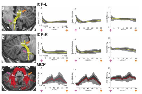
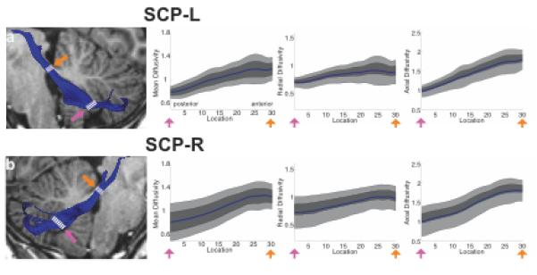
Intact development of cerebellar connectivity is essential for healthy neuromotor and neurocognitive development. To date, limited knowledge about the microstructural properties of the cerebellar peduncles, the major white matter tracts of the cerebellum, is available for children and adolescents. Such information would be useful as a comparison for studies of normal development, clinical conditions, or associations of cerebellar structures with cognitive and motor functions. The goal of the present study was to evaluate the variability in diffusion measures of the cerebellar peduncles within individuals and within a normative sample of healthy children. Participants were 19 healthy children and adolescents, aged 9-17 years, mean age 13.0 ± 2.3. We analyzed diffusion magnetic resonance imaging (dMRI) data with deterministic tractography. We generated tract profiles for each of the cerebellar peduncles by extracting four diffusion properties (fractional anisotropy (FA) and mean, radial, and axial diffusivity) at 30 equidistant points along each tract. We were able to identify the middle cerebellar peduncle and the bilateral inferior and superior cerebellar peduncles in all participants. The results showed that within each of the peduncles, the diffusion properties varied along the trajectory of the tracts. However, the tracts showed consistent patterns of variation across individuals; the coefficient of variation for FA across individual profiles was low (≤20%) for each tract. We observed no systematic variation of the diffusion properties with age. These cerebellar tract profiles of the cerebellar peduncles can serve as a reference for future studies of children across the age range and for children and adolescents with clinical conditions that affect the cerebellum.
Keywords: Cerebellum; Development; Diffusion magnetic resonance imaging (dMRI); Premature birth; Tractography; White matter.
Figures
Similar articles
-
Cerebellar white matter pathways are associated with reading skills in children and adolescents.Hum Brain Mapp. 2015 Apr;36(4):1536-53. doi: 10.1002/hbm.22721. Epub 2014 Dec 12. Hum Brain Mapp. 2015. PMID: 25504986 Free PMC article.
-
Age-Dependent White Matter Characteristics of the Cerebellar Peduncles from Infancy Through Adolescence.Cerebellum. 2019 Jun;18(3):372-387. doi: 10.1007/s12311-018-1003-9. Cerebellum. 2019. PMID: 30637673
-
Diffusion Tensor Tractography of the Cerebellar Peduncles in Prematurely Born 7-Year-Old Children.Cerebellum. 2017 Apr;16(2):314-325. doi: 10.1007/s12311-016-0796-7. Cerebellum. 2017. PMID: 27255706 Free PMC article.
-
Probing the neuroanatomy of the cerebellum using tractography.Handb Clin Neurol. 2018;154:235-249. doi: 10.1016/B978-0-444-63956-1.00014-X. Handb Clin Neurol. 2018. PMID: 29903442 Review.
-
Functional tracts of the cerebellum-essentials for the neurosurgeon.Neurosurg Rev. 2021 Feb;44(1):273-278. doi: 10.1007/s10143-020-01242-1. Epub 2020 Feb 13. Neurosurg Rev. 2021. PMID: 32056026 Free PMC article.
Cited by
-
Abnormal white matter properties in adolescent girls with anorexia nervosa.Neuroimage Clin. 2015 Oct 23;9:648-59. doi: 10.1016/j.nicl.2015.10.008. eCollection 2015. Neuroimage Clin. 2015. PMID: 26740918 Free PMC article.
-
Local-to-distant development of the cerebrocerebellar sensorimotor network in the typically developing human brain: a functional and diffusion MRI study.Brain Struct Funct. 2019 Apr;224(3):1359-1375. doi: 10.1007/s00429-018-01821-5. Epub 2019 Feb 7. Brain Struct Funct. 2019. PMID: 30729998 Free PMC article.
-
Diffusion weighted imaging evidence of extra-callosal pathways for interhemispheric communication after complete commissurotomy.Brain Struct Funct. 2019 Jun;224(5):1897-1909. doi: 10.1007/s00429-019-01864-2. Epub 2019 May 7. Brain Struct Funct. 2019. PMID: 31062161 Free PMC article.
-
Spatiotemporal changes in along-tract profilometry of cerebellar peduncles in cerebellar mutism syndrome.Neuroimage Clin. 2022;35:103000. doi: 10.1016/j.nicl.2022.103000. Epub 2022 Mar 30. Neuroimage Clin. 2022. PMID: 35370121 Free PMC article.
-
Procedural and declarative memory brain systems in developmental language disorder (DLD).Brain Lang. 2020 Jun;205:104789. doi: 10.1016/j.bandl.2020.104789. Epub 2020 Mar 30. Brain Lang. 2020. PMID: 32240854 Free PMC article.
References
-
- Ito M. Control of mental activities by internal models in the cerebellum. Nature Reviews Neuroscience. 2008;9(4):304–13. PubMed PMID: 18319727. - PubMed
-
- Hyam JA, Owen SL, Kringelbach ML, Jenkinson N, Stein JF, Green AL, et al. Contrasting connectivity of the ventralis intermedius and ventralis oralis posterior nuclei of the motor thalamus demonstrated by probabilistic tractography. Neurosurgery. 2012;70(1):162–9. discussion 9. PubMed PMID: 22158304. - PubMed
-
- Schmahmann JD. An emerging concept. The cerebellar contribution to higher function. Archives of Neurology. 1991;48(11):1178–87. PubMed PMID: 1953406. - PubMed
-
- Schmahmann JD, Sherman JC. The cerebellar cognitive affective syndrome. Brain. 1998;121(Pt 4):561–79. PubMed PMID: 9577385. - PubMed
-
- Schmahmann JD. Disorders of the cerebellum: ataxia, dysmetria of thought, and the cerebellar cognitive affective syndrome. Journal of Neuropsychiatry & Clinical Neurosciences. 2004;16(3):367–78. PubMed PMID: 15377747. - PubMed
Publication types
MeSH terms
Grants and funding
LinkOut - more resources
Full Text Sources
Other Literature Sources
