. 2015 Mar 27;290(13):8154-65.
doi: 10.1074/jbc.M114.615922. Epub 2015 Feb 3.

The intracellular domain of teneurin-1 induces the activity of microphthalmia-associated transcription factor (MITF) by binding to transcriptional repressor HINT1

Affiliations

The intracellular domain of teneurin-1 induces the activity of microphthalmia-associated transcription factor (MITF) by binding to transcriptional repressor HINT1

Jonas Schöler et al. J Biol Chem. .

Abstract

Teneurins are large type II transmembrane proteins that are necessary for the normal development of the CNS. Although many studies highlight the significance of teneurins, especially during development, there is only limited information known about the molecular mechanisms of function. Previous studies have shown that the N-terminal intracellular domain (ICD) of teneurins can be cleaved at the membrane and subsequently translocates to the nucleus, where it can influence gene transcription. Because teneurin ICDs do not contain any intrinsic DNA binding sequences, interaction partners are required to affect transcription. Here, we identified histidine triad nucleotide binding protein 1 (HINT1) as a human teneurin-1 ICD interaction partner in a yeast two-hybrid screen. This interaction was confirmed in human cells, where HINT1 is known to inhibit the transcription of target genes by directly binding to transcription factors at the promoter. In a whole transcriptome analysis of BS149 glioblastoma cells overexpressing the teneurin-1 ICD, several microphthalmia-associated transcription factor (MITF) target genes were found to be up-regulated. Directly comparing the transcriptomes of MITF versus TEN1-ICD-overexpressing BS149 cells revealed 42 co-regulated genes, including glycoprotein non-metastatic b (GPNMB). Using real-time quantitative PCR to detect endogenous GPNMB expression upon overexpression of MITF and HINT1 as well as promoter reporter assays using GPNMB promoter constructs, we could demonstrate that the teneurin-1 ICD binds HINT1, thus switching on MITF-dependent transcription of GPNMB.

Keywords: BEX1; GPNMB; Glioblastoma; MACF1; Microarray; ODZ; Protein-Protein Interaction; Receptor; TENM1; Transcription Repressor.


Figures

FIGURE 1.
FIGURE 1.
Novel interaction partners of teneurin-1 ICD identified by yeast two-hybrid screen. A, DUALhunter yeast two-hybrid screen. 1, when the teneurin-1 ICD (bait protein) and the prey protein do not interact, the reporter genes are not expressed. 2, once an interaction takes place, the C- and N-terminal parts of split ubiquitin are united. 3, a ubiquitin-specific protease releases the attached transcription factor. 4, the transcription factor switches on expression of the HIS3/LacZ reporters. B, interaction partners identified by the screen. Left, serial dilutions of yeast strain NMY51 transformed with the prey-NubG plasmid or an empty prey-NubG plasmid (Control), together with either the membrane-anchored bait plasmid TEN1-ICD-Cub-LexA-VP16 (+) or the membrane-bound empty bait plasmid Cub-LexA-VP16 (−) grown on −Trp/−Leu medium containing histidine. Center, the same dilutions of the transformed NMY51 yeast as shown on the left, grown on medium lacking histidine, thus showing growth only if an interaction between bait and prey proteins took place. Right, β-galactosidase assay of yeast transformed with the bait TEN1-ICD-Cub-LexA-VP16 together with the prey plasmids indicated or with an empty prey plasmid (Control).
FIGURE 2.
FIGURE 2.
Overexpression of the teneurin-1 ICD through a modified tet system in BS149 cells, a cell line expressing endogenous teneurin-1. A, left, quantitative RT-PCR screen of teneurin-1 expression in glioblastoma cell lines. Values are normalized to TBP, using the comparative CT method. Right, quantitative RT-PCR results of teneurin-1 expression comparing the glioblastoma cell line BS149 with different brain tissues. Values are normalized to TBP, using the comparative CT method. Note that teneurin-1 transcript levels in cerebellum are 181.8-fold higher than in BS149 cells. B, scheme of the modified tet system. Through the addition of Dex and Dox, the tet activator construct (irtTA/VP16/GBD) is released from HSP90 in the cytosol and induces expression of the ICD through the tet-CMV promoter. C, Western blot with anti-GFP showing the expression of the control construct GFP-His and the teneurin-1 ICD-GFP-His 24 h after the addition of Dex and Dox. Anti-vinculin is the internal control for equal loading. D, quantitative RT-PCR results show increased levels of the ICD-GFP fusion construct (using teneurin-1 ICD-GFP primers) compared with endogenous teneurin-1 levels (using teneurin-1 ECD primers) in BS149 cells overexpressing the ICD-GFP construct by a modified tet system, using cDNA prepared from the RNA used in the microarray. Values were normalized to TBP, using the comparative CT method. Note that teneurin-1 ICD transcript levels are 214.9 times higher than endogenous teneurin-1, which is in the range of the endogenous teneurin-1 present in cerebellum. Error bars, S.D.
FIGURE 3.
FIGURE 3.
Teneurin-1 ICD overexpression affects MITF target genes. IPA identified all of its eight MITF target genes in our microarray to be differentially regulated, six up-regulated (red) and two down-regulated (green). The arrows indicate that all genes are directly activated or inhibited by MITF. The regulation of these target genes is consistent with the database for all genes except for NGFR, which according to IPA is up-regulated by MITF but is down-regulated in our microarray.
FIGURE 4.
FIGURE 4.
Microarray identifies nine up-regulated MITF target genes due to teneurin-1 ICD overexpression. A, volcano plot showing the 430 differentially regulated genes when the teneurin-1 ICD is overexpressed in BS149 cells compared with BS149 cells with overexpression of GFP only (-fold change >1.5; p < 0.05). MITF target genes are marked in the plot. B, detailed view of the expression levels of the 11 MITF target genes in a heat map, where bright green means little or no expression and bright red means high expression. Nine genes are up-regulated, and two genes are down-regulated, with the -fold change indicated. C, quantitative RT-PCR confirmed the up-regulation of some of the MITF target genes. Values are normalized to TBP, and -fold change values compare overexpression of teneurin-1 ICD-GFP-His with GFP-His. Error bars, S.D.
FIGURE 5.
FIGURE 5.
Proximity ligation assay confirms the interaction between HINT1 and the TEN1-ICD. A, immunocytochemistry (ICC) images of COS-7 cells co-transfected with TEN1ICD-HA and HINT1-MYC stained with anti-HA (red) and anti-MYC (green) and the merged channels showing co-expression in yellow. B, proximity ligation assay images of COS-7 cells co-transfected with TEN1ICD-HA and HINT1-MYC (left and center images) and COS-7 cells transfected only with HINT1-MYC as a negative control (right image); nuclei are stained with DAPI; white bar, 50 μm.
FIGURE 6.
FIGURE 6.
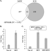
MITF-induced overexpression of GPNMB is attenuated by co-transfection of HINT1. A, a total of 497 genes are differentially regulated when MITF is overexpressed in BS149 cells. Of these genes, 42 are either up- or down-regulated due to both MITF and teneurin-1 ICD overexpression. One gene that is up-regulated in both microarrays is MITF target gene GPNMB (-fold change after MITF induction in parentheses). Overexpression of MITF-RFP-HA is compared with that of RFP-HA. Biological triplicates were used for the microarray. B, quantitative RT-PCR confirmed the up-regulation of GPNMB due to MITF overexpression. Values are normalized to TBP; -fold change value compares overexpression of MITF-RFP-HA with that of RFP-HA. C, quantitative RT-PCR results show a reduction of MITF-dependent transcription of GPNMB in the presence of HINT1. Values are normalized to TBP; -fold change values are compared with co-transfection of RFP-HA and CFP-C1 constructs (Neg. ctrl); the graph is representative of four separate experiments. The inhibition of GPNMB due to HINT1 compares the co-transfection of MITF-RFP-HA and HINT1-CFP-MYC with the co-transfection of MITF-RFP-HA and CFP-C1. Co-transfected cells were FACS-sorted before RNA isolation. Error bars, S.D.
FIGURE 7.
FIGURE 7.
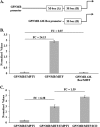
The GPNMB promoter is induced by MITF in BS149 cells and further increased by overexpression of the teneurin-1 ICD. A, diagram of the full-length GPNMB promoter and the GPNMB ΔM-box promoter, missing the M-box required for MITF-dependent regulation. B, SEAP assays show that MITF can induce the full-length GPNMB promoter activity, whereas the GPNMB ΔM-box promoter is not inducible. The transfection efficiency was normalized to luciferase assay values; -fold change values indicated by FC above the bars are compared with the full-length GPNMB promoter, co-transfected with empty pcDNA3.1 vector. C, SEAP assays show an increase of full-length GPNMB promoter activity when the teneurin-1 ICD is co-transfected with MITF, compared with MITF co-transfected with an empty pcDNA3.1 vector. The transfection efficiency was normalized to luciferase assay values, and -fold change values between the different conditions are indicated above the bars. Error bars, S.D.
FIGURE 8.
FIGURE 8.
The ICD of teneurin-1 regulates MITF-dependent GPNMB expression by competing for HINT1. A, teneurin-1 ECD is not bound to an interaction partner, and the intracellular domain is not released from the plasma membrane. HINT1 in the nucleus inhibits MITF at the promoter of GPNMB. B, 1, teneurin-1 interacts with a transmembrane protein of another cell. 2, both the extracellular and intracellular domains are released from the membrane. The ICD translocates to the nucleus. 3, the ICD competes for HINT1 binding, thus switching on MITF-dependent transcription of GPNMB.

References

    1. Baumgartner S., Chiquet-Ehrismann R. (1993) Tena, a Drosophila gene related to tenascin, shows selective transcript localization. Mech. Dev. 40, 165–176 - PubMed
    1. Baumgartner S., Martin D., Hagios C., Chiquet-Ehrismann R. (1994) Tenm, a Drosophila gene related to tenascin, is a new pair-rule gene. EMBO J. 13, 3728–3740 - PMC - PubMed
    1. Levine A., Bashan-Ahrend A., Budai-Hadrian O., Gartenberg D., Menasherow S., Wides R. (1994) Odd Oz: a novel Drosophila pair rule gene. Cell 77, 587–598 - PubMed
    1. Minet A. D., Chiquet-Ehrismann R. (2000) Phylogenetic analysis of teneurin genes and comparison to the rearrangement hot spot elements of E. coli. Gene 257, 87–97 - PubMed
    1. Oohashi T., Zhou X. H., Feng K., Richter B., Mörgelin M., Perez M. T., Su W. D., Chiquet-Ehrismann R., Rauch U., Fässler R. (1999) Mouse ten-m/Odz is a new family of dimeric type II transmembrane proteins expressed in many tissues. J. Cell Biol. 145, 563–577 - PMC - PubMed