. 2015 Mar 3;54(8):1681-93.
doi: 10.1021/bi501171r. Epub 2015 Feb 19.

The membrane proximal external regions of gp41 from HIV-1 strains HXB2 and JRFL have different sensitivities to alanine mutation

Affiliations

The membrane proximal external regions of gp41 from HIV-1 strains HXB2 and JRFL have different sensitivities to alanine mutation

Hyun Ah Yi et al. Biochemistry. .

Abstract

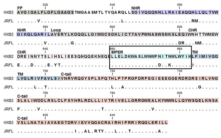
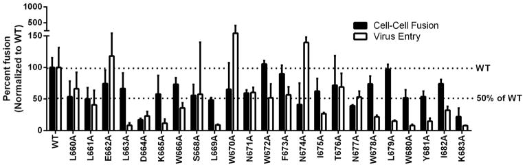
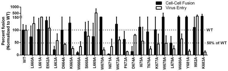
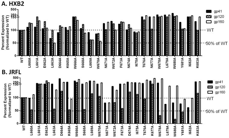
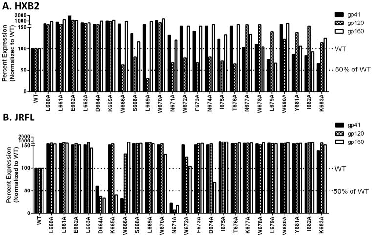
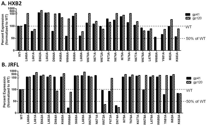
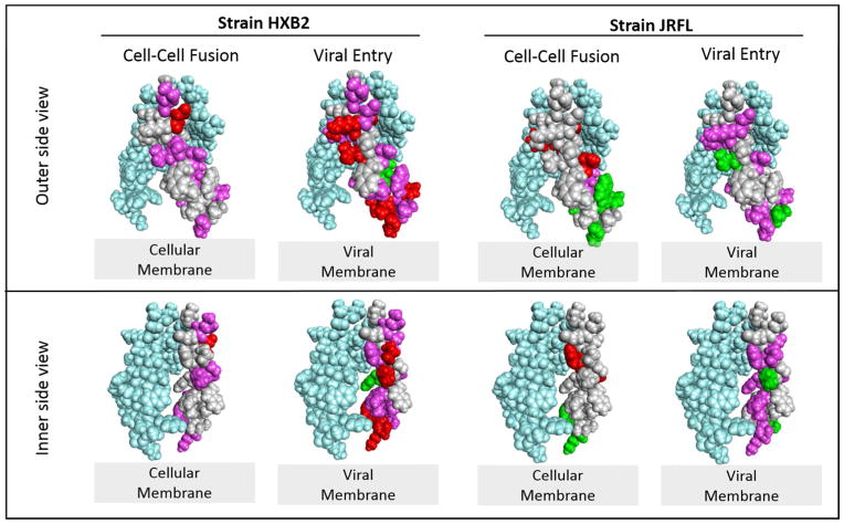
The transmembrane subunit (gp41) of the HIV envelope protein complex (Env) mediates the viral fusion step of HIV entry. The membrane proximal external region (MPER), one of the functional domains of gp41, has been the focus of a great deal of research because it is a target for neutralizing antibodies. In this study, we examined 23 amino acid residues in the MPER (660-683) in both a CXCR4 coreceptor-utilizing strain (HXB2) and a CCR5-utilizing strain (JRFL) by alanine scanning mutagenesis. Despite the high degree of gp41 sequence conservation, the effects of alanine mutation in the MPER were different between the two strains. Most mutations in HXB2 had fusogenicity and protein expression levels not less than 50% of that of the wild type in the case of cell-cell fusion. However, ∼30% of the mutants in HXB2 showed a severe defect in fusogenicity in viral entry. Mutations in the MPER of strain JRFL had more dramatic effects than that in HXB2 in cell-cell fusion and viral entry. The fact that there are large differences in the effects of mutation between two strains suggests the potential for the interaction of the MPER with nonconserved sequences such as the fusion peptide and/or other NHR domains as well as potential long-range structural effects on the conformational changes that occur with the Env complex during membrane fusion.


Figures

Figure 1
Figure 1. Alignment of gp41 amino acid sequence highlighting the MPER from the HXB2 and the JRFL strains
Gp41 begins at the N-terminus with the fusion peptide (FP) followed by the N-terminal heptad repeat (NHR), a loop, the C-terminal heptad repeat (CHR), a membrane proximal external region (MPER), and a transmembrane domain (TM) followed by a C-terminal cytoplasmic region (C-tail). The alignment of gp41 amino acid sequence was done using CLC Main Workbench 7.5.1. The numbering is shown based on HXB2 strain and the matching residues shown as dots. The boxed area indicates the MPER region. We made 23 alanine substitutions in both strain HXB2 and strain JRFL.
Figure 2
Figure 2. Effects of alanine substitution on HXB2 Env mediated cell-cell fusion and viral entry
For cell-cell fusion (black bars), each mutant plasmid was transfected into 293T cells along with plasmids expressing the tat and rev proteins. The transfected cells were then co-incubated with the receptor cell line TZM-bl for 24 hours. Fusion activity was measured by luciferase assay. For viral entry (white bars), each mutant was transfected into 293T cells along with the HIV backbone plasmid and virus was harvested after 48 hours. The amount of virus was determined by a p24 antigen capture ELISA assay, and virus stocks with an equivalent amount of p24 protein (100 ng) were added to each well of TZM-bl cells. Viral entry levels were determined by luciferase assay at 48 hours post infection. The WT level and the 50% of WT fusion are indicated with a dotted line.
Figure 3
Figure 3. Effects of alanine substitution on JRFL Env mediated cell-cell fusion and viral entry
Cell-cell fusion (black bars) and viral entry levels (white bars) were measured as described in Figure 2 using JRFL Env mutants.
Figure 4
Figure 4. Protein expression level in the cell lysates used for cell-cell fusion
293T cells transfected with Env, Tat, and Rev expressing plasmids. The cells were lysed and the total protein concentration was quantified by BCA assay. An equal amount of protein from each transfected cell sample was loaded onto an SDS-PAGE gel followed by Western blot analysis using Chessie-8 (for gp41, and gp160) and anti-gp120 antibodies. Each membrane was scanned and band intensity was quantified. The band intensities of gp41, gp160, and gp120 were normalized to the WT level of each protein. A) Protein expression level of HXB2 Env. B) Protein expression level of JRFL Env.
Figure 5
Figure 5. Protein expression level in the lysates of the virus-producing cells
293T cells were used for producing virus. These cells were lysed and the protein concentration was quantified by BCA assay. Subsequent Western blot analysis was performed using Chessie-8 and anti-gp120 antibodies followed by band quantification as described in Figure 4. Each membrane was reprobed with anti-p24 antibodies and band intensity was quantified. Each band intensity of gp41, gp160, and gp120 was normalized to the p24 level and then normalized again to the WT level of each protein. A) Protein expression level of HXB2 virus producing cells. B) Protein expression level of JRFL virus producing cells.
Figure 6
Figure 6. Protein levels on the virus
Concentrated virus with each mutant Env were lysed followed by Western blot analysis was performed as described above. A) Protein level on HXB2 virus. B) Protein level on JRFL virus.
Figure 7
Figure 7. Molecular models highlighting the functionality of mutations in the HIV gp41 MPER
A membrane associated MPER trimer structure was used to diagram the residues that are either diminished or enhanced in function upon mutation to alanine . For simplicity, mutations are indicated only on one of the monomers (light gray) and the other two monomers making up the trimer structure are shown in light blue. The top panel is a view of the outer side of the MPER. The bottom panel is a view that is rotated to display the inner side of the MPER. Amino acid residues without highlighting (light gray) have levels 80–120% of WT. Those colored pink have levels diminished to 20–50% of the WT level. Those colored red have levels diminished to < 20% of the WT level. The residues colored green have levels increased above 120% of the WT level. The structure was rendered with Discovery Studio Visualizer 4.0 based upon the coordinates from the Protein Data Bank entry 2LP7.

Similar articles

Cited by

References

    1. Caffrey M. HIV envelope: challenges and opportunities for development of entry inhibitors. Trends Microbiol. 2011;19:191–197. - PMC - PubMed
    1. Muster T, Steindl F, Purtscher M, Trkola A, Klima A, Himmler G, Ruker F, Katinger H. A conserved neutralizing epitope on gp41 of human immunodeficiency virus type 1. J Virol. 1993;67:6642–6647. - PMC - PubMed
    1. Salzwedel K, West JT, Hunter E. A conserved tryptophan-rich motif in the membrane-proximal region of the human immunodeficiency virus type 1 gp41 ectodomain is important for Env-mediated fusion and virus infectivity. J Virol. 1999;73:2469–2480. - PMC - PubMed
    1. Muñoz-Barroso I, Salzwedel K, Hunter E, Blumenthal R. Role of the membrane-proximal domain in the initial stages of human immunodeficiency virus type 1 envelope glycoprotein-mediated membrane fusion. Journal of Virology. 1999;73:6089–6092. - PMC - PubMed
    1. Montero M, van Houten NE, Wang X, Scott JK. The membrane-proximal external region of the human immunodeficiency virus type 1 envelope: dominant site of antibody neutralization and target for vaccine design. Microbiology and molecular biology reviews : MMBR. 2008;72:54–84. table of contents. - PMC - PubMed

Publication types

LinkOut - more resources