. 2015 Apr 1;75(7):1366-75.
doi: 10.1158/0008-5472.CAN-14-1849. Epub 2015 Feb 3.

BH3 Inhibitor Sensitivity and Bcl-2 Dependence in Primary Acute Lymphoblastic Leukemia Cells

Affiliations

BH3 Inhibitor Sensitivity and Bcl-2 Dependence in Primary Acute Lymphoblastic Leukemia Cells

Sarah E Alford et al. Cancer Res. .

Abstract

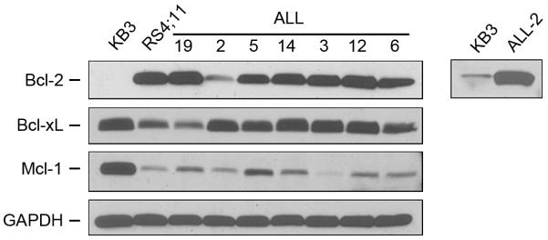
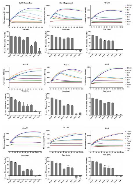
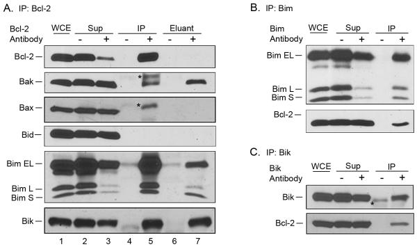
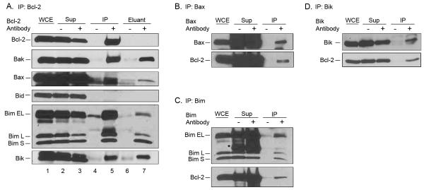
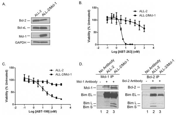
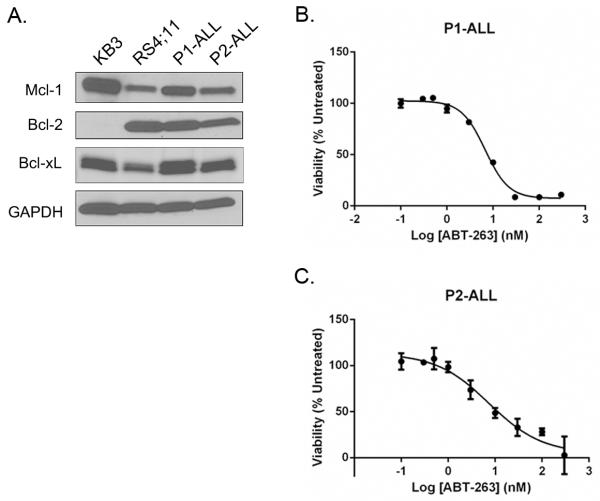
BH3 mimetic drugs may be useful to treat acute lymphoblastic leukemia (ALL) but the sensitivity of primary tumor cells has not been fully evaluated. Here, B-lineage ALL cell cultures derived from a set of primary tumors were studied with respect to sensitivity to the BH3 mimetics ABT-263 and ABT-199 and to Bcl-2 dependence and function. These ALL cells each expressed high levels of Bcl-2 and exhibited great sensitivity to ABT-263 and ABT-199, which induced rapid apoptotic cell death. BH3 profiling indicated that the ALL cultures were Bcl-2 dependent. Coimmunoprecipitation studies revealed a multifaceted role for Bcl-2 in binding proapoptotic partners including Bax, Bak, Bik, and Bim. ABT-263 disrupted Bcl-2:Bim interaction in cells. Mcl-1 overexpression rendered ALL cells resistant to ABT-263 and ABT-199, with Mcl-1 assuming the role of Bcl-2 in binding Bim. Freshly isolated pediatric ALL blasts also expressed high levels of Bcl-2 and exhibited high sensitivity to Bcl-2 inhibition by the BH3 mimetic compounds. Overall, our results showed that primary ALL cultures were both more sensitive to BH3 mimetics and more uniform in their response than established ALL cell lines that have been evaluated previously. Furthermore, the primary cell model characterized here offers a powerful system for preclinical testing of novel drugs and drug combinations to treat ALL.


Figures

Fig. 1
Fig. 1. Primary ALL cells express high levels of Bcl-2
Extracts were made from KB3 or RS4;11 cell lines or from the indicated primary ALL cell cultures and subjected to immunoblotting for Bcl-2, Bcl-xL or Mcl-1. GAPDH was used as a loading control. The right panel shows an overexposure (20 × duration) of the indicated lanes.
Fig. 2
Fig. 2. Primary ALL cells are Bcl-2-dependent
Mcl-1-dependent or Bcl-2-dependent leukemia cell lines, RS4;11 cells, or primary ALL cultures, as indicated, were subjected to whole cell BH3 profiling with JC-1 dye, as described in Materials and Methods. The traces shown in the top of each panel represent an average of triplicates, and are color coded according to the key on the right of each set. The bar graphs in the lower portion of each panel represent mean ± s.d. (n = 3), normalized to that of FCCP (100% loss of mitochondrial membrane potential) and are derived from readings obtained at 120 min. Note that Noxa and Hrk peptides sometimes gave fluorescence readings (top panels) that were slightly greater than DMSO control, and these appear as small negative values in the bar graph representation in the lower panels.
Fig. 3
Fig. 3. Bcl-2 interacts with multiple pro-apoptotic Bcl-2 family proteins in RS4;11 cells
A. Bcl-2 was immunoprecipitated from RS4;11 cells, and subjected to immunoblotting for Bcl-2 and the indicated pro-apoptotic Bcl-2 family members. WCE, whole cell extract; Sup, supernatant; IP, immunoprecipitate; eluant, detergent eluant of immunoprecipitate. Samples were obtained from immunoprecipitations performed in the absence (−) or presence (+) of Bcl-2 antibody. Asterisks indicate non-specific bands. B. Bim was immunoprecipitated from RS4;11 cells, and subjected to immunoblotting for Bim and Bcl-2. C. Bik was immunoprecipitated from RS4;11 cells, and subjected to immunoblotting for Bik and Bcl-2.
Fig. 4
Fig. 4. Bcl-2 interacts with multiple pro-apoptotic Bcl-2 family proteins in primary ALL cells
A. Bcl-2 was immunoprecipitated from ALL-19 cells and subjected to immunoblotting for Bcl-2 and the indicated pro-apoptotic Bcl-2 family members. WCE, whole cell extract; Sup, supernatant; IP, immunoprecipitate; eluant, detergent eluant of immunoprecipitate. Samples were obtained from immunoprecipitations performed in the absence (−) or presence (+) of Bcl-2 antibody. Asterisks indicate non-specific bands. B, C, and D. Bax (panel B), Bim (panel C) or Bik (panel D) were immunoprecipitated from ALL-19 cells, and subjected to immunoblotting for the proteins indicated.
Fig. 5
Fig. 5. Mcl-1 overexpression confers resistance to ABT-263 and ABT-199
ALL-2 cells overexpressing Mcl-1 (ALL-2/Mcl-1) were prepared by retroviral transduction, as described in Materials and Methods. A. Extracts were made from ALL-2 and ALL-2/Mcl-1 cells and subjected to immunoblotting for Bcl-2, Bcl-xL or Mcl-1. GAPDH was used as a loading control. B. ALL-2 or ALL-2/Mcl-1 cells were treated with vehicle (100% viability) or increasing concentrations of ABT-263 for 72 h and cell viability was assessed by MTT assay as described in Materials and Methods. Results are given as mean ± s.d. (n = 6). C. ALL-2 or ALL-2/Mcl-1 cells were treated with vehicle (100% viability) or increasing concentrations of ABT-199 for 72 h and cell viability was assessed by MTT assay as described in Materials and Methods. Results are given as mean ± s.d. (n = 6). D. Overexpressed Mcl-1 sequesters Bim. Extracts from ALL-2 (lane 2) or ALL-2/Mcl-1 (lane 3) cells were immunoprecipitated with either Mcl-1 antibody (left panel) or Bcl-2 antibody (right panel) as indicated and subjected to immunoblotting for Mcl-1 and Bim (left) or Bcl-2 and Bim (right). Mock immunoprecipitations in the absence of primary antibody were also performed, using extracts from ALL-2 cells (lane 1 in each panel). The asterisk indicates a non-specific band.
Fig. 6
Fig. 6. Leukemic blasts from pediatric ALL patients express high levels of Bcl-2 and are highly sensitive to ABT-263
Blasts from two patients, termed P1-ALL and P2-ALL, were isolated and cultured, as described in Materials and Methods. A. Extracts were made from KB3 or RS4;11 cell lines or from P1-ALL and P2-ALL cultures as indicated and subjected to immunoblotting for Bcl-2, Bcl-xL or Mcl-1. GAPDH was used as a loading control. B, C. Cell viability was assessed by MTT assay as described in Materials and Methods. P1-ALL or P2-ALL cells as indicated were treated with vehicle (100% viability) or increasing concentrations of ABT-263 for 72 h. Results are given as mean ± s.d. (n = 6).

References

    1. Youle RJ, Strasser A. The BCL-2 protein family: opposing activities that mediate cell death. Nature reviews Molecular cell biology. 2008;9(1):47–59. - PubMed
    1. Brunelle JK, Letai A. Control of mitochondrial apoptosis by the Bcl-2 family. Journal of cell science. 2009;122(Pt 4):437–41. - PMC - PubMed
    1. Chipuk JE, Moldoveanu T, Llambi F, Parsons MJ, Green DR. The BCL-2 family reunion. Molecular cell. 2010;37(3):299–310. - PMC - PubMed
    1. Shamas-Din A, Kale J, Leber B, Andrews DW. Mechanisms of action of Bcl-2 family proteins. Cold Spring Harbor perspectives in biology. 2013;5(4):a008714. - PMC - PubMed
    1. Czabotar PE, Lessene G, Strasser A, Adams JM. Control of apoptosis by the BCL-2 protein family: implications for physiology and therapy. Nature reviews Molecular cell biology. 2014;15(1):49–63. - PubMed

Publication types

MeSH terms