. 2015 Feb 4:8:70.
doi: 10.1186/s13071-015-0675-y.

The expression dynamics of transforming growth factor-β/Smad signaling in the liver fibrosis experimentally caused by Clonorchis sinensis

Affiliations

The expression dynamics of transforming growth factor-β/Smad signaling in the liver fibrosis experimentally caused by Clonorchis sinensis

Chao Yan et al. Parasit Vectors. .

Abstract

Background: Liver fibrosis is a hallmark of clonorchiasis suffered by millions people in Eastern Asian countries. Recent studies showed that the activation of TGF-β/Smad signaling pathway can potently regulate the hepatic fibrogenesis including Schistosoma spp. and Echinococcus multilocularis-caused liver fibrosis. However, little is known to date about the expression of transforming growth factor-β (TGF-β) and other molecules in TGF-β/Smad signaling pathway which may play an important role in hepatic fibrosis caused by C. sinensis.

Methods: A total of 24 mice were individually infected orally with 45 metacercariae, both experimental mice and mocked-infected control mice were anesthetized at 4 week post-infection (wk p.i.), 8 wk p.i. and 16 wk p.i., respectively. For each time-point, the liver and serum from each animal were collected to analyze histological findings and various fibrotic parameters including TGF-β₁, TGF-β receptors and down-stream Smads activation, as well as fibrosis markers expression.

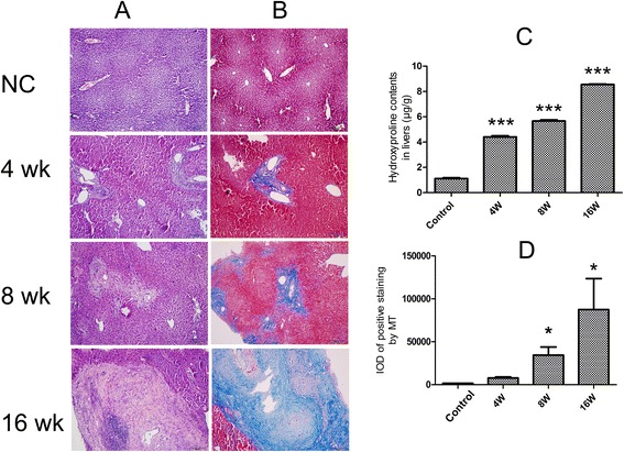
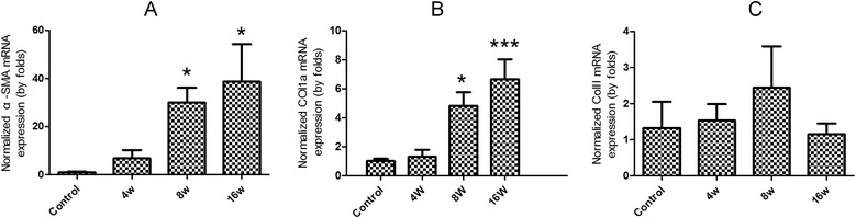
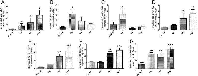
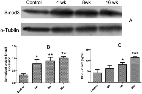
Results: The results showed that collagen deposition indicated by hydroxyproline content and Masson's trichrome staining was increased gradually with the development of infection. The expression of collagen type α1 (Col1a) mRNA transcripts was steadily increased during the whole infection. The mRNA levels of Smad2, Smad3 as well as the protein of Smad3 in the liver of C. sinensis-infected mice were increased after 4 wk p.i. (P < 0.05, compared with normal control) whereas the TGF-β₁, TGF-β type I receptor (TGFβRI) and TGF-β type II receptor (TGFβRII) mRNA expression in C. sinensis-infected mice were higher than those of normal control mice after 8 wk p.i. (P < 0.05). However, the gene expression of Smad4 and Smad7 were peaked at 4 wk p.i. (P < 0.05), and thereafter dropped to the basal level at 8 wk p.i., and 16 wk p.i., respectively. The concentrations of TGF-β₁ in serum in the C. sinensis-infected mice at 8 wk p.i. and 16 wk p.i (P < 0.05) were significantly higher than those in the control mice.

Conclusions: The results of the present study indicated for the first time that the activation of TGF-β/Smad signaling pathway might contribute to the synthesis of collagen type I which leads to liver fibrosis caused by C. sinensis.


Figures

Figure 1
Figure 1
Evaluation of hepatic fibrosis in mice caused by Clonorchis sinensis at 4 week post-infection (wk p.i.), 8 wk p.i. and 16 wk p.i. (A) Histological examination of liver tissues from C. sinensis-infected mice and normal control mice at different time-points as indicated. (B) Collagen depositions were specifically stained by Masson’s trichrome at different time-points as indicated. (C) Hydroxyproline (Hyp) concentration was measured in liver homogenate (0.1 g) in fibrotic or normal livers of BALB/c mice at indicated time-points. (D) Collagen depositions from each specimen were semi-quantified using Image-Pro Plus software. Data were presented as mean ± SEM from 8 C. sinensis-infected mice and 5 normal control (NC) mice at each time-point, * = P < 0.05, ** = P < 0.01, *** = P < 0.001 versus control mice.
Figure 2
Figure 2
The mRNA expression of the pro-fibrotic molecular markers in livers of mice during Clonorchis sinensis infection. Mice were orally infected with 45 metacercariae of C. sinensis, livers were collected from infected and non-infected mice at indicated time-points and mRNA levels were determined by qPCR (normalized to beta-actin transcript levels). (A) α-SMA; (B) Col1a; (C) Col III. Data were presented as mean ± SEM from 8 C. sinensis-infected mice and 5 normal control mice at each time-point, * = P < 0.05, ** = P < 0.01, *** = P < 0.001 versus control mice.
Figure 3
Figure 3
Gene expression of TGF-β/Smad signaling in the liver fibrosis caused by C. sinensis . Mice were orally infected with 45 metacercariae of C. sinensis, livers were collected from infected and non-infected mice at indicated time-points and mRNA levels were determined by qPCR (normalized to beta-actin transcript levels). (A) Smad3; (B) Smad4; (C) Smad7; (D) TGF-β1; (E) TGFβRI; (F) TGFβRII; (G) Smad2. Data were presented as mean ± SEM from 8 C. sinensis-infected mice and 5 normal control mice at each time-point, * = P < 0.05, ** = P < 0.01, *** = P < 0.001 versus control mice.
Figure 4
Figure 4
The protein expression of Smad3 in livers and dynamic changes of serous TGF-β 1 in sera from C. sinensis -infected and non-infected mice . Mice were orally infected with 45 metacercariae of C. sinensis, livers and sera were collected from infected and non-infected mice at indicated time-points and the protein expression levels of Smad3 (A&B) in the livers and TGF-β1 (C) in the sera from mice were determined by western-blot and ELISA, respectively. Data were presented as mean ± SEM from 8 C. sinensis-infected mice and 5 normal control mice at each time-point, * = P < 0.05, ** = P < 0.01, *** = P < 0.001 versus control mice.

Similar articles

Cited by

References

    1. Fang YY, Chen YD, Li XM, Wu J, Zhang QM, Ruan CW. Current prevalence of Clonorchis sinensis infection in endemic areas of China. ZhongguoJi Sheng Chong Xue Yu Ji Sheng Chong Bing Za Zhi 2008, 26:99–103, 109. - PubMed
    1. Hong ST, Fang Y. Clonorchis sinensis and clonorchiasis, an update. Parasitol Int. 2012;61:17–24. doi: 10.1016/j.parint.2011.06.007. - DOI - PubMed
    1. Li S, Chung YB, Chung BS, Choi MH, Yu JR, Hong ST. The involvement of the cysteine proteases of Clonorchis sinensis metacercariae in excystment. Parasitol Res. 2004;93:36–40. doi: 10.1007/s00436-004-1097-5. - DOI - PubMed
    1. Kim TI, Yoo WG, Kwak BK, Seok JW, Hong SJ. Tracing of the Bile-chemotactic migration of juvenile Clonorchis sinensis in rabbits by PET-CT. PLoS Negl Trop Dis. 2011;5:e1414. doi: 10.1371/journal.pntd.0001414. - DOI - PMC - PubMed
    1. Chen J, Xu MJ, Zhou DH, Song HQ, Wang CR, Zhu XQ. Canine and feline parasitic zoonoses in China. Parasit Vectors. 2012;5:152. doi: 10.1186/1756-3305-5-152. - DOI - PMC - PubMed

MeSH terms

Substances