Review
. 2015 Jul;13(3):261-76.
doi: 10.1007/s12021-015-9260-y.

Automatic Detection of White Matter Hyperintensities in Healthy Aging and Pathology Using Magnetic Resonance Imaging: A Review

Affiliations
Review

Automatic Detection of White Matter Hyperintensities in Healthy Aging and Pathology Using Magnetic Resonance Imaging: A Review

Maria Eugenia Caligiuri et al. Neuroinformatics. 2015 Jul.

Abstract

White matter hyperintensities (WMH) are commonly seen in the brain of healthy elderly subjects and patients with several neurological and vascular disorders. A truly reliable and fully automated method for quantitative assessment of WMH on magnetic resonance imaging (MRI) has not yet been identified. In this paper, we review and compare the large number of automated approaches proposed for segmentation of WMH in the elderly and in patients with vascular risk factors. We conclude that, in order to avoid artifacts and exclude the several sources of bias that may influence the analysis, an optimal method should comprise a careful preprocessing of the images, be based on multimodal, complementary data, take into account spatial information about the lesions and correct for false positives. All these features should not exclude computational leanness and adaptability to available data.


Figures

Fig. 1
Fig. 1
Graphical scheme of FLAIR-histoseg method. Top panels: two different axial slices of a FLAIR image and corresponding results of the segmentation. Bottom panel: histogram of the FLAIR image, intensities (arbitrary units) on the abscissa, number of voxels on the ordinate. For each FLAIR image, the thresholds used to segment the histogram in the three domains were defined by regression equations (for details see Jack et al. 2001); normal brain voxels are green; WMH voxels are red; CSF voxels are blue
Fig. 2
Fig. 2
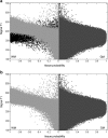
a: signal-to-probability maps of subject with WMH. The GM probability is shown in dark gray and the WM probability is shown in light gray. WM probability values are multiplied by −1 for display purposes. Voxels classified as WMH are shown in black. b: after removing WMH voxels, the signal-to-probability maps of the patient are comparable to those of a normal brain (both GM and WM tissues are no longer contaminated by the abnormalities). From Seghier et al. (2008)
Fig. 3
Fig. 3
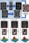
The WHASA method. Top panel shows the computation of the contrast parameter used for non-linear diffusion. Bottom panel illustrates the segmentation of the FLAIR image using non-linear diffusion and watershed. The third row shows a 3D visualization of the enlarged image part, where color and height indicate intensity values. Reproduced from Samaille et al. (2012)

Similar articles

Cited by

References

    1. Admiraal-Behloul F, van den Heuvel DMJ, Olofsen H, van Osch MJP, van der Grond J, van Buchem M, Reiber JHC. Fully automatic segmentation of white matter hyperintensities in MR images of the elderly. NeuroImage. 2005;28(3):607–17. doi: 10.1016/j.neuroimage.2005.06.061. - DOI - PubMed
    1. Anbeek P, Vincken KL, van Osch MJP, Bisschops RHC, van der Grond J. Probabilistic segmentation of white matter lesions in MR imaging. NeuroImage. 2004;21(3):1037–44. doi: 10.1016/j.neuroimage.2003.10.012. - DOI - PubMed
    1. Anitha, M., Selvy, P. T., & Palanisamy, V. (2012). WML detection of brain images using fuzzy and possibilistic approach in feature space, WSEAS TRANSACTIONS on COMPUTERS, E-ISSN, 2224–2872.
    1. Bastianello, S., Bozzao, A., Paolillo, A., Giugni, E., Gasperini, C., Koudriavtseva, T., … Bozzao, L. (1997). Fast spin-echo and fast fluid-attenuated inversion-recovery versus conventional spin-echo sequences for MR quantification of multiple sclerosis lesions. American Journal of Neuroradiology, 18 (4), 699–704. - PMC - PubMed
    1. Bastos Leite AJ, van Straaten EC, Scheltens P, Lycklama G, Barkhof F. Thalamic lesions in vascular dementia: low sensitivity of fluid-attenuated inversion recovery (FLAIR) imaging. Stroke. 2004;35(2):415–9. doi: 10.1161/01.STR.0000109226.67085.5A. - DOI - PubMed