

A peripheral giant cell granuloma with extensive osseous metaplasia or a hybrid peripheral giant cell granuloma-peripheral ossifying fibroma: a case report
- PMID: 25649957
- PMCID: PMC4417193
- DOI: 10.1186/1752-1947-9-14
A peripheral giant cell granuloma with extensive osseous metaplasia or a hybrid peripheral giant cell granuloma-peripheral ossifying fibroma: a case report
Abstract
Introduction: Peripheral giant cell granuloma and peripheral ossifying fibroma are clinicopathologically distinct gingival lesions. Both are included in clinical differential diagnoses of common benign and reactive gingival epulides in humans. It is often impossible to make a clinical distinction between the two entities, thereby making definitive diagnosis dependent on histopathologic features. While our search of the English literature revealed several reports of peripheral giant cell granuloma with 'bone formation', we were unable to identify any reports of hybrid peripheral ossifying fibroma-peripheral giant cell granulomas.
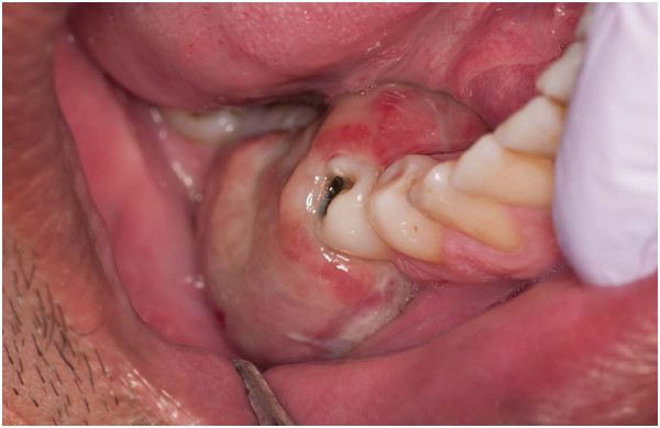
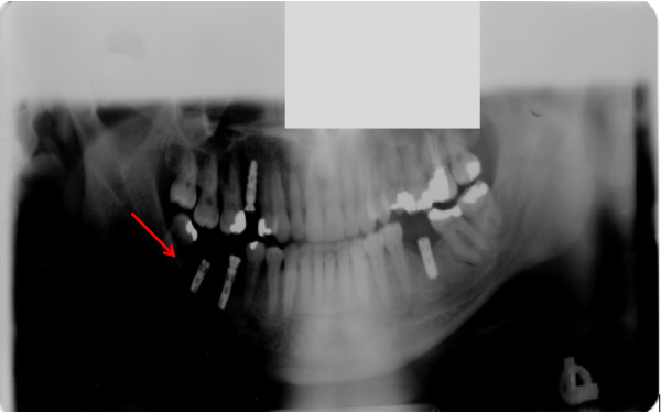
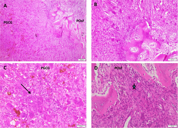
Case presentation: We report a case of a 44-year-old Caucasian man presenting with a three-month history of swelling of his right posterior mandible, related to an area of previous dental implant restoration. A clinical examination revealed modest extraoral facial swelling of his right posterior mandible, while an intraoral examination showed a 45 × 25 × 15 mm sessile, lobular soft tissue mass of the right posterior mandibular gingiva. The mucosal covering of the lesion exhibited focal surface ulceration. A panoramic radiograph showed two implants at the vicinity of the lesion with no other significant findings. An excisional biopsy of the lesion followed by histopathologic examination of the biopsy specimen revealed salient and distinctive features of peripheral giant cell granuloma and of peripheral ossifying fibroma, estimated at near equal proportions. This raises the possibility of a hybrid odontogenic lesion.
Conclusion: The presentation of this lesion, with areas of peripheral giant cell granuloma along with a distinct area of extensive osseous formation and stroma reminiscent of a peripheral ossifying fibroma, justifies consideration of this as a possible hybrid lesion. Although the biologic behavior of a combined lesion is not anticipated to deviate significantly from that of either of the single entities, this case resurrects an enduring debate as to whether peripheral giant cell granuloma and peripheral ossifying fibroma are simply parts of a disease spectrum, or whether some of these lesions represent true hybrid lesions. It is therefore recommended that more cases with histopathologic features similar to the lesion in our case be reported in the literature to further elucidate the histogenesis of these lesions.
Figures
Similar articles
-
Long-term follow-up of peripheral ossifying fibroma: report of three cases.Periodontal Clin Investig. 2001;23(1):11-4. Periodontal Clin Investig. 2001. PMID: 11575108
-
Peripheral ossifying fibroma: a clincal report.Compend Contin Educ Dent. 2011 Jun;32(5):E86-90. Compend Contin Educ Dent. 2011. PMID: 23738938
-
Case Report: rare hybrid lesion of a central giant cell granuloma within a juvenile ossifying fibroma.F1000Res. 2019 Jul 30;8:1218. doi: 10.12688/f1000research.19891.1. eCollection 2019. F1000Res. 2019. PMID: 31632653 Free PMC article.
-
A giant peripheral ossifying fibroma of the maxilla with extreme difficulty in clinical differentiation from malignancy: a case report and review of the literature.J Med Case Rep. 2024 May 4;18(1):220. doi: 10.1186/s13256-024-04529-9. J Med Case Rep. 2024. PMID: 38702820 Free PMC article. Review.
-
Hybrid tumor of central giant cell granuloma and trabecular juvenile ossifying fibroma of the mandible: A rare event in the oral cavity with a review on pathogenesis.Indian J Pathol Microbiol. 2024 Jul 1;67(3):638-640. doi: 10.4103/ijpm.ijpm_623_22. Epub 2023 Jul 10. Indian J Pathol Microbiol. 2024. PMID: 38391302 Review.
Cited by
-
Atypical Peripheral Ossifying Fibroma of the Mandible.Dent J (Basel). 2022 Jan 6;10(1):9. doi: 10.3390/dj10010009. Dent J (Basel). 2022. PMID: 35049607 Free PMC article.
-
Complete excision and soft tissue augmentation after recurrence of a peripheral ossifying fibroma as a pyogenic granuloma: A case report.J Adv Periodontol Implant Dent. 2020 Dec 5;12(2):95-99. doi: 10.34172/japid.2020.016. eCollection 2020. J Adv Periodontol Implant Dent. 2020. PMID: 35919751 Free PMC article.
-
Surgical resection of a giant peripheral ossifying fibroma in mouth floor managed with fiberscopic intubation.Clin Case Rep. 2020 Nov 6;9(1):180-184. doi: 10.1002/ccr3.3494. eCollection 2021 Jan. Clin Case Rep. 2020. PMID: 33489156 Free PMC article.
-
An analysis of the prevalence of peripheral giant cell granuloma and pyogenic granuloma in relation to a dental implant.BMC Oral Health. 2021 Apr 23;21(1):204. doi: 10.1186/s12903-021-01566-4. BMC Oral Health. 2021. PMID: 33892689 Free PMC article.
-
Peripheral giant cell granuloma associated with a dental implant: A case report.J Clin Exp Dent. 2021 Oct 1;13(10):e1049-e1052. doi: 10.4317/jced.57189. eCollection 2021 Oct. J Clin Exp Dent. 2021. PMID: 34667501 Free PMC article.
References
Publication types
MeSH terms
LinkOut - more resources
Full Text Sources
Other Literature Sources
