Review
. 2015 Mar;43(3):819-31.
doi: 10.1007/s10439-015-1263-1. Epub 2015 Feb 4.

Scaffolds for tendon and ligament repair and regeneration

Affiliations
Review

Scaffolds for tendon and ligament repair and regeneration

Anthony Ratcliffe et al. Ann Biomed Eng. 2015 Mar.

Abstract

Enhanced tendon and ligament repair would have a major impact on orthopedic surgery outcomes, resulting in reduced repair failures and repeat surgeries, more rapid return to function, and reduced health care costs. Scaffolds have been used for mechanical and biologic reinforcement of repair and regeneration with mixed results. This review summarizes efforts made using biologic and synthetic scaffolds using rotator cuff and ACL as examples of clinical applications, discusses recent advances that have shown promising clinical outcomes, and provides insight into future therapy.


Figures

Figure 1
Figure 1
Images of arthroscopic surgery of rotator cuff tear (a), repair of the tendon back to the bone (b) and reinforcement with a synthetic patch (c). Figure 1a shows the rotator cuff tendon detached (white arrows) from the top of the humerus exposing the bone attachment site (black arrow). Figure 1b shows the surgical repair where the tendon has been reattached to the bone using sutures attached to suture anchors. Figure 1c shows a reinforcement patch overlaid on the surgical repair and fixed in place medially to the tendon and laterally to the bone.
Figure 2
Figure 2
Defining functional design criteria for tendon and ligament repairs. A relative load-displacement curve for tendons and ligaments is depicted with load on the y-axis and displacement on the x-axis displayed as percentage of maximum load and displacement, respectively. When the tissue is first elongated (origin of curve) the force slowly develops in the so-called nonlinear “toe region.” With further elongation the tissue experiences a constant slope, called the linear stiffness. Upon further elongation, the tissue undergoes partial failures before achieving the highest point on the failure curve (the maximum force or strength). Tendons and ligaments typically do not experience loads approaching the failure region during normal activities. Instead they experience much lower loads that define the functional region. We have labeled peak in vivo forces recorded in the goat patellar tendon (PT), rabbit flexor digitorum profundus (FDP) tendon, rabbit Achilles tendon (AT), rabbit PT, and goat anterior cruciate ligament (ACL) via horizontal bars on the plot. These peak in vivo loads range anywhere from 10% (goat ACL) up to 40% (goat PT) of maximum load. Therefore, researchers should design their repairs to match the shape of this loading curve (red arrow) up to and beyond peak in vivo forces to provide an additional safety factor.
Figure 3
Figure 3
Force-displacement curves are shown for the normal uninjured rabbit patellar tendon compared to the a) naturally healing PT and one tissue-engineered repair of a central, full-length defect in the PT (autologous mesenchymal stem cells in a collagen gel) and b) two tissue-engineered repairs (autologous MSCs in collagen sponge scaffolds exposed to mechanical preconditioning) at 12 weeks post surgery. a) Note that the naturally healing tendon and the tissue engineered repair fail both criteria as they neither exceed peak in vivo forces nor match the slope or “tangent stiffness” of the normal patellar tendon. b) By contrast, the tissue engineered repairs using autologous bone marrow-derived progenitor cells meet both criteria: using one collagen scaffold, the repair curve matches the tangent stiffness of the normal patellar tendon up to 25% beyond peak in vivo forces. When a stiffer collagen scaffold is used, the repair curve matches the tangent stiffness of the normal curve to 50% beyond peak IVFs. Figures adapted from Butler et al,.
Figure 4
Figure 4
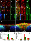
Paratenon progenitor cells expressing SMA contribute to patellar tendon healing. Triple transgenic mice containing 1) SMA-CreERT2 construct to drive Cre enzyme expression in a cell population in the paratenon, 2) Ai9-tdTomato Cre-reporter mice that once activated by Cre will express constitutively express tdTomato fluorescence, and 3) ScxGFP construct that is expressed in tendon cells. A lineage trace was conducted where tamoxifen was delivered to the mice prior to injury in order to label the SMA+ population (SMA9 in red). The healing process was assessed at 1, 2, and 5 weeks post-injury. The SMA9 cells form an anterior bridge over the defect space at 1 week (A-J) but only 10% of these cells are ScxGFP+ (V). These cells differentiate into ScxGFP+ cells at 2 weeks (K-T) with over 60% of SMA9+ cells are also ScxGFP+. These SMA9 cells reduce their level of Scx expression by 5 weeks (V), which corresponds with a plateau in mechanical properties. Scale bars = 100 m. *significantly different than 1 and 5 weeks, ^significantly different than 1 week (p<0.05). Error bars denote SD. Figure adapted from Dyment et al.
Figure 5
Figure 5
Stress-strain curves for human and canine infraspinatus tendon (IFS) of the rotator cuff, and for products used for reinforcement of rotator cuff surgical repair. Note that the human and canine IFS tendons demonstrate steep stress-strain curves indicating they undergo only a small amount of strain under these loads. In contrast some of the reinforcement products undergo substantial stain when under load. Adapted from Derwin et al,.

References

    1. Aurora A, Mccarron JA, Van Den Bogert AJ, Gatica JE, Iannotti JP, Derwin KA. The biomechanical role of scaffolds in augmented rotator cuff tendon repairs. J Shoulder Elbow Surg. 2012;21:1064–1071. - PubMed
    1. Baker AR, Mccarron JA, Tan CD, Iannotti JP, Derwin KA. Does augmentation with a reinforced fascia patch improve rotator cuff repair outcomes? Clin Orthop Relat Res. 2012;470:2513–2521. - PMC - PubMed
    1. Barber FA, Burns JP, Deutsch A, Labbe MR, Litchfield RB. A prospective, randomized evaluation of acellular human dermal matrix augmentation for arthroscopic rotator cuff repair. Arthroscopy. 2012;28:8–15. - PubMed
    1. Beimers L, Lam PH, Murrell GA. The biomechanical effects of polytetrafluoroethylene suture augmentations in lateral-row rotator cuff repairs in an ovine model. J Shoulder Elbow Surg. 2014;23:1545–1552. - PubMed
    1. Bishop J, Klepps S, Lo IK, Bird J, Gladstone JN, Flatow EL. Cuff integrity after arthroscopic versus open rotator cuff repair: a prospective study. J Shoulder Elbow Surg. 2006;15:290–299. - PubMed

Publication types