Review
. 2015:2015:745613.
doi: 10.1155/2015/745613. Epub 2015 Jan 20.

Seizure-induced oxidative stress in temporal lobe epilepsy

Affiliations
Review

Seizure-induced oxidative stress in temporal lobe epilepsy

Sreekanth Puttachary et al. Biomed Res Int. 2015.

Abstract

An insult to the brain (such as the first seizure) causes excitotoxicity, neuroinflammation, and production of reactive oxygen/nitrogen species (ROS/RNS). ROS and RNS produced during status epilepticus (SE) overwhelm the mitochondrial natural antioxidant defense mechanism. This leads to mitochondrial dysfunction and damage to the mitochondrial DNA. This in turn affects synthesis of various enzyme complexes that are involved in electron transport chain. Resultant effects that occur during epileptogenesis include lipid peroxidation, reactive gliosis, hippocampal neurodegeneration, reorganization of neural networks, and hypersynchronicity. These factors predispose the brain to spontaneous recurrent seizures (SRS), which ultimately establish into temporal lobe epilepsy (TLE). This review discusses some of these issues. Though antiepileptic drugs (AEDs) are beneficial to control/suppress seizures, their long term usage has been shown to increase ROS/RNS in animal models and human patients. In established TLE, ROS/RNS are shown to be harmful as they can increase the susceptibility to SRS. Further, in this paper, we review briefly the data from animal models and human TLE patients on the adverse effects of antiepileptic medications and the plausible ameliorating effects of antioxidants as an adjunct therapy.


Figures

Figure 1
Figure 1
Biochemical reactions of ROS/RNS and their elimination by cellular endogenous antioxidants. Components in blue represent nonenzymatic antioxidants; green represents oxidative and antioxidant enzymes; and small red explosion sign represents generation of free radicals. NOX is the key enzymatic source of ROS. It reduces oxygen to superoxide anion and hydrogen peroxide. O2 forms H2O2 which is the most reactive radical among its group that is produced via Fenton reaction. OH leads to lipid peroxidation by producing harmful metabolites such as MDA and 4-HNE leading to mitochondrial dysfunction and cell death. It also produces HOCl and PhO which are extremely toxic oxidants that disrupt tight junctions and increase paracellular permeability. H2O2 is eliminated by CAT, in peroxisomes, and GPx (location varies). At rapid rates, superoxide anions compete with NO which results in the formation of highly reactive molecule called peroxynitrite (ONOO), in cytoplasm, leading to increased ROS production, oxidation of DNA, RNA, and proteins, ion channel dysfunction, and loss of bioactive NO. Peroxynitrite inactivates Mn-SOD, thereby increasing the flux of superoxide anions available to react with NO. SOD catalyzes the reduction of superoxide anions into H2O2, in mitochondria in the presence of enzymes GPx and CAT; H2O2 gets converted into water and oxygen. Antioxidant enzymes such as GPx oxidize GSH to GSSG and GSHred recycles GSH back from GSSG. NADPH gets reduced to NADP by GSHred. GSH/GSSG is a commonly used biomarker of oxidative stress in biological systems. However, GPx also catalyzes H2O2 into H2O by using reduced TRXred. Antioxidant defense against toxic oxygen intermediates comprises an intricate network which is heavily influenced by nutrition (vitamins A, E, and C and fatty acids). CGS plays an important role in glutathione metabolism and acts as an antioxidant in glial cells such as astrocytes. Extracellular oxidized cysteine is reduced to cysteine by thioredoxin reductase or glutathione that helps to maintain the steady state balance between antioxidants and ROS [24, 41, 80]. ROS, reactive oxygen species; NADPH, nicotinamide adenine dinucleotide phosphate; NOX, NADPH oxidase; SOD, superoxide dismutase (Cu/Zn—copper/zinc, Mn—manganese); CAT, catalase; O2 −•, superoxide anion; H2O2, hydrogen peroxide; NO, nitric oxide; ONOO, peroxynitrite; HOCl, hypochlorous acid; PhO, phenoxy radical; OH, hydroxyl radical; GSH, glutathione; GSSG, oxidized glutathione; TRXox/red, thioredoxin reduced and oxidized; TRXred, thioredoxin reductase; GSHred, glutathione reductase; GPx, glutathione peroxidase; CGS, cystine/glutamate antiporter system.
Figure 2
Figure 2
Post-SE pathways in neurodegeneration. SE increases glutamate receptors subunits interactions (NMDA, AMPA, and metabotropic), receptor turn-over, and their trafficking to the postsynaptic membrane. This leads to rapid calcium influx and calcium overload. As a result of this, several calcium dependent enzymes get activated in uncontrolled manner. This results in the activation of several signaling pathways that causes mitochondrial swelling, decrease in ATP, and increase in ROS, which results in oxidization of protein, lipid, and DNA, causing neuronal death. In addition, hypermetabolism, overwhelming glycolysis, and TCA cycle during SE further increase ROS/RNS. High production of lactate can cause cerebral lactic acidosis thereby increasing the production of ROS causing further damage due to mitochondrial dysfunction. Excessive calcium and ROS leads to the collapse of mitochondrial membrane potential, activation of mitochondrial matrix enzymes, and opening of mitochondrial permeability transition pores, decreasing ATP production. ROS are produced in mitochondria through the activity of ETC as a by-product of oxidative phosphorylation. CoASH/CoASSG and GSH/GSSG (described in Figure 3) ratio also decrease in brain tissues during this process and following SE, due to increased oxidative stress [, –84]. TCA: tricarboxylic acid cycle; ETC: electron transport chain; mtDNA: mitochondrial DNA; Cyt C NAD: cytochrome NADH reductase; CoASH: coenzyme A; CoASSG: coenzyme A glutathione disulfide; SE: status epilepticus.
Figure 3
Figure 3
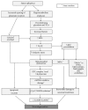
Sequence of events during status epilepticus, latent period, and SRS. The damage to the specific areas of the brain during SE can initiate varying period of neurobiological changes that can lead to the development of SRS. The enzymes free radicals and the pathways involved in these disorders are common in all types of insult (SE, latent period, and SRS), as described in Figures 1 and 2, but with subtle differences. The concentration of antioxidant enzymes rises after an initial insult (imitating their protective role) such as glutamine synthetase in SE; later it reduces which may or may not recover after few days/weeks depending upon the severity of the insult. Latent period is generally characterized by a series of slow neurodegenerative changes in the brain leading to epileptogenesis. The concentration of GST falls during latency that affects glutamate metabolism. High levels of the glutamate in the extracellular and intracellular space can lead to neuronal excitability through activated calcium signaling, as described in Figure 2. Levels of H2O2 return to the basal levels with mtDNA repair and low GSH/GSSG and CoASH/CoASSG ratio. During latent period, nitrosylation of protein fragments and posttranslational modifications of ion channels and transporters will further lead to hyperexcitability of neurons [24, 48, 82, 83, 85, 86]. SRS: spontaneous recurrent seizures; GST: glutamine synthetase; mtDNA: mitochondrial DNA; X: reverse reaction does not occur; Li-Pilo: lithium- pilocarpine model.
Figure 4
Figure 4
Schematic representation of a synapse, with postsynaptic ionotropic glutamate receptors (NMDA, AMPA, and KA/GLUR6), its associated glial cells, and extrasynaptic effects of a seizure. First seizure due to hyperexcitability of neurons (as evident from increased Fos expression in the hippocampus) induces reactive gliosis at a later stage, which produces inflammatory cytokines and iNOS that are mediated by NFκB transcription. These in turn sensitize postsynaptic neurons and decrease their seizure threshold. Reactive astrocytes also downregulate glutamate uptake, thus increasing the concentration of glutamate at the synapse. These events contribute to further hyperexcitability of neurons as evident from increased spiking activity on EEG. These changes in turn lead to neurodegenerative changes after 3 days following the first seizure (Fluoro-Jade-B (FJB)+, neuronal nuclei protein (NeuN), the markers used to detect neurodegeneration) [30, 35, 87, 88].
Figure 5
Figure 5
Immunohistochemistry (IHC) of the brain sections from kainate mouse and rat models of epilepsy at 2 h, 24 h, and 7 days after SE. (a) c-Fos ((A), (B)) expression was more widespread in the hippocampal formation at 2 h after SE (B). More than 3-4-fold increased expression (quantified data not shown) of c-Fos in CA3 pyramidal cell layer was observed (B). CCR2 ((C), (D)) and astrocytic NFκB expression ((F), orange) at 24 hours after SE. (b) By 7 days after SE, there was increased astrogliosis ((H), GFAP, green) and microgliosis ((J), IB1A is marker for microglia, green) compared to controls ((G), (I)). SE induced neurodegeneration (FJB +ve neurons) was observed in CA3 of hippocampus (L). There were increased FJB +ve cells in CA3 of hippocampus (green label in (L), all scale bars 100 μm). The same area was invaded by reactive astrocytes and microglia (green cells in (H) and (J)). Hematoxylin and Eosin stained hippocampal sections ((M), (N)) with pyknotic nucleus and shrunken cytoplasm are evident due to SE-induced changes at 7 days post-SE.

References

    1. Delorenzo R. J., Sun D. A., Deshpande L. S. Cellular mechanisms underlying acquired epilepsy: the calcium hypothesis of the induction and maintainance of epilepsy. Pharmacology & Therapeutics. 2005;105(3):229–266. - PMC - PubMed
    1. ILAE Commission Report. The epidemiology of the epilepsies: future directions. International league against Epilepsy. Epilepsia. 1997;38(5):614–618. - PubMed
    1. Panayiotopoulos C. P. The Epilepsies: Seizures, Syndromes and Management. 2005. - PubMed
    1. Martinc B., Grabnar I., Vovk T. The role of reactive species in epileptogenesis and influence of antiepileptic drug therapy on oxidative stress. Current Neuropharmacology. 2012;10(4):328–343. doi: 10.2174/157015912804499447. - DOI - PMC - PubMed
    1. DeLorenzo R. J., Pellock J. M., Towne A. R., Boggs J. G. Epidemiology of status epilepticus. Journal of Clinical Neurophysiology. 1995;12(4):316–325. doi: 10.1097/00004691-199512040-00003. - DOI - PubMed

Publication types

LinkOut - more resources