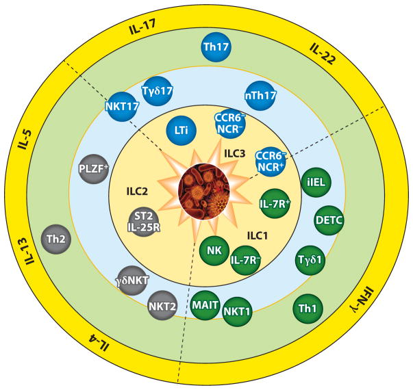
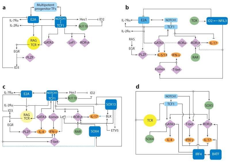
Review
. 2015:33:505-38.
doi: 10.1146/annurev-immunol-032414-112025. Epub 2015 Jan 30.

Transcription factor networks directing the development, function, and evolution of innate lymphoid effectors

Affiliations
Review

Transcription factor networks directing the development, function, and evolution of innate lymphoid effectors

Joonsoo Kang et al. Annu Rev Immunol. 2015.

Abstract

Mammalian lymphoid immunity is mediated by fast and slow responders to pathogens. Fast innate lymphocytes are active within hours after infections in mucosal tissues. Slow adaptive lymphocytes are conventional T and B cells with clonal antigen receptors that function days after pathogen exposure. A transcription factor (TF) regulatory network guiding early T cell development is at the core of effector function diversification in all innate lymphocytes, and the kinetics of immune responses is set by developmental programming. Operational units within the innate lymphoid system are not classified by the types of pathogen-sensing machineries but rather by discrete effector functions programmed by regulatory TF networks. Based on the evolutionary history of TFs of the regulatory networks, fast effectors likely arose earlier in the evolution of animals to fortify body barriers, and in mammals they often develop in fetal ontogeny prior to the establishment of fully competent adaptive immunity.

Keywords: T cells; cytokines; evolution of immunity; fetal lymphopoiesis; transcription factor regulatory network; γδ T cells.


Figures

Figure 1
Figure 1
Three overlapping layers of T cell and T cell–like immunity and representative effector subsets in each layer. A pathogen breach of the body barrier activates each layer in succession, with the fast-acting innate lymphocytes arrayed most proximal to the barrier surfaces. The subsets are described in the text. The effector repertoire of each layer is similar, broadly divided into three wedges united by the type of cytokines each can produce. Heterogeneity of functional units (at the cell lineage level) within the fast inner layers is complex, whereas for the slow outer adaptive ring it is more limited, although inflammations can drive conversions of established Th subsets. Conversely, clonal variations at the outer ring are extremely high because individual cells express distinct antigen receptors, whereas the subsets in the inner rings often express canonical antigen receptors and/or pathogen-associated pattern receptors. The middle ring contains innate effectors that express either αβTCR or γδTCR. For the latter, those found in lymphoid tissues (such as Tγδ1) are mostly naive, express clonal TCRs, and primarily exhibit default IFN-γ production, similar to NK cells. Their numbers in mammals are dwarfed by those of the innate γδT cells in tissues, owing to the vast surface areas of mucosal tissues such as the skin and the gut. Type 1 cytokine–producing cells are marked as IFN-γ enabled, ranging from cNK cells in the inner ring to the adaptive Th1 cells in the outer ring. Cells in the inner rings are mostly tissue tropic, although some (such as cNK and Tγδ1 cells) are circulatory. The number of type 2 cytokine (IL-4/5/13)-producing cell subsets is the smallest, and IL-4 production in peripheral tissues is constrained. Type 3 cytokine–producing fast effector cells exhibit distinct profiles of IL-17 and IL-22 secretion, with the innermost fast effectors biased toward IL-22 production, whereas in the middle ring IL-22 production is tightly regulated, with skin Tγδ17 cells primed for IL-17, but not IL-22, secretion. For simplicity, the classification of cytokine types shown is not comprehensive; some cytokines are not listed (an example is IL-9, which is considered a type 2 cytokine) or detailed (for example, the IL-17 family has six members, some with unique activities). Also, additional Th subsets, such as Bcl6+ T follicular helper (Tfh) cells and PU.1+ Th9 cells, are not depicted because clear corollary populations among ILE cells have not been established. NKT and γδT cells can secrete cytokines made by these Th subsets (IL-9, IL-10, and IL-21) and can promote B cell maturation and antibody production, but it is unclear whether corresponding specialized subsets exist. ST2 (Il1rl1) and IL-25R (IL-17RB) are receptors for two epithelial cytokines, IL-33 and IL-17E (IL-25), respectively, and both are activators of various type 2 cytokine–producing cells. Abbreviations: DETC, dendritic epidermal T cell; iIEL, intestinal intraepithelial lymphocyte; LTi, lymphoid tissue–inducer cell; MAIT, mucosal-associated invariant T cell; NCR, NK cell receptor; PLZF, promyelocytic leukemia zinc finger.
Figure 2
Figure 2
The basic TF network circuitry of T cell development and variations incorporated into it generate lymphoid effector cell types. Minimal components of each circuit are depicted. Cytokine receptor–signaling inputs controlling TF expression are not shown. (a) The foundation regulatory TF circuit of T cell development is the NOTCH-TCF1 hub (rectangles) that intimately integrates with the E2A regulatory domain and induces central genes (circles) necessary for T cell development from ETPs. This process operates in conjunction with TFs already expressed in LMPPs or CLPs that are active at multiple states of T cells (Ikaros, Myb, Runx1, Gfi1, Sox4, and others belong to this cluster). E2A induces the expression of IL-7R and RAG proteins required for the V(D)J recombination of Tcr genes. E2A and TCF1 control chromatin accessibility of the Tcr loci and regulate their ordered gene rearrangements. TCR signaling, along with E2A, can promote PLZF expression. But in adaptive T cells, PLZF is under an autoregulatory feedback loop involving ID3, induced by TCR signaling that in turn inhibits E2A to maintain a naive state of mature thymocytes. Absence of TCF1 in mice leads to a complete loss of T cell–committed DN2 precursor cells, and TCF1 controls the expression of one of the markers of DN2, IL-2R α-chain. TCR signaling, NOTCH, and TCF1 cooperate to turn on the effector lineage controlling TFs GATA3, LEF1, and RORγt (diamonds), but during intrathymic αβT cell development, they do not access the target cytokine gene loci. NOTCH and TCF1 target genes, most notably Bcl11b, inhibit genes active in non–T cell lymphoid lineages, such as ID2, which is required for ILE development. (b) Three sequentially incorporated ILE TFs, NFIL3 (nuclear factor, IL-3 regulated), ID2, and TOX (thymocyte selection–associated high-mobility group box), constitute the ILC hub. The high level of ID2 expression, induced by NFIL3, possibly in concert with TOX, leads to a muted activity of E2A that in turn impedes the cooperative NOTCH-E2A outputs. The consequences are lack of antigen receptor expression and varied dependence on NOTCH signaling for ILC differentiation. NFIL3 can inhibit Rorc expression in Th17 cells (dashed lines represent connections by function only; direct gene-to-gene interactions have not been confirmed in indicated cell types), and conversely, it turns on Eomes in NK cells to regulate IFN-γ (ovals, effector cytokines). In other ILC1s, T-bet, not EOMES, drives IFN-γ production. For ILC2s, TCF1 regulates Gata3 and cytokine receptor genes critical for ILC2 development. RORα is essential for ILC2 differentiation, but its upstream inducers and downstream targets are unmapped. PLZF is transiently induced, perhaps by RAS signaling in conjunction with E2A, and is also required for ILC2s, but its target genes are unmapped in ILCs. As in T cells, TCF1 inhibits IL-17 production and promotes IL-2R α-chain expression in ILC3s. TCF1 may contribute to Rorc transcription in ILCs directly, as in T cells, or indirectly, by promoting IL-7R expression, which in turn enhances RORγt expression (not depicted). RAR and other positive regulators of RORγt further entrench the ILC3 program. (c) Incorporation of two additional HMG TFs, SOX13 and SOX4, institutes the TCR+ ILE program. Activities of TCF1 are modulated by its interacting partners SOX13 and SOX4, which together turn on Rorc, with additional inputs from RAR and ETV5. SOX13 also induces Etv5, Blk, and Il7r and inhibits Lef1 to specify the Tγδ17 cell program. SOX4 regulates iNKT cell development, potentially by calibrating TCR signals, but the molecular mechanism remains to be ascertained. PLZF, ID3, and T-bet induction is initiated by TCR signaling. TCF1 positively regulates Gata3, to generate IL-4-producing cells, and Eomes and Lef1, to control IFN-γ synthesis. (d) A simplified adaptive Th effector circuit illustrates the dominance of TCR signaling as the arbiter of functional specialization. TFs belonging to the IRF-BATF axis, along with others induced by TCR and cytokine receptor signaling, constitute the hub. IRF4 and BATF cooperatively induce RORγt. IRF4 further controls Gata3 expression, whereas BATF amplifies IL-17 synthesis. The ILE HMG TFs still exert modulatory functions as NOTCH enhances expression of effector cell type–specific TFs and promotes cytokine gene expression. Conversely, TCF1 negatively regulates IFN-γ and IL-17 production, but its activity is downmodulated by TCR signaling. SOX4 and SOX5, two additional HMG TFs, modulate Th2 and Th17 cell differentiation, respectively. SOX4 inhibits GATA3 function, whereas SOX5 promotes Rorc expression. Abbreviations: CLP, clonogenic lymphoid precursor; ETP, early thymic progenitor; HMG, high-mobility group; ILC, innate lymphoid cell; ILE, innate lymphoid effector; iNKT cell, invariant NKT cell; LMPP, lymphoid-primed multipotent progenitor; RAG, recombination activating gene; RAR, retinoic acid receptor; TF, transcription factor.
Figure 3
Figure 3
A simplified phylogenetic tree of TFs and cytokines directing lymphoid effector subset development and function. Depicted are the major phyla (ovals); the kingdoms they belong to (double ovals) and subphyla, classes, or genera they include (bold, referred to in the text); and extant organisms (species) of Opisthokonta (domain Eukaryota, animals and fungi supergroup, no taxonomic rank). Only the holozoan (animals and related unicellular organisms) branch that generated animals (metazoans) is detailed, and the tree is drawn not to a timescale but according to the sequence in which the phyla emerged. Only the clear-cut orthologs of rodent TFs and cytokines (rectangles) are noted in species where they first appear, and for early genes that are not clearly defined as homologs (because a member is missing key functional domains, for instance), they are denoted as ancestral and preceded by the letter a. Lymphocytes only exist in deuterostomes, but it is clear that immune mechanisms, such as the complement cascades and pathogen-associated molecular patterns, predate the emergence of mobile cell types mediating immune responses. Filastereans (Capsaspora owczarzaki), single-cell eukaryotes, and the earliest clade of the holozoan branch are already endowed with intracellular mediators of innate sensors, such as NF-κB, the STAT TF that is activated by cytokine and growth factor receptors; and an ancestral Runx gene linked to the mammalian Runx1, which is essential for definitive hematopoiesis. The closest extant unicellular relatives of animals are choanoflagellates, and they provide evidence for the emergence of sequence-specific HMG TFs, related to Capicua (Cic) and SoxF/H members (Sox17, 18, or 30), ~900 Mya. A significant expansion of the repertoire of immune-related TFs is observed in Porifera (sponges), and a large family of invertebrate IL-17 is found in oysters (Protostomia). A Tbr gene is annotated in the genome of Lottia gigantea, a protostome mollusk, but not in the genome of flies. An ancestral gene for Rag is present in Nematostella vectensis (sea anemone) and further innovated in Strongylocentrotus purpuratus (purple sea urchin), where orthologs of Rag1/2 emerged. Definitive evidence for a discrete ILE subset is first obtained in lampreys, where Il17-expressing SOX13+ lymphocytes that home to mucosal tissues have been discovered. The diversification of mammalian cytokines and central antigen receptor–activated TFs controlling αβT cell effector lineage specification initiates in vertebrates, first in cartilaginous fishes (Callorhinchus milii). The emergence of type 2 cytokines appears to lag behind the emergence of type 1 cytokines, as a single Il4/13 gene and an Il5 homolog are catalogued in teleosts, with only an ancestral Il4 gene in C. milii. Among the central modulators of Th cell specification, the mammalian inducers of type 3 cytokines RORγt and IL-23 appear last in teleosts. Abbreviations: HMG, high-mobility group; ILE, innate lymphoid effector; TF, transcription factor.

References

    1. Spits H, Di Santo JP. The expanding family of innate lymphoid cells: regulators and effectors of immunity and tissue remodeling. Nat Immunol. 2011;12:21–27. - PubMed
    1. Sumaria N, Roediger B, Ng LG, Qin J, Pinto R, et al. Cutaneous immunosurveillance by self-renewing dermal γΔT cells. J Exp Med. 2011;208:505–18. - PMC - PubMed
    1. Kastenmuller W, Torabi-Parizi P, Subramanian N, Lammermann T, Germain RN. A spatially-organized multicellular innate immune response in lymph nodes limits systemic pathogen spread. Cell. 2012;150:1235–48. - PMC - PubMed
    1. Sheridan BS, Lefrançois L. Regional and mucosal memory T cells. Nat Immunol. 2011;12:485–91. - PMC - PubMed
    1. Mueller SN, Gebhardt T, Carbone FR, Heath WR. Memory T cell subsets, migration patterns, and tissue residence. Annu Rev Immunol. 2013;31:137–61. - PubMed

Publication types

Substances

LinkOut - more resources