Aqueous cell differentiation in anterior uveitis using Fourier-domain optical coherence tomography
- PMID: 25650415
- PMCID: PMC4347308
- DOI: 10.1167/iovs.14-15118
Aqueous cell differentiation in anterior uveitis using Fourier-domain optical coherence tomography
Abstract
Purpose: The differential diagnosis of a patient presenting with anterior uveitis is broad and can present a diagnostic challenge. In this study, we evaluate the characteristic findings of inflammatory cells on optical coherence tomography (OCT) both in vitro and in vivo.
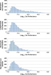
Methods: Blood from two healthy volunteers was prepared using standardized methods for cell sorting with a flow cytometer (FASCAria). Neutrophils, lymphocytes, monocytes, and red blood cells were placed in suspension and scanned with a 26-kHz Fourier-domain OCT system (RTVue) with 5-μm axial resolution. Custom software algorithms were used to identify cells based on their reflectance distribution. These algorithms were then applied to OCT images obtained from uveitis patients with active anterior chamber inflammation.
Results: On OCT images the cells appeared as hyperreflective spots. In vitro, cell reflectance was statistically significantly different between all of the cell types (neutrophils, monocytes, lymphocytes, and red blood cells, P < 0.001, Mann-Whitney test). In vivo, the relationship between underlying disease and cell type imaged on OCT was highly statistically significant, with human leukocyte antigen (HLA)-B27-associated uveitis patients having a predominantly polymorphonuclear pattern on OCT and sarcoidosis and inflammatory bowel disease patients having a predominantly mononuclear pattern on OCT (P < 0.001, Fisher's exact test).
Conclusions: These in vitro and in vivo data demonstrate the potential of OCT to evaluate cells in the anterior chamber of patients noninvasively. Optical coherence tomography may be a useful adjunct to guide the diagnosis and treatment of ocular inflammatory conditions.
Keywords: Fourier-domain optical coherence tomography; anterior uveitis; image analysis; mononuclear leukocytes; polymorphonuclear leukocytes.
Copyright 2015 The Association for Research in Vision and Ophthalmology, Inc.
Figures


Similar articles
-
The Feasibility of Spectral-Domain Optical Coherence Tomography Grading of Anterior Chamber Inflammation in a Rabbit Model of Anterior Uveitis.Invest Ophthalmol Vis Sci. 2016 Jul 1;57(9):OCT184-8. doi: 10.1167/iovs.15-18883. Invest Ophthalmol Vis Sci. 2016. PMID: 27409471 Free PMC article.
-
Objective Quantification of Anterior Chamber Inflammation: Measuring Cells and Flare by Anterior Segment Optical Coherence Tomography.Ophthalmology. 2017 Nov;124(11):1670-1677. doi: 10.1016/j.ophtha.2017.05.013. Epub 2017 Jun 16. Ophthalmology. 2017. PMID: 28625685
-
High-speed optical coherence tomography for imaging anterior chamber inflammatory reaction in uveitis: clinical correlation and grading.Am J Ophthalmol. 2009 Mar;147(3):413-416.e3. doi: 10.1016/j.ajo.2008.09.024. Epub 2008 Dec 3. Am J Ophthalmol. 2009. PMID: 19054493
-
Application of anterior segment optical coherence tomography in glaucoma.Surv Ophthalmol. 2014 May-Jun;59(3):311-27. doi: 10.1016/j.survophthal.2013.06.005. Epub 2013 Oct 15. Surv Ophthalmol. 2014. PMID: 24138894 Review.
-
Instrument-based Tests for Measuring Anterior Chamber Cells in Uveitis: A Systematic Review.Ocul Immunol Inflamm. 2020 Aug 17;28(6):898-907. doi: 10.1080/09273948.2019.1640883. Epub 2019 Aug 16. Ocul Immunol Inflamm. 2020. PMID: 31418609 Free PMC article.
Cited by
-
The Feasibility of Spectral-Domain Optical Coherence Tomography Grading of Anterior Chamber Inflammation in a Rabbit Model of Anterior Uveitis.Invest Ophthalmol Vis Sci. 2016 Jul 1;57(9):OCT184-8. doi: 10.1167/iovs.15-18883. Invest Ophthalmol Vis Sci. 2016. PMID: 27409471 Free PMC article.
-
Ocular Sarcoidosis.Clin Chest Med. 2015 Dec;36(4):669-83. doi: 10.1016/j.ccm.2015.08.009. Clin Chest Med. 2015. PMID: 26593141 Free PMC article. Review.
-
Ocular anterior chamber blood cell population differentiation using spectroscopic optical coherence tomography.Biomed Opt Express. 2019 Jun 13;10(7):3281-3300. doi: 10.1364/BOE.10.003281. eCollection 2019 Jul 1. Biomed Opt Express. 2019. PMID: 31467779 Free PMC article.
-
Development of fully automated anterior chamber cell analysis based on image software.Sci Rep. 2021 May 21;11(1):10670. doi: 10.1038/s41598-021-89794-0. Sci Rep. 2021. PMID: 34021183 Free PMC article.
-
Artificial Intelligence in Uveitis: Innovations in Diagnosis and Therapeutic Strategies.Clin Ophthalmol. 2024 Dec 14;18:3753-3766. doi: 10.2147/OPTH.S495307. eCollection 2024. Clin Ophthalmol. 2024. PMID: 39703602 Free PMC article. Review.
References
-
- Lightman S, Chan CC. Immune mechanisms in choroido-retinal inflammation in man. Eye. 1990; 4: 345–353. - PubMed
-
- Cote MA, Rao NA. The role of histopathology in the diagnosis and management of uveitis. Int Ophthalmol. 1990; 14: 309–316. - PubMed
-
- Rao NA. Pathology of Vogt-Koyanagi-Harada disease. Int Ophthalmol. 2007; 27: 81–85. - PubMed
-
- Chan CC, Wetzig RP, Palestine AG, et al. Immunohistopathology of ocular sarcoidosis. Report of a case and discussion of immunopathogenesis. Arch Ophthalmol. 1987; 105: 1398–1402. - PubMed
-
- Chang JH, McCluskey PJ, Wakefield D. Acute anterior uveitis and HLA-B27. Surv Ophthalmol. 2005; 50: 364–388. - PubMed
Publication types
MeSH terms
Grants and funding
LinkOut - more resources
Full Text Sources
Other Literature Sources
Research Materials

