. 2015 Feb 3;8(362):ra12.
doi: 10.1126/scisignal.2005654.

FOXP1 potentiates Wnt/β-catenin signaling in diffuse large B cell lymphoma

Affiliations

FOXP1 potentiates Wnt/β-catenin signaling in diffuse large B cell lymphoma

Matthew P Walker et al. Sci Signal. .

Abstract

The transcription factor FOXP1 (forkhead box protein P1) is a master regulator of stem and progenitor cell biology. In diffuse large B cell lymphoma (DLBCL), copy number amplifications and chromosomal translocations result in overexpression of FOXP1. Increased abundance of FOXP1 in DLBCL is a predictor of poor prognosis and resistance to therapy. We developed a genome-wide, mass spectrometry-coupled, gain-of-function genetic screen, which revealed that FOXP1 potentiates β-catenin-dependent, Wnt-dependent gene expression. Gain- and loss-of-function studies in cell models and zebrafish confirmed that FOXP1 was a general and conserved enhancer of Wnt signaling. In a Wnt-dependent fashion, FOXP1 formed a complex with β-catenin, TCF7L2 (transcription factor 7-like 2), and the acetyltransferase CBP [CREB (adenosine 3',5'-monophosphate response element-binding protein)-binding protein], and this complex bound the promoters of Wnt target genes. FOXP1 promoted the acetylation of β-catenin by CBP, and acetylation was required for FOXP1-mediated potentiation of β-catenin-dependent transcription. In DLBCL, we found that FOXP1 promoted sensitivity to Wnt pathway inhibitors, and knockdown of FOXP1 or blocking β-catenin transcriptional activity slowed xenograft tumor growth. These data connect excessive FOXP1 with β-catenin-dependent signal transduction and provide a molecular rationale for Wnt-directed therapy in DLBCL.


Figures

Figure 1
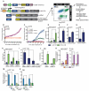
Figure 1. Identification of FOXP1 as a promoter of Wnt signaling (A)
Schematic of the CDt/MS approach. IRES: internal ribosomal entry site, LTR: long terminal repeat. (B,C) A375 cells harboring a β-catenin activated reporter (BAR), which reports mCherry protein expression, were transfected with FOXP1 (splice isoform 1) or GFP control; mCherry fluorescence intensity quantitation is shown in (B). At 24 hours post-transfection, cells were treated with control or Wnt3a conditioned media (C). Fluorescence intensity was quantified every hour using the Incucyte Cell Player. Error bars represent standard error between 3 biological replicate wells. (D) Quantification of BAR-driven luciferase reporter assay in HEK293T cells transfected with FOXP1 or control and treated with Wnt3a or control conditioned media. (E-G) Quantification of β-catenin-dependent reporter activity in HCT116, DLD1, and DB cells after FOXP1 overexpression. (H-J) Quantification of BAR activity in the indicated stable BAR/Renilla infected cell line after transfection with control, FOXP1-A, or FOXP1-B siRNA. Cells were treated with control or Wnt3a conditioned media, as indicated. (K,L) Quantification of AXIN2 and NKD1 mRNA following FOXP1 overexpression. (M,N) AXIN2 and NKD1 mRNA quantification following transfection with the indicated siRNAs, as determined by qRT-PCR. (B-N) Significance was determined by Wilcoxon Signed Rank across a minimum of three independent experiments. * < 0.05; **<0.005; ***< 0.0005. Error bars represent SEM.
Figure 2
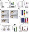
Figure 2. FOXP1 potentiates Wnt signaling downstream of the destruction complex
(A) Quantification of BAR activity after FOXP1 overexpression in the presence or absence of CT99021 treatment. (B) Western blot of β-catenin in Concanavalin A (CONA) stripped lysates. HEK293T cells were transfected with control or FOXP1 before treatment with control or Wnt3a conditioned media. (C) Quantification of BAR luciferase reporter assay in HEK293T cells transfected with FOXP1 or control and then treated with Vehicle (VEH), C59, rhDKK1, or XAV939. (D) Quantification of BAR activity in HEK293T cells transfected with control or FOXP1 and control or β-catenin siRNA. (E) Representative images of different classifications of Wnt or foxp1b overexpressing embryos at 24 hours post fertilization (hpf). The classes represent increasing severity of anterior truncations (reduction and loss of eyes and midbrain-hindbrain-boundary, arrow in classes 1-3) and reduction in dorsal mesoderm derivatives (notochord in class 4, arrow). (F) Quantification of foxp1b injected embryos binned into classes representing severity of Wnt phenotype. Embryos were injected with foxp1b (n=63) or control RNA (n=55). (G) Quantification of foxp1b or control injected embryos overexpressing wnt8. 25pg of foxp1b or control RNA was injected in hs:wnt8 transgenic embryos and wnt8 expression induced by heat-shock during gastrulation. Control (n=70), foxp1b (n=64), control+wnt8 (n=49), and foxp1b + wnt8 (n=79) (H) At 8 hpf, expression of GFP RNA in 6×Tcf/Lef-miniP:d2EGFPisi04 transgenic embryos is expanded in embryos injected with 100 pg foxP1b RNA (n=52 embryos, 100% of the embryos show this phenotype) compared to embryos injected with equimolar amounts of control RLuc RNA (n=69 embryos). (I) Quantification of Wnt target genes in foxp1b injected embryos 24 hours post fertilization. Target genes were normalized to GAPDH gene and presented as fold-induction over GFP control. (N=4 embryos per condition) (J) HEK293T cells were transfected with the indicated transcriptional reporter, CMV-Renilla and either GFP control or FOXP1. Cells were then treated with the indicated recombinant protein before luciferase quantitation. (A-D,F-G, I-J) Significance was determined by Wilcoxon Signed Rank across a minimum of three independent experiments. * < 0.05; **<0.005; ***< 0.0005. Error bars represent SEM.
Figure 3
Figure 3. FOXP1 promotes acetylation of β-catenin by the acetyltransferase CBP
(A) β-catenin reporter quantitation in HCT 116 cells following FOXP1 overexpression in the presence or absence of Anacardic Acid, CPTH2, or ICG-001. (B) Quantification of BAR activity in HEK293T cells transfected with control or FOXP1, and control, β-catenin, CBP-A, or CBP-B siRNA. Inset: Western blot showing CBP protein abundance following siRNA transfection. (C) Western blot analysis of β-catenin acetylated at Lys49 and total β-catenin after transfection with increasing amounts of FOXP1 plasmid. LI-COR-based quantification is shown above. (D) Western blot analysis of β-catenin acetylated at Lys49 and total β-catenin in nuclear and cytosolic extracts from HEK293T cells expressing control or FLAG-FOXP1. (E) HEK293T cells transfected with control or FOXP1-specific siRNAs, treated with Wnt3a and TSA, and Western blotted for the indicated proteins. (F) DB cells stably expressing control or FOXP1 shRNA were analyzed by Western blot for FOXP1, β-catenin, and acetylated-β-catenin. (G) Western blot of the acetylation of β-catenin at Lys49 in HEK293T cells after transfection with control or FOXP1 plasmid and the indicated siRNAs. (N=3 biological replicates) (H) Quantification of BAR luciferase reporter after transfection with β-catenin T41A, β-catenin T41A, K49R, or GFP control and FOXP1 or control. (C-F) LI-COR-based quantification of the Western blots are shown as a ratio of acetyl-β-catenin/total β-catenin. Error bars represent S.E.M. of tubulin-normalized biological triplicates. (A-H) Significance was determined by Wilcoxon Signed Rank across a minimum of three independent experiments. * < 0.05; **<0.005; ***< 0.0005. Error bars represent SEM.
Figure 4
Figure 4. FOXP1 co-complexes with TCF7L2, β-catenin, and CBP
(A) HEK293T cells were transiently transfected with FLAG-FOXP1 or FLAG-HC-RED as control before protein extraction and FLAG immunoprecipitation. Co-complexed endogenous proteins were detected by Western blot analysis. (N=3 biological replicates) (B) Immunoprecipitation and Western blot analysis of endogenous FOXP1 in HCT116 cells revealed association with endogenous β-catenin, acetylated β-catenin, CBP, and TCF7L2. (N=3 biological replicates) (C) Immunoprecipitation and Western blot analysis of endogenous FOXP1 in HEK293T cells revealed association with endogenous β-catenin and acetylated β-catenin, but only after 2 hours of Wnt3a treatment. (N=3 biological replicates) (D) ChIP analysis of the AXIN2 promoter Wnt response element (WRE) or AXIN2 open reading frame (ORF) in HEK293T cells after treatment with control or Wnt3a conditioned media. Control IgG, FOXP1 or β-catenin ChIP antibodies were used. (E) ChIP analysis of stable knockdown DB cells using FOXP1 or β-catenin antibodies. (F,G) ChIP analysis on AXIN2 WRE and AXIN2 ORF of either β-catenin (F) or CBP (G) following FOXP1 knockdown in HCT116 cells. (H) Quantification of BAR activity in HEK293T cells transfected with equal amounts of control or FOXP1 and/or TCF7L2 or LEF1. Significance was determined by paired T-tests across a minimum of three independent experiments. * < 0.05; Error bars represent SEM. (I) Quantification of GAL4 reporter in cells transfected with GAL4-DBD-β-catenin and Control, FOXP1, or EP300. Data is presented as fold induction over GAL4-DBD alone. (J) Quantification of BAR-luciferase transfected with control, FOXP1-WT, or FOXP1 DNA binding mutant. (D-J) All experiments were performed in biological triplicate and p-values were determined by Wilcoxon Signed Rank test. * < 0.05; ** < 0.005. Error bars represent SEM. (K) Relative distances between TCF7L2 (blue) and FOXP1 (red) motifs were calculated. The observed distribution of the relative distance between the two motifs was plotted (red dot) together with relative distribution after randomization (grey dot). p<2.2×10⁻¹⁶ Kolmogorov–Smirnov test. (L,M) All HOMER derived motifs were searched in the set of TCF7L2 ChIP-seq peaks. The significance of the association for each motif was plotted by rank order. FOXP1 is highlighted (red). Inset shows the fraction of TCF7L2 motifs (blue) and FOXP1 motifs (green) in the set of regions enriched by TCF7L2 ChIP. A randomized background is shown for control.
Figure 5
Figure 5. FOXP1 potentiates Wnt signaling in DLBCL
(A) Cell lysates were Western blotted for FOXP1, β-catenin, and acetylated-β-catenin. (N=3 biological replicates) (B,C) Viability of cell lines treated with XAV938 or rhDKK. (D) Cell viability was determined in stable knockdown DB cells treated with rhDKK or XAV939. Viability was normalized to DMSO control. p-value = *<0.05 was measured between the 2 stable cells lines for indicated treatment. (E-G) Cell viability of the indicated cell lines treated with XAV939 and increasing doses of doxorubicin. p-value < 0.05 between treatments at given doxorubicin dose. (H) Quantification of tumor size in DB cell mouse xenograft in 2 independent experiments with 6 mice per group per replicate (N=12). P=0.07 as determined by linear mixed models. (I) RNA was isolated from resected tumors and FOXP1 gene expression was determined by qRT-PCR analysis. p-values were determined by Wilcoxon test on all mouse tumors. *p<0.05 (N=6 mice per group). (J) RNA was isolated from dnTCF4 stable cell lines treated with DMSO or CT99021. (N=3 biological replicates) (K) Quantification of tumor size in DB cell mouse xenograft in 2 independent experiments with 7 or 9 mice per group per replicate (N=16). Significance was determined by linear mixed models. (A-G) were performed in biological triplicate and p-values were determined by Wilcoxon Signed Rank test. *<0.05; **<0.005; ***< 0.0005. Error bars represent SEM.

Similar articles

Cited by

References

    1. Anastas JN, Moon RT. WNT signalling pathways as therapeutic targets in cancer. Nature reviews. Cancer. 2013;13:11–26. - PubMed
    1. MacDonald BT, Tamai K, He X. Wnt/beta-catenin signaling: components, mechanisms, and diseases. Dev Cell. 2009;17:9–26. - PMC - PubMed
    1. Kim SE, Huang H, Zhao M, Zhang X, Zhang A, Semonov MV, MacDonald BT, Garcia Abreu J, Peng L, He X. Wnt stabilization of beta-catenin reveals principles for morphogen receptor-scaffold assemblies. Science. 2013;340:867–870. - PMC - PubMed
    1. Vogelstein B, Papadopoulos N, Velculescu VE, Zhou S, Diaz LA, Jr., Kinzler KW. Cancer genome landscapes. Science. 2013;339:1546–1558. - PMC - PubMed
    1. Polakis P. Wnt signaling in cancer. Cold Spring Harbor perspectives in biology. 2012;4 - PMC - PubMed

Publication types

MeSH terms

LinkOut - more resources