. 2015 Apr 15;6(4):615-31.
doi: 10.1021/cn500337u. Epub 2015 Feb 24.

Quantitative proteomics analysis of CaMKII phosphorylation and the CaMKII interactome in the mouse forebrain

Affiliations

Quantitative proteomics analysis of CaMKII phosphorylation and the CaMKII interactome in the mouse forebrain

Anthony J Baucum 2nd et al. ACS Chem Neurosci. .

Abstract

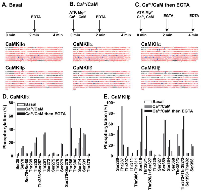
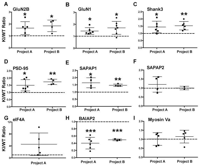
Ca(2+)/calmodulin-dependent protein kinase IIα (CaMKIIα) autophosphorylation at Thr286 and Thr305/Thr306 regulates kinase activity and modulates subcellular targeting and is critical for normal synaptic plasticity and learning and memory. Here, a mass spectrometry-based approach was used to identify Ca(2+)-dependent and -independent in vitro autophosphorylation sites in recombinant CaMKIIα and CaMKIIβ. CaMKII holoenzymes were then immunoprecipitated from subcellular fractions of forebrains isolated from either wild-type (WT) mice or mice with a Thr286 to Ala knock-in mutation of CaMKIIα (T286A-KI mice) and analyzed using the same approach in order to characterize in vivo phosphorylation sites in both CaMKII isoforms and identify CaMKII-associated proteins (CaMKAPs). A total of six and seven autophosphorylation sites in CaMKIIα and CaMKIIβ, respectively, were detected in WT mice. Thr286-phosphorylated CaMKIIα and Thr287-phosphorylated CaMKIIβ were selectively enriched in WT Triton-insoluble (synaptic) fractions compared to Triton-soluble (membrane) and cytosolic fractions. In contrast, Thr306-phosphorylated CaMKIIα and Ser315- and Thr320/Thr321-phosphorylated CaMKIIβ were selectively enriched in WT cytosolic fractions. The T286A-KI mutation significantly reduced levels of phosphorylation of CaMKIIα at Ser275 across all subcellular fractions and of cytosolic CaMKIIβ at Ser315 and Thr320/Thr321. Significantly more CaMKAPs coprecipitated with WT CaMKII holoenzymes in the synaptic fraction compared to that in the membrane fraction, with functions including scaffolding, microtubule organization, actin organization, ribosomal function, vesicle trafficking, and others. The T286A-KI mutation altered the interactions of multiple CaMKAPs with CaMKII, including several proteins linked to autism spectrum disorders. These data identify CaMKII isoform phosphorylation sites and a network of synaptic protein interactions that are sensitive to the abrogation of Thr286 autophosphorylation of CaMKIIα, likely contributing to the diverse synaptic and behavioral deficits of T286A-KI mice.

Keywords: Synaptic plasticity; autophosphorylation; mass spectrometry; postsynaptic density; protein−protein interactions; proteomics; subcellular fractionation.


Figures

Figure 1
Figure 1. Identification of in vitro CaMKIIα and CaMKIIβ phosphorylation sites
A–C. Sequence coverage (red) and phosphorylation-site detection (blue, underlined) in purified CaMKIIα or CaMKIIβ following either: A, a control incubation (Basal), B, phosphorylation in the presence of Ca2+/calmodulin alone (Ca2+/CaM), or C, sequential phosphorylation in the presence of Ca2+/calmodulin and then EGTA (Ca2+/CaM then EGTA) (see Methods). D, E. The AUCs of XICs were used to compare relative levels of phosphorylation of CaMKIIα at 16 different phosphorylation sites (D) or CaMKIIβ at 15 different phosphorylation sites (E) in each sample.
Figure 2
Figure 2. Identification of phosphorylation sites in mouse forebrain CaMKIIα and CaMKIIβ
A. Cytosolic (S1), membrane (S2), and synaptic (S3) fractions were immunoprecipitated using control or anti-CaMKII IgG, and immune complexes were analyzed by SDS-PAGE followed by staining with Sypro Ruby. B. Semi-quantitative analysis of relative levels of CaMKIIα phosphorylation at 6 different sites in each fraction normalized to the highest level for each site. The synaptic fraction was selectively enriched for Thr286 phosphorylation, whereas the cytosolic fraction was selectively enriched for Thr306 phosphorylation. C. Immunoblot analysis of subcellular fractions confirms that the synaptic (S3) fraction is significantly enriched in Thr286 phosphorylated CaMKIIα. D. Semi-quantitative analysis of relative levels of CaMKIIβ phosphorylation at 6 different sites in each fraction normalized to the highest level for each site. The cytosolic fraction was selectively enriched for phosphorylation at Ser315 and Thr320/1. *p<0.05; ***p<0.001; ****p<0.0001; in comparison to the highest level at that site. E. Summary of CaMKII phosphorylation sites. Horizontal bars indicate aligned domain structures of the canonical CaMKIIα and CaMKIIβ isoforms based on a sequence alignment (accession numbers: P11798 and P28652, respectively). Sequence alignments of selected regions are indicated above the bars, whereas CaMKIIβ-specific sequences of the actin-binding domain (ABD) are indicated below. All phosphorylation sites detected in this study (in vitro or in vivo) are indicated by red dots adjacent to amino acid sequences or by residues in red font adjacent to the domain bars. Black dots and fonts indicate additional residues detected in prior global phospho-proteomics studies. Yellow highlighted labels indicate phosphorylation sites identified in only one study.
Figure 3
Figure 3. Effect of T286A-KI mutation on CaMKIIα and CaMKIIβ phosphorylation at other sites
Levels of phosphorylation of CaMKIIα (A–E) and CaMKIIβ (F–K) at the indicated sites were compared across subcellular fractions isolated in parallel from WT and T286A-KI mice. Data are the mean from 4 (A–E) or 3 (F–K) biological replicates after normalization to the estimated level in the WT S1 fraction within each replicate. A 2-way ANOVA revealed a significant genotype effect on phosphorylation of CaMKIIα at Ser275 (A; F(1,15) = 157.8; p<0.0001) and Ser314 (C; F(1,15) = 22.42; p=0.0003) and a significant fractionation effect on Thr306 phosphorylation (B; F(2,15) = 35.41; p<0.0001). For CaMKIIβ there were significant fractionation and genotype effects and an interaction effect on the phosphorylation at Ser315 (E; Fractionation effect (F(2,9) = 153.8, p<0.0001), Genotype effect (F(1,9) = 49.4, p<0.0001), Interaction (F(2,9) = 33.55, p<0.0001) and Thr320/Thr321(F; Fractionation effect (F(2,9) = 58.31, p<0.0001), Genotype effect (F(1,9) = 9.115, p=0.0145), Interaction (F(2,9) = 6.159, p=0.0206). Significant differences revealed by post-hoc analyses are coded as follows: *, compared to S1 WT. $, compared to S2 WT. @, compared to S3 WT. #, compared to S1 KI. Single, double, triple and quadruple symbols indicate p<0.05, p<0.01, p<0.001, and p<0.0001, respectively.
Figure 4
Figure 4. Comparative levels of CaMKII and CaMKAPs in WT S2 and S3 fractions
A. Fewer total spectral counts derived from all CaMKII isoforms were detected in the S3 fraction compared to S2 fraction. B. The total number of spectral counts derived from proteins other than CaMKII was larger in the S3 fraction that in the S2 fraction. C. More proteins other than CaMKII were detected in S3 complexes compared to S2 complexes. These data are derived from project A; relative differences between fractions are representative of two other independent biological replicates (see Figure S4).
Figure 5
Figure 5. A CaMKAP interaction map
The STRING-db identifies interactions utilizing genomic context, high-throughput experiments, co-expression, and previous knowledge. Default parameters were used to generate this map. Associations from multiple sources and/or contexts have thicker lines. Proteins were arbitrarily assigned to 14 different groups based on protein function and interaction data.
Figure 6
Figure 6. Effect of T286A-KI mutation of CaMKIIα on the association of selected CaMKAPs with CaMKII holoenzymes in the S3 fraction
Relative levels of each CaMKAP in WT and T286A-KI samples were estimated from the AUC of XICs for multiple peptides, normalized to the relative levels of CaMKIIα (similarly estimated from AUCs for 9 peptides) and then expressed as a ratio (KI/WT). Data from 1 or 2 independent biological replicates (“projects”) are shown. Each data point is the ratio calculated for a single CaMKAP derived peptide, with the mean and S.E.M indicated. A. GluN2B, B. GluN1, C. Shank3, D. PSD-95, E. SAPAP1, F. SAPAP2, G. EIF4A, H. BAIAP2, I. Myosin Va. Data for each project were compared to a theoretical value of 1 (no difference; dashed line) using a one-column t-test. *p<0.05, **p<0.01, ***p<0.001. ****p<0.0001.

References

    1. Erondu NE, Kennedy MB. Regional distribution of type II Ca2+/calmodulin-dependent protein kinase in rat brain. J Neurosci. 1985;5:3270–3277. - PMC - PubMed
    1. van Woerden GM, Hoebeek FE, Gao Z, Nagaraja RY, Hoogenraad CC, Kushner SA, Hansel C, De Zeeuw CI, Elgersma Y. betaCaMKII controls the direction of plasticity at parallel fiber-Purkinje cell synapses. Nat Neurosci. 2009;12:823–825. - PubMed
    1. Fink CC, Bayer KU, Myers JW, Ferrell JE, Jr, Schulman H, Meyer T. Selective regulation of neurite extension and synapse formation by the beta but not the alpha isoform of CaMKII. Neuron. 2003;39:283–297. - PubMed
    1. Bingol B, Wang CF, Arnott D, Cheng D, Peng J, Sheng M. Autophosphorylated CaMKIIalpha acts as a scaffold to recruit proteasomes to dendritic spines. Cell. 2010;140:567–578. - PubMed
    1. Silva AJ, Paylor R, Wehner JM, Tonegawa S. Impaired spatial learning in alpha-calcium-calmodulin kinase II mutant mice. Science. 1992;257:206–211. - PubMed

Publication types

MeSH terms

LinkOut - more resources