Meta-Analysis
. 2015 Apr;72(4):305-15.
doi: 10.1001/jamapsychiatry.2014.2206.

Identification of a common neurobiological substrate for mental illness

Affiliations
Meta-Analysis

Identification of a common neurobiological substrate for mental illness

Madeleine Goodkind et al. JAMA Psychiatry. 2015 Apr.

Abstract

Importance: Psychiatric diagnoses are currently distinguished based on sets of specific symptoms. However, genetic and clinical analyses find similarities across a wide variety of diagnoses, suggesting that a common neurobiological substrate may exist across mental illness.

Objective: To conduct a meta-analysis of structural neuroimaging studies across multiple psychiatric diagnoses, followed by parallel analyses of 3 large-scale healthy participant data sets to help interpret structural findings in the meta-analysis.

Data sources: PubMed was searched to identify voxel-based morphometry studies through July 2012 comparing psychiatric patients to healthy control individuals for the meta-analysis. The 3 parallel healthy participant data sets included resting-state functional magnetic resonance imaging, a database of activation foci across thousands of neuroimaging experiments, and a data set with structural imaging and cognitive task performance data.

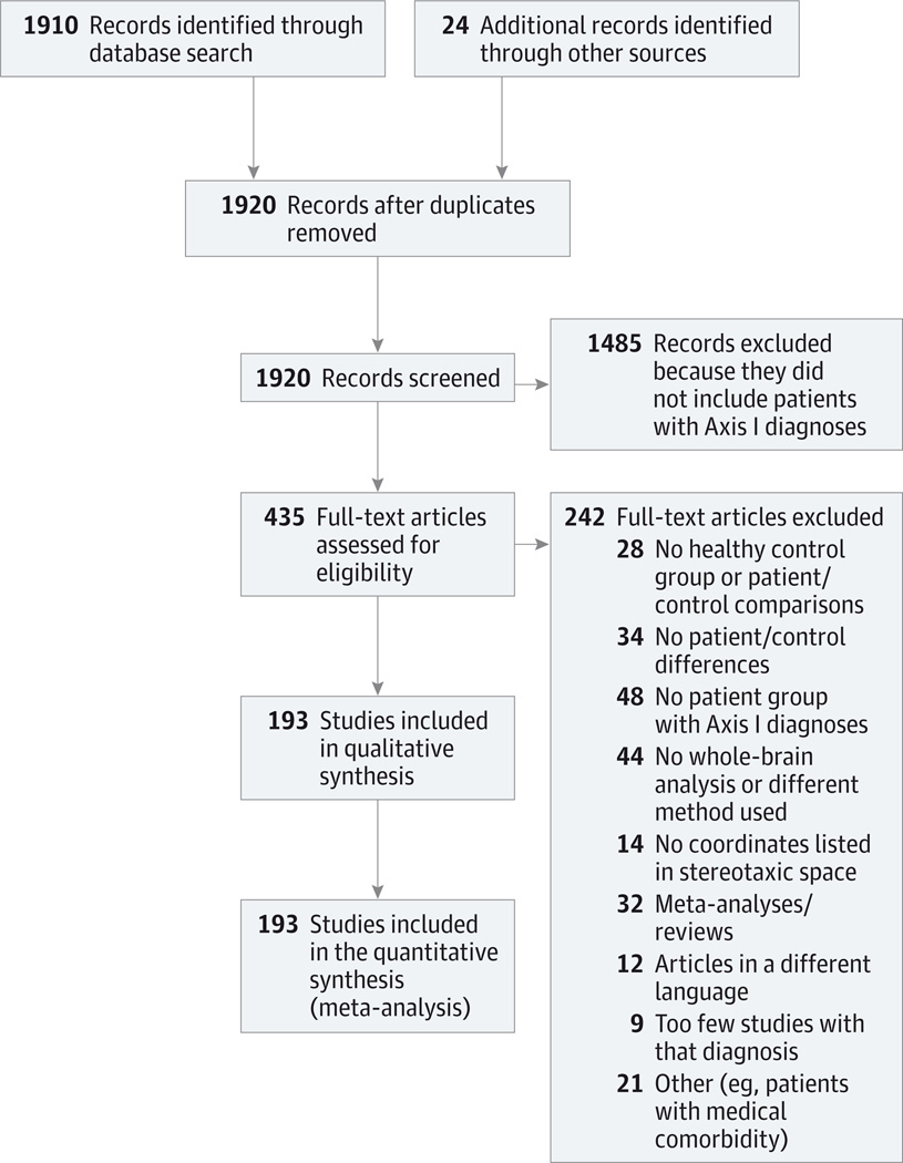
Data extraction and synthesis: Studies were included in the meta-analysis if they reported voxel-based morphometry differences between patients with an Axis I diagnosis and control individuals in stereotactic coordinates across the whole brain, did not present predominantly in childhood, and had at least 10 studies contributing to that diagnosis (or across closely related diagnoses). The meta-analysis was conducted on peak voxel coordinates using an activation likelihood estimation approach.

Main outcomes and measures: We tested for areas of common gray matter volume increase or decrease across Axis I diagnoses, as well as areas differing between diagnoses. Follow-up analyses on other healthy participant data sets tested connectivity related to regions arising from the meta-analysis and the relationship of gray matter volume to cognition.

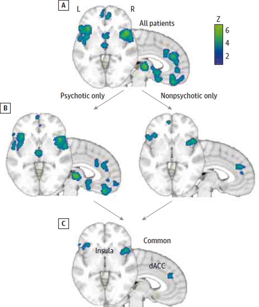
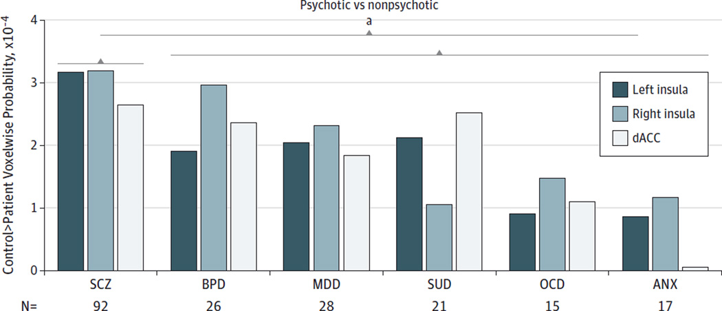
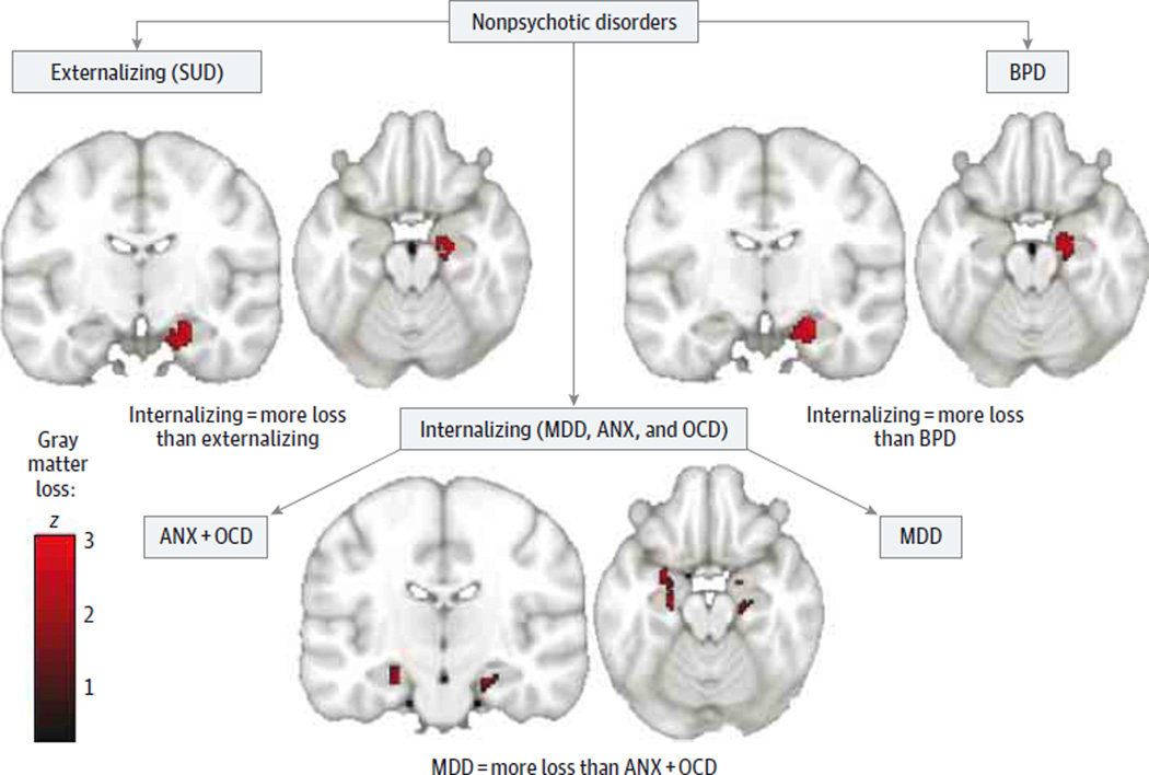
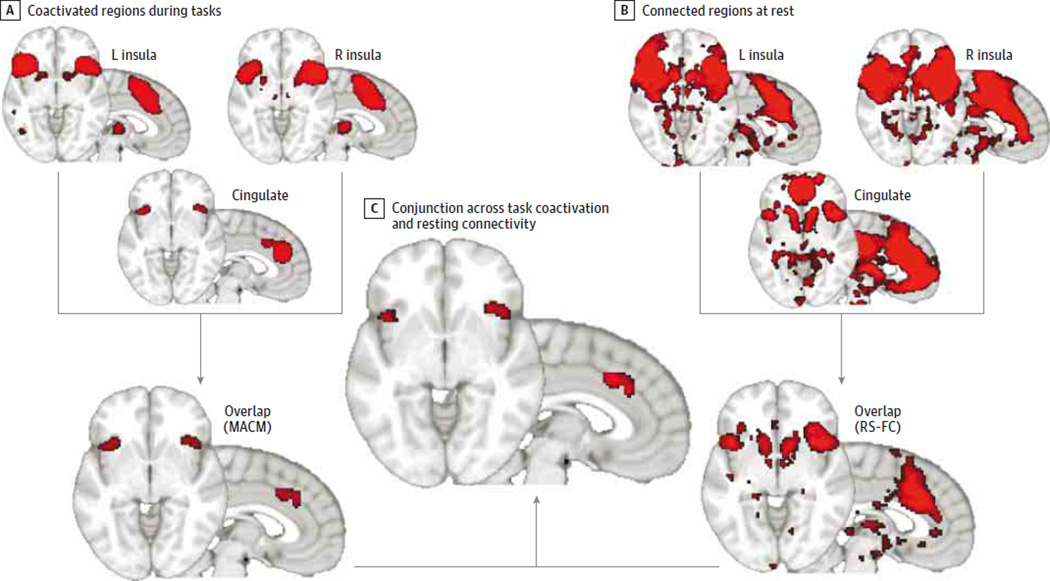
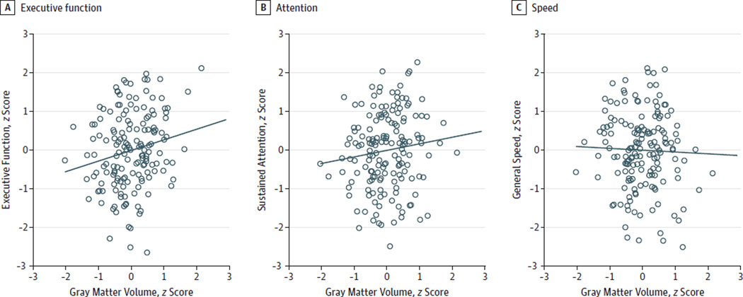
Results: Based on the voxel-based morphometry meta-analysis of 193 studies comprising 15 892 individuals across 6 diverse diagnostic groups (schizophrenia, bipolar disorder, depression, addiction, obsessive-compulsive disorder, and anxiety), we found that gray matter loss converged across diagnoses in 3 regions: the dorsal anterior cingulate, right insula, and left insula. By contrast, there were few diagnosis-specific effects, distinguishing only schizophrenia and depression from other diagnoses. In the parallel follow-up analyses of the 3 independent healthy participant data sets, we found that the common gray matter loss regions formed a tightly interconnected network during tasks and at resting and that lower gray matter in this network was associated with poor executive functioning.

Conclusions and revelance: We identified a concordance across psychiatric diagnoses in terms of integrity of an anterior insula/dorsal anterior cingulate-based network, which may relate to executive function deficits observed across diagnoses. This concordance provides an organizing model that emphasizes the importance of shared neural substrates across psychopathology, despite likely diverse etiologies, which is currently not an explicit component of psychiatric nosology.


Conflict of interest statement

Conflict of Interest Disclosures: None reported.

Figures

Figure 1
Figure 1
Flow Diagram of Study Selection
Figure 2
Figure 2. Shared Patterns of Decreased Gray Matter From the Voxel-Based Morphometry Meta-analysis
Results are from patient vs healthy participant comparisons for studies pooled across all diagnoses (A), separately by psychotic or nonpsychotic diagnosis studies (B), and from a conjunction across the psychotic and nondiagnosis diagnosis group maps in panel B (C). Results show common gray matter loss across diagnoses in the anterior insula and dorsal anterior cingulate (dACC). The z score is for the activation likelihood estimation analysis for gray matter loss. L indicates left; and r, right.
Figure 3
Figure 3. Extracted per-Voxel Probabilities of Decreased Gray Matter in the Voxel-Based Morphometry Meta-analysis, Separated by Individual Diagnosis and Common Gray Matter Loss Region (Left and Right Anterior Insula)
Values represent the probability of identifying a gray matter abnormality for an average voxel within the region of interest, derived from the modeled activation maps. ANX indicates anxiety disorders; BPD, bipolar disorder; dACC, dorsal anterior cingulate; MDD, major depressive disorder; OCD, obsessive-compulsive disorder; SCZ, schizophrenia; and SUD, substance use disorder. aP < .05 for comparison of the psychotic with the nonpsychotic disorders.
Figure 4
Figure 4. Voxel-Based Morphometry Meta-analysis Contrasts Between Subdivisions of the Nonpsychotic Disorder Studies Group, Broken Down by Internalizing, Externalizing, and Bipolar Disorder (BPD) Groupings
Internalizing disorders show greater gray matter loss in the anterior hippocampus and amygdala when compared with either externalizing or BPD diagnoses. This effect is driven by the major depressive disorder (MDD) group, which shows greater gray matter loss in these regions than the other diagnoses within the internalizing grouping (anxiety disorder [ANX] and obsessive-compulsive disorder [OCD]). The z score is for the activation likelihood estimation analysis for gray matter loss. SUD indicates substance use disorder.
Figure 5
Figure 5. Common Gray Matter Loss Regions From the Voxel-Based Morphometry Meta-analysis Are Part of an Interconnected Brain Network
A, Meta-analytic coactivation maps (MACMs) showing regions coactivated with each of the common gray matter loss regions in healthy participant task-based activation studies in the BrainMap database, as well as a conjunction across all 3 MACM maps. B, Resting-state (RS) functional connectivity (FC) in healthy individuals seeded by each of the common gray matter loss regions, as well as a conjunction across all RS-FC maps. C, Conjunction across all of the MACMs and RS-FC map demonstrates that each of the common gray matter loss regions shows both task-dependent and task-independent FC with the bilateral anterior insula and dorsal anterior cingulate (the regions showing consistent gray matter changes) as well as the thalamus. L indicates left; and R, right.
Figure 6
Figure 6. Relationship Between Gray Matter Volume in the Common Gray Matter Loss Regions and Performance on a Computerized Battery of Behavioral Cognitive Tests
Based on a principal components analysis, cognitive test performance was reduced to 3 components: general executive function, the specific domain of sustained attention, and general cognitive performance and performance speed. Lower voxel-based morphometry–measured gray matter volume in these regions is associated with worse executive functioning (A) and a trend for worse sustained attention (B) but not general performance and speed (C).

References

    1. Spitzer RL, Endicott J, Robins E. Research diagnostic criteria: rationale and reliability. Arch Gen Psychiatry. 1978;35(6):773–782. - PubMed
    1. Fischer BA. A review of American psychiatry through its diagnoses: the history and development of the Diagnostic and Statistical Manual of Mental Disorders. J Nerv Ment Dis. 2012;200(12):1022–1030. - PubMed
    1. Ellison-Wright I, Bullmore E. Anatomy of bipolar disorder and schizophrenia: a meta-analysis. Schizophr Res. 2010;117(1):1–12. - PubMed
    1. Etkin A, Wager TD. Functional neuroimaging of anxiety: a meta-analysis of emotional processing in PTSD, social anxiety disorder, and specific phobia. Am J Psychiatry. 2007;164(10):1476–1488. - PMC - PubMed
    1. Hamilton JP, Etkin A, Furman DJ, Lemus MG, Johnson RF, Gotlib IH. Functional neuroimaging of major depressive disorder: a meta-analysis and new integration of base line activation and neural response data. Am J Psychiatry. 2012;169(7):693–703. - PMC - PubMed

Publication types