. 2015 Jun;39(6):813-25.
doi: 10.1097/PAS.0000000000000389.

Dichotomy of Genetic Abnormalities in PEComas With Therapeutic Implications

Affiliations

Dichotomy of Genetic Abnormalities in PEComas With Therapeutic Implications

Narasimhan P Agaram et al. Am J Surg Pathol. 2015 Jun.

Abstract

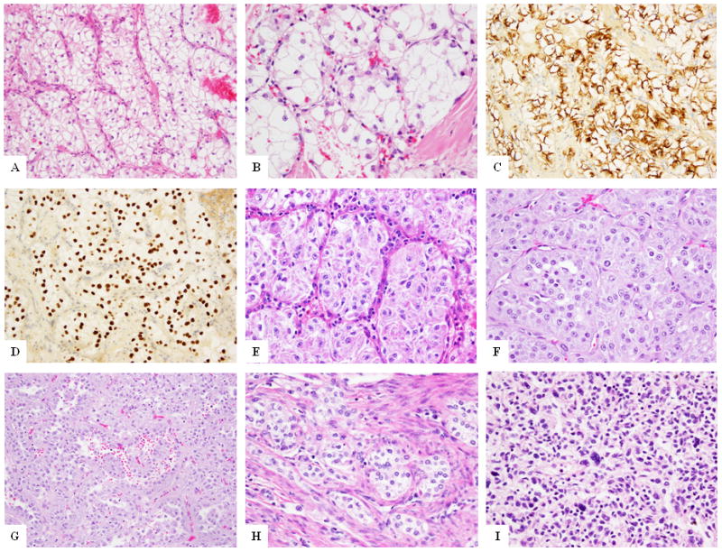
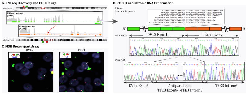
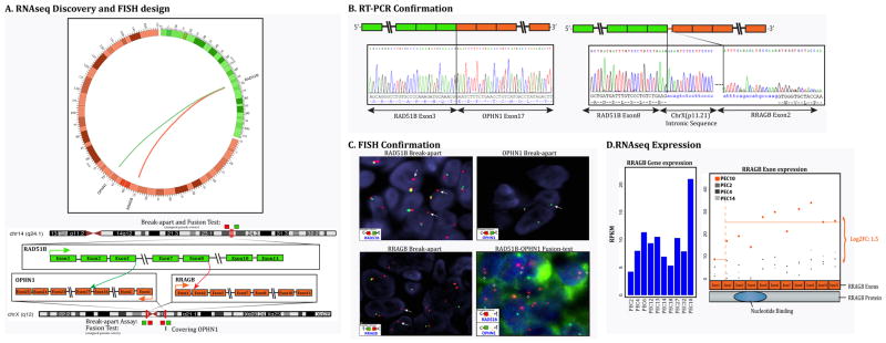
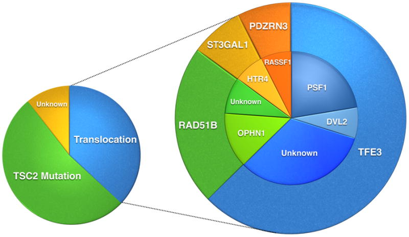
Perivascular epithelioid cell neoplasms (PEComa) are a family of rare mesenchymal tumors with hybrid myo-melanocytic differentiation. Although most PEComas harbor loss-of-function TSC1/TSC2 mutations, a small subset were reported to carry TFE3 gene rearrangements. As no comprehensive genomic study has addressed the molecular classification of PEComa, we sought to investigate by multiple methodologies the incidence and spectrum of genetic abnormalities and their potential genotype-phenotype correlations in a large group of 38 PEComas. The tumors were located in soft tissue (11 cases) and visceral sites (27) including uterus, kidney, liver, lung, and urinary bladder. Combined RNA sequencing and fluorescence in situ hybridization analysis identified 9 (23%) TFE3 gene-rearranged tumors, with 3 cases showing an SFPQ/PSF-TFE3 fusion and 1 case showing a novel DVL2-TFE3 gene fusion. The TFE3-positive lesions showed a distinctive nested/alveolar morphology and were equally distributed between soft tissue and visceral sites. In addition, novel RAD51B gene rearrangements were identified in 3 (8%) uterine PEComas, which showed a complex fusion pattern and were fused to RRAGB/OPHN1 genes in 2 cases. Other nonrecurrent gene fusions, HTR4-ST3GAL1 and RASSF1-PDZRN3, were identified in 2 cases. Targeted exome sequencing using the IMPACT assay was used to address whether the presence of gene fusions is mutually exclusive from TSC gene abnormalities. TSC2 mutations were identified in 80% of the TFE3 fusion-negative cases tested. Coexistent TP53 mutations were identified in 63% of the TSC2-mutated PEComas. Our results showed that TFE3-rearranged PEComas lacked coexisting TSC2 mutations, indicating alternative pathways of tumorigenesis. In summary, this comprehensive genetic analysis significantly expands our understanding of molecular alterations in PEComas and brings forth the genetic heterogeneity of these tumors.


Conflict of interest statement

Conflict of interest: none

Figures

Figure 1
Figure 1. Morphologic appearance of TFE3 fusion positive PEComas
PEComa with PSF-TFE3 gene fusion (Case 1) at low (A) and higher magnification (B) showing the characteristic epithelioid cells in a nested architecture. HMB45 (C) and TFE3 (D) immunostains show diffuse positivity of the tumor cells. PEComa with PSF-TFE3 gene fusion (Case 3) showing similar morphology (E) of epithelioid cells with clear to granular cytoplasm in a nested appearance. PEComa with DVL2-TFE3 gene fusion (Case 4) showing epithelioid cells in a nested pattern (F) and other areas of pseudo-papillary arrangement (G). Uterine PEComa (Case 6) showing nested epithelioid cells in infiltrating the myometrium (H). Pulmonary PEComa (Case 8) showing spindle to ovoid cells with scattered atypical cells (I).
Figure 2
Figure 2. PEComa with DVL2-TFE3 gene fusion
(Case 4). (A) Schematic representation of the fusion of DVL2 located on 17p13.1 with TFE3 on Xp11.2, resulting in a t(X;17)(p11.2;p13.1) translocation. (B) RT-PCR validation showing fusion of the DVL2 exon 4 to TFE3 exon 7 (top right), followed by DNA PCR confirming the fusion of DVL2 exon 5 with TFE3 intron 6 with anti-parallel sequence of TFE3 exon 6 and TFE3 intron 5 in-between (bottom right). (C) FISH break-apart assays showing unbalanced rearrangements of DVL2 (arrows) with loss of telomeric signal (red) and trisomy of Xp11.2 locus with TFE3 rearrangements (arrows) (t-telomeric; c-centromeric).
Figure 3
Figure 3. RAD51B-associated gene fusion in uterine PEComa (Case 10)
(A) Schematic representation of the two fusion transcript candidates identified by RNAseq, involving the RAD51B locus on 14q24.1 with RRAGB located on xp11.2, resulting in a t(x;14)(p11.2;q24.1) translocation, and the other involving the RAD51B with OPHN1 gene on xq12 resulting in a t(x;14)(q12;q24.1); (B) Fusion candidates were validated by RT-PCR showing fusion of the RAD51B exon 8 to RRAGB exon 2 with an intervening intronic sequence of Xp11.21, and the fusion of RAD51B exon 3 with OPHN1 exon 17. (C) FISH break-apart assays showing unbalanced rearrangement of RAD51B gene (arrows) with loss of the telomeric signal (upper left). FISH break-apart assay showing unbalanced rearrangement of RRAGB gene (arrows) on Xp11.21 with loss of the telomeric signal (lower left); FISH for OPHN1 gene on Xq12 showing loss of one copy of signals, indicating a larger deletion at this locus (upper right). The RAD51B-OPHN1 fusion assay (lower right) showing fusion of the RAD51B (red) with a small fragment of OPHN1 (green) gene (arrows), suggesting a deletion in the OPHN1 gene locus. (t-telomeric; c-centromeric) (D) Bar chart showing increased expression of RRAGB in PEC10 compared to other PEComas. The dot plot further shows the differential exon expression of RRAGB (case 9, after the exon 2 breakpoint) compared to other PEComas.
Figure 4
Figure 4. Morphologic spectrum of PEComas with non-TFE3 fusions
RAD51B-rearranged tumors (A-C): epithelioid cells in a sheet-like arrangement (A, case 10); or nested pattern (B, case 11), or spindle cells in fascicles in a hemangiopericytoma-like branching vascular pattern (C, case 12); (D) PEComa with HTR4-ST3GAL1 fusion (Case 13) showing predominantly spindle cells in fascicles, reminiscent of smooth muscle neoplasm; (E) PEComa with RASSF1-PDZRN3 gene fusion (Case 14) showing epithelioid to spindle cells with clear cytoplasm in a nested and fascicular pattern; (F-I) PEComas with no known gene fusions: epithelioid cells with clear cytoplasm in sheets (F, case 32), spindle to epithelioid cells and scattered pleomorphic cells (G, case 16); epithelioid neoplasm with scattered pleomorphic cells (H, case 19), and fascicles of spindle cells in a densely sclerotic background (I, case 28).
Figure 5
Figure 5. Schematic pie chart representing the genetic findings in PEComas
The larger pie chart demonstrates the different translocations identified in PEComas. The smaller pie on the left shows the different genetic subgroups in PEComa with a predominant group showing TSC2 mutations, a second group with translocations, and a smaller group with unknown genetic alterations.

Similar articles

Cited by

References

    1. Fletcher C, Bridge JA, Hogendoorn PC, et al. WHO Classification of Tumours of Soft Tissue and Bone. IARC; Lyon: 2013.
    1. Folpe AL, Mentzel T, Lehr HA, et al. Perivascular epithelioid cell neoplasms of soft tissue and gynecologic origin: a clinicopathologic study of 26 cases and review of the literature. Am J Surg Pathol. 2005;29(12):1558–75. - PubMed
    1. Pan CC, Chung MY, Ng KF, et al. Constant allelic alteration on chromosome 16p (TSC2 gene) in perivascular epithelioid cell tumour (PEComa): genetic evidence for the relationship of PEComa with angiomyolipoma. J Pathol. 2008;214(3):387–93. - PubMed
    1. Qin W, Bajaj V, Malinowska I, et al. Angiomyolipoma have common mutations in TSC2 but no other common genetic events. PLoS One. 2011;6(9):e24919. - PMC - PubMed
    1. Kenerson H, Folpe AL, Takayama TK, et al. Activation of the mTOR pathway in sporadic angiomyolipomas and other perivascular epithelioid cell neoplasms. Hum Pathol. 2007;38(9):1361–71. - PMC - PubMed

Publication types