

Resting-State Functional Connectivity in Patients with Long-Term Remission of Cushing's Disease
- PMID: 25652248
- PMCID: PMC4839512
- DOI: 10.1038/npp.2015.38
Resting-State Functional Connectivity in Patients with Long-Term Remission of Cushing's Disease
Abstract
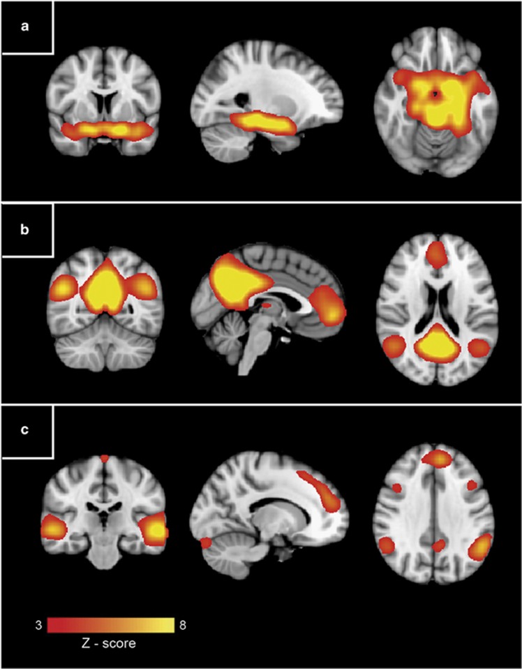
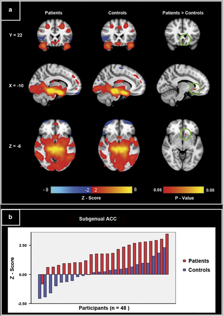
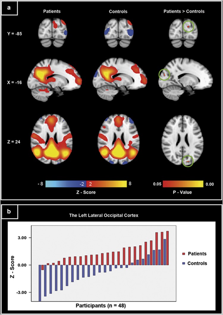
Glucocorticoid disturbance can be a cause of psychiatric symptoms. Cushing's disease represents a unique model for examining the effects of prolonged exposure to high levels of endogenous cortisol on the human brain as well as for examining the relation between these effects and psychiatric symptomatology. This study aimed to investigate resting-state functional connectivity (RSFC) of the limbic network, the default mode network (DMN), and the executive control network in patients with long-term remission of Cushing's disease. RSFC of these three networks of interest was compared between patients in remission of Cushing's disease (n=24; 4 male, mean age=44.96 years) and matched healthy controls (n=24; 4 male, mean age=46.5 years), using probabilistic independent component analysis to extract the networks and a dual regression method to compare both groups. Psychological and cognitive functioning was assessed with validated questionnaires and interviews. In comparison with controls, patients with remission of Cushing's disease showed an increased RSFC between the limbic network and the subgenual subregion of the anterior cingulate cortex (ACC) as well as an increased RSFC of the DMN in the left lateral occipital cortex. However, these findings were not associated with psychiatric symptoms in the patient group. Our data indicate that previous exposure to hypercortisolism is related to persisting changes in brain function.
Figures
Similar articles
-
Altered neural processing of emotional faces in remitted Cushing's disease.Psychoneuroendocrinology. 2015 Sep;59:134-46. doi: 10.1016/j.psyneuen.2015.05.001. Epub 2015 May 13. Psychoneuroendocrinology. 2015. PMID: 26092780
-
Smaller grey matter volumes in the anterior cingulate cortex and greater cerebellar volumes in patients with long-term remission of Cushing's disease: a case-control study.Eur J Endocrinol. 2013 Oct 21;169(6):811-9. doi: 10.1530/EJE-13-0471. Print 2013 Dec. Eur J Endocrinol. 2013. PMID: 24031092
-
Dysregulation of resting-state functional connectivity in patients with Cushing's disease.Neuroradiology. 2019 Aug;61(8):911-920. doi: 10.1007/s00234-019-02223-y. Epub 2019 May 17. Neuroradiology. 2019. PMID: 31101946
-
Structural brain abnormalities in Cushing's syndrome.Curr Opin Endocrinol Diabetes Obes. 2018 Aug;25(4):285-289. doi: 10.1097/MED.0000000000000414. Curr Opin Endocrinol Diabetes Obes. 2018. PMID: 29746308 Review.
-
Structural and functional brain alterations in Cushing's disease: A narrative review.Front Neuroendocrinol. 2022 Oct;67:101033. doi: 10.1016/j.yfrne.2022.101033. Epub 2022 Sep 17. Front Neuroendocrinol. 2022. PMID: 36126747 Review.
Cited by
-
Trends in regional morphological changes in the brain after the resolution of hypercortisolism in Cushing's disease: a complex phenomenon, not mere partial reversibility.Endocr Connect. 2021 Oct 25;10(11):1377-1386. doi: 10.1530/EC-21-0385. Endocr Connect. 2021. PMID: 34596577 Free PMC article.
-
Altered microstructural pattern of white matter in Cushing's disease identified by automated fiber quantification.Neuroimage Clin. 2021;31:102770. doi: 10.1016/j.nicl.2021.102770. Epub 2021 Jul 24. Neuroimage Clin. 2021. PMID: 34332193 Free PMC article.
-
A voxel-level brain-wide association study of cortisol at 8 a.m.: Evidence from Cushing's disease.Neurobiol Stress. 2021 Oct 25;15:100414. doi: 10.1016/j.ynstr.2021.100414. eCollection 2021 Nov. Neurobiol Stress. 2021. PMID: 34786440 Free PMC article.
-
Dynamic functional connectivity changes associated with psychiatric traits and cognitive deficits in Cushing's disease.Transl Psychiatry. 2023 Oct 5;13(1):308. doi: 10.1038/s41398-023-02615-y. Transl Psychiatry. 2023. PMID: 37798280 Free PMC article.
-
Cortical thickness abnormalities in long-term remitted Cushing's disease.Transl Psychiatry. 2020 Aug 21;10(1):293. doi: 10.1038/s41398-020-00980-6. Transl Psychiatry. 2020. PMID: 32826851 Free PMC article.
References
-
- Andela CD, van der Werff SJ, Pannekoek JN, van den Berg SM, Meijer OC, van Buchem MA et al (2013). Smaller grey matter volumes in the anterior cingulate cortex and greater cerebellar volumes in patients with long-term remission of Cushing's disease: a case-control study. Eur J Endocrinol 169: 811–819. - PubMed
-
- Beck AT, Epstein N, Brown G, Steer RA (1988). An inventory for measuring clinical anxiety: psychometric properties. J Consult Clin Psychol 56: 893–897. - PubMed
Publication types
MeSH terms
LinkOut - more resources
Full Text Sources
Other Literature Sources
