. 2015 May 15;24(10):2884-98.
doi: 10.1093/hmg/ddv050. Epub 2015 Feb 4.

MBTPS1/SKI-1/S1P proprotein convertase is required for ECM signaling and axial elongation during somitogenesis and vertebral development†

Affiliations

MBTPS1/SKI-1/S1P proprotein convertase is required for ECM signaling and axial elongation during somitogenesis and vertebral development†

Annita Achilleos et al. Hum Mol Genet. .

Abstract

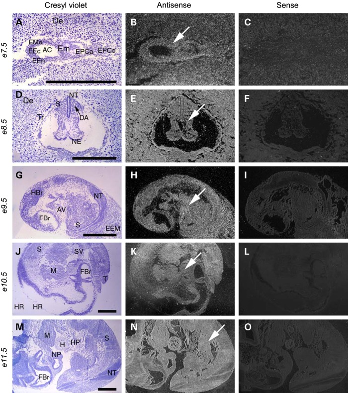
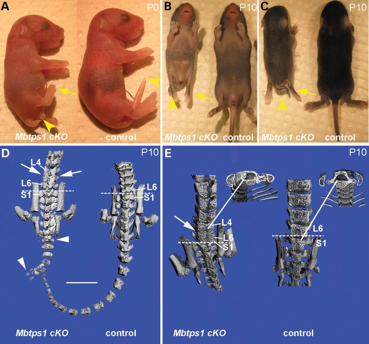
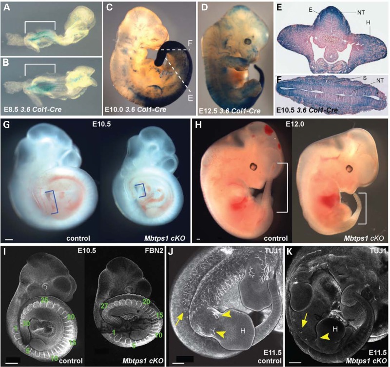
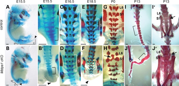
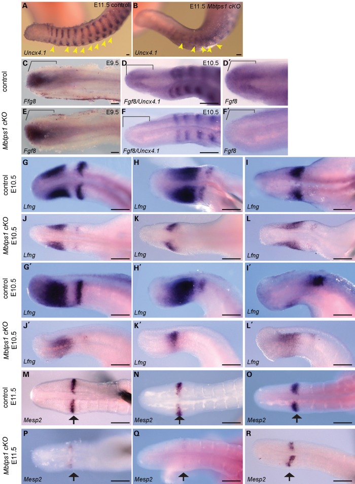
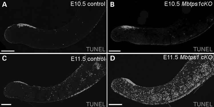
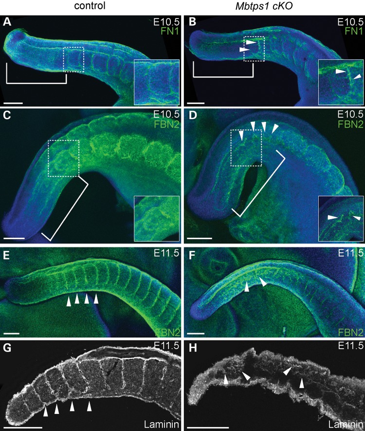
Caudal regression syndrome (sacral agenesis), which impairs development of the caudal region of the body, occurs with a frequency of about 2 live births per 100 000 newborns although this incidence rises to 1 in 350 infants born to mothers with gestational diabetes. The lower back and limbs can be affected as well as the genitourinary and gastrointestinal tracts. The axial skeleton is formed during embryogenesis through the process of somitogenesis in which the paraxial mesoderm periodically segments into bilateral tissue blocks, called somites. Somites are the precursors of vertebrae and associated muscle, tendons and dorsal dermis. Vertebral anomalies in caudal regression syndrome may arise through perturbation of somitogenesis or, alternatively, could result from defective bone formation and patterning. We discovered that MBTPS1/SKI-1/S1P, which proteolytically activates a class of transmembrane transcription factors, plays a critical role in somitogenesis and the pathogenesis of lumbar/sacral vertebral anomalies. Conditional deletion of Mbtps1 yields a viable mouse with misshapen, fused and reduced number of lumbar and sacral vertebrae, under-developed hind limb bones and a kinky, shortened tail. We show that Mbtps1 is required to (i) maintain the Fgf8 'wavefront' in the presomitic mesoderm that underpins axial elongation, (ii) sustain the Lfng oscillatory 'clock' activity that governs the periodicity of somite formation and (iii) preserve the composition and character of the somitic extracellular matrix containing fibronectin, fibrillin2 and laminin. Based on this spinal phenotype and known functions of MBTPS1, we reason that loss-of-function mutations in Mbtps1 may cause the etiology of caudal regression syndrome.


Figures

Figure 1.
Figure 1.
Mbtps1 is expressed ubiquitously in the post-implantation and mid-gestation mouse embryo. Left column shows embryo sections stained with Cresyl Violet; middle column shows the same sections with comparative antisense hybridization labeling seen as bright under darkfield illumination (arrows) and right column shows control sense hybridization. (A–C) Comparison of in situ hybridization with sense and antisense probes reveals a broad expression of Mbtps1 in the uterine decidua and in the embryo at E7.5 (arrow). E7.5 early embryo sagittal section seen within uterine cavity near site of implantation. Presence of Mbtps1 mRNA labeling is evident within embryo, ectoplacental cone and decidua. (DF) E8.5 embryo coronal section. (GI) E9.5 embryo sagittal section. (JL) E10.5 embryo sagittal section. (MO) E11.5 embryo sagittal section. AC, amniotic cavity; AV, atrioventricular canal; DA, dorsal aorta, primordium; De, decidua; EEc, embryonic ectoderm; EEM, extraembryonic membranes; EEn, embryonic endoderm; Em, embryo; EMe, embryonic mesenchyma; EPCa, ectoplacental cavity; EPCo, ectoplacental cone; FBr, forebrain; H, heart; HBr, hindbrain; HP, hepatic primordium; HR, hinbrain roof; M, mandible; NE, neuroepithelium; NP, nasal pit; NT, neural tube; S, somite; SV, sinus venosum; T, tail; and Tr, trophoblasts. Magnifications: (A–C) ×36; (D–F) ×24; (G–I) ×15 and (J–O) ×10. Scale bar: 1 mm.
Figure 2.
Figure 2.
Mbtps1cKO mice exhibit a severe caudal truncation and changes in vertebrae patterning. (A) At P0, Mbtps1cKO mice exhibit caudal axial truncation (arrowheads) and hypotrophy of the hind limbs (arrows). (B and C) At P10, Mbtps1cKO mice are smaller than normal littermates and the caudal axial truncation, as well as the hypotrophy of the hind limbs, are even more evident. B, ventral and C, dorsal view. (D and E) Micro-CT images of control and Mbtps1cKO mice at P10. (Each micro-CT image represents a separate mouse.). (D) Posterior comparison of Mbtps1cKO spine with that for control littermate. Arrowheads denote abnormal tail vertebrae and arrows demark abnormal bone patterning and fusion of lower lumbar vertebrae. Scale bar: 5 mm. (E) Sacral-lumbar comparison. Arrow denotes region with abnormal bone patterning and fusion of L4-L6 vertebrae in Mbtps1cKO. Solid lines identify regions from which respective cross-sections were taken. Dashed lines denote the sacral-lumbar border. S, sacral. L, lumbar.
Figure 3.
Figure 3.
The Cre recombinase expression in 3.6 Col1-Cre mice nicely correlates with the caudal phenotype of the Mbtps1cKO mice. (AD) Whole-mount LacZ staining of 3.6 Col1-Cre;ROSA26 mice at E8.5 (A,B), E10.0 (C) and E12.5 (D) reveals expression of the Cre recombinase prominently in the posterior tail region (brackets). (E and F) LacZ stained transverse sections of E10.5 3.6 Col- Cre: ROSA26 embryos showing strong expression of the Cre recombinase in the somites (S), the neural tube (NT), the hind limb buds (H) and the ectoderm (E). Sections were taken at the axial levels denoted in 3C. (G and H) A caudal axial truncation is evident in homozygous Mbtps1cKO embryos (right) by E10.5 compared to their wild type (left) littermates (G, brackets). The caudal axial truncation becomes more prevalent by E12.0 (H, brackets). (I) At E10.5, Fibrillin2 (FBN2) antibody staining reveals a reduced number of somites in Mbtps1cKO embryos (right) compared to their littermate control (left) (27 instead of 30 in these embryos). (J and K) Whole mount antibody staining of neuron-specific class III β tubulin (TUJ1) at E11.5 in Mbtps1cKO embryo (K) and control littermate (J) reveals a general failure of neuronal projections in the Mbtps1cKO embryos (arrows). Interestingly, no neuronal projections are seen in the hind limb bud (H) and spines of Mbtps1cKO embryos (arrowheads). Scale bar: 500 μm.
Figure 4.
Figure 4.
Skeletal preparations show that Mbtps1 deficiency disrupts the development of the lumbar/sacral spine. Alcian blue and Alizarin red staining reflect cartilage and mineralized bone, respectively. (A and B) Control (A) and Mbtps1cKO (B) skeletons at E15.5 reveal a severe caudal axial truncation (arrowheads). Brackets mark the sacral (S) and coccygeal (C) vertebrae. (A’ and B’) Dorsal views of higher magnification of the caudal region of control and Mbtps1cKO embryos at E15.5. Note the fused vertebrae precursors that resemble “stacking” (B’, bracket) and the short tail (B’ arrowhead) in the Mbtps1cKOembryo. (CF) Dorsal views of E16.5 (C and D) and E18.5 (E and F) control and Mbtps1cKO skeletons reveal delayed mineralization of sacral vertebrae (compare the alizarin red staining). Brackets denote the sacral (S) vertebrae. (G and H) Fusion of L6 and S1 vertebrae in Mbtps1cKO embryos is observed at P0 (arrow). (I and J) Tails of Mbtps1cKO mice routinely exhibited multiple fused vertebrae (compare bracketed regions). (I’ and J’) Some S1 vertebrae in Mbtps1cKO spines displayed asymmetric abnormal differentiation (see arrow) resembling that for L6. Scale bar: 2 mm.
Figure 5.
Figure 5.
Expression of key markers of somitogenesis including Fgf8 and Lunatic Fringe (Lfng) are dramatically decreased in the presomitic mesoderm of Mbtps1cKO embryos. Whole mount in situ hybridization was performed at E9.5 (C and E), E10.5 (D, F and GL) or E11.5 (A, B and MR) in control and Mbtps1cKO embryos. (A and B) Expression of Uncx4.1 reveals a general disorganization of the somites including loss of somite polarity in Mbtps1cKO embryos, 3 days after the onset of the 3.6 Col1-Cre recombinase. (C–F) Expression of Fgf8, normally restricted to the posterior-most region of the PSM at E9.5 and E10.5 (brackets) (C and D), is absent in Mbtps1cKO embryos at E10.5 (F), but still present a day earlier in Mbtps1cKO embryos at E9.5 (E). (G–L) Lfng expression is greatly reduced and its cyclic expression disturbed in E10.5 Mbtps1cKO embryos as demonstrated by dorsal (G–L) and lateral (G’–L’) views of the non-segmented PSM. (M–R) As illustrated by a representative sampling of three embryos, Mesp2 expression displays a range of expression in the determination front of Mbtps1cKO embryos which is consistently less than that for controls. Scale bar: 100 µm.
Figure 6.
Figure 6.
Cell death is evident in the caudal region of Mbtps1cKO embryos. (AD) Whole mount TUNEL staining reveals increased cell death in the caudal region of the Mbtps1cKO embryos at E11.5 (C and D), but no change at E10.5 (A and B). Scale bar: 200 µm.
Figure 7.
Figure 7.
Mbtps1 deficiency disrupts the organized distributions of Fibronectin, Fibrillin2, and Laminin, within the extracellular matrix of the developing spine. Immunofluorescence staining was carried out in whole mount embryos at E10.5 (AD) or E11.5 (E and F) and in sagittal sections of E11.5 mice (G and H). (A and B) Fibronectin (FN1) is normally localized in the non-segmented PSM (bracket) and around each formed somite (A). In Mbtps1cKO embryos, FN1 is reduced in the PSM (bracket) and a general disorganization of the ECM is observed (arrowheads). The inserts are higher magnifications of the boxed area. (C and D) Fibrillin2 (FBN2) normally surrounds each formed somite (C), but it is abnormally expressed and distributed in the Mbtps1cKO embryos (arrowheads). A reduction of FBN2 is also observed in the somites of Mbtps1cKOembryos. The inserts are higher magnifications of the boxed area. The insert in D was enhanced to emphasize the disorganization of the fibrils. (E and F), A day later, expression of FBN2 reveals an even more disorganized extracellular matrix in the Mbtps1cKO embryos (arrowheads, F). (G and H) Laminin normally demarcates the somite boundaries (G), but its localization pattern is greatly disturbed in the Mbtps1cKO embryos (H). Arrowheads demark the positions of somite boundaries within the developing tail (G). Arrowheads also point at disturbed patterns of Laminin in the PSM and the presumptive somites (H). Scale bar: 200 µm.

References

    1. Boulas M.M. (2009) Recognition of caudal regression syndrome. Adv. Neonatal. Care 9, 61–69 quiz 70-61. - PubMed
    1. Thottungal A.D., Charles A.K., Dickinson J.E., Bower C. (2010) Caudal dysgenesis and sirenomelia-single centre experience suggests common pathogenic basis. Am. J. Med. Genet. A, 152A, 2578–2587. - PubMed
    1. Pourquie O. Vertebrate segmentation: from cyclic gene networks to scoliosis. Cell, 145, 650–663. - PMC - PubMed
    1. Dias A.S., de Almeida I., Belmonte J.M., Glazier J.A., Stern C.D. (2014) Somites without a clock. Science, 343, 791–795. - PMC - PubMed
    1. Sparrow D.B., Chapman G., Wouters M.A., Whittock N.V., Ellard S., Fatkin D., Turnpenny P.D., Kusumi K., Sillence D., Dunwoodie S.L. (2006) Mutation of the LUNATIC FRINGE gene in humans causes spondylocostal dysostosis with a severe vertebral phenotype. Am. J. Hum. Genet., 78, 28–37. - PMC - PubMed

Publication types

MeSH terms

Substances

Supplementary concepts