. 2015 May 15;24(10):2808-25.
doi: 10.1093/hmg/ddv042. Epub 2015 Feb 4.

Analysis of hedgehog signaling in cerebellar granule cell precursors in a conditional Nsdhl allele demonstrates an essential role for cholesterol in postnatal CNS development

Affiliations

Analysis of hedgehog signaling in cerebellar granule cell precursors in a conditional Nsdhl allele demonstrates an essential role for cholesterol in postnatal CNS development

David Cunningham et al. Hum Mol Genet. .

Abstract

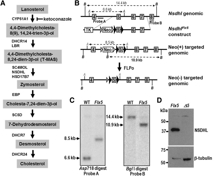
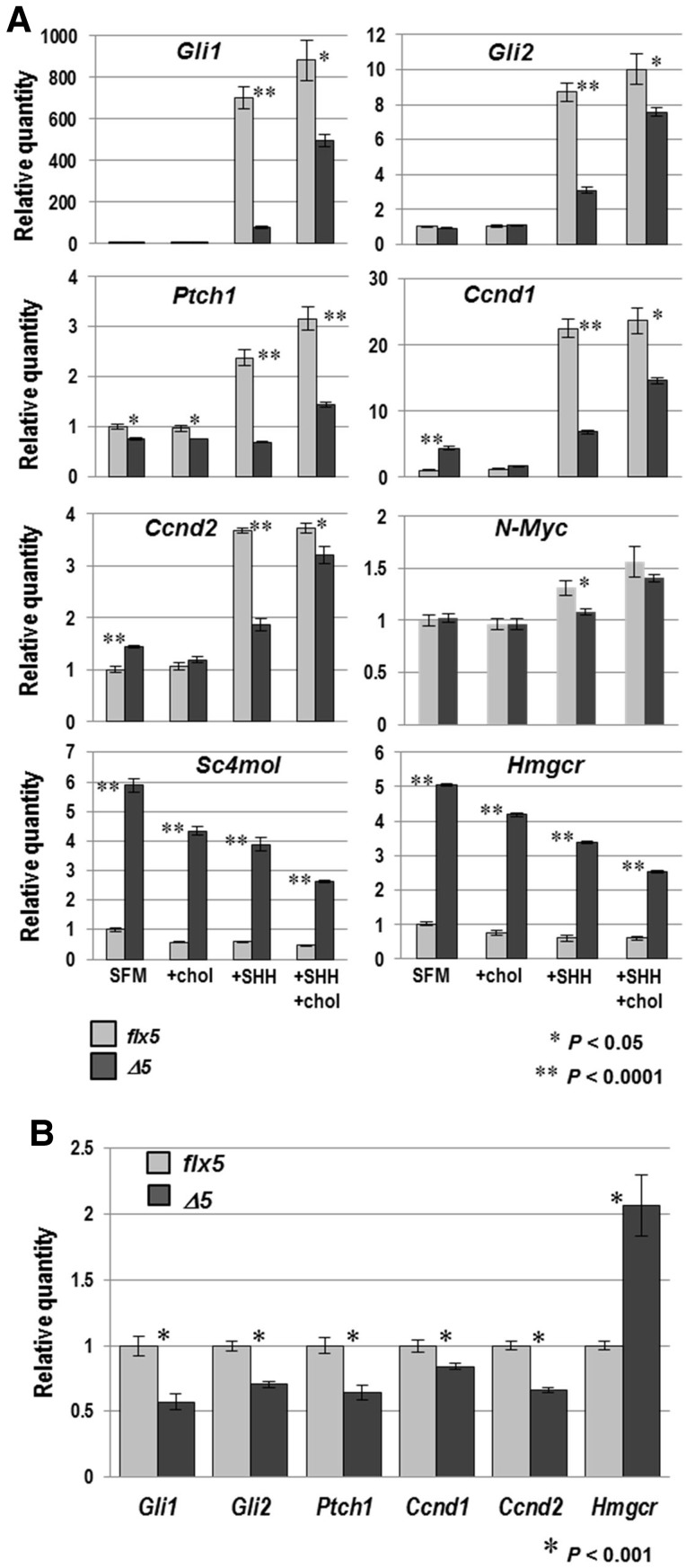
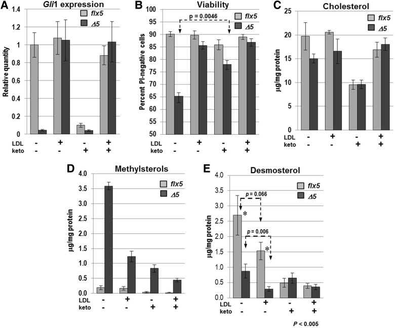
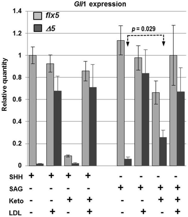
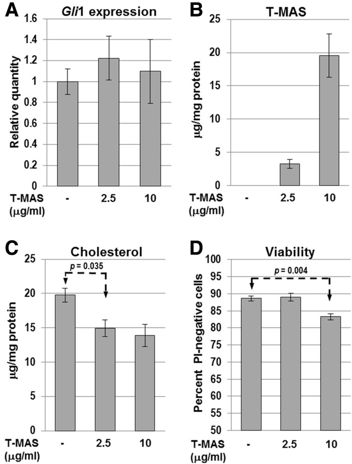
NSDHL is a 3β-hydroxysterol dehydrogenase that is involved in the removal of two C-4 methyl groups in one of the later steps of cholesterol biosynthesis. Mutations in the gene encoding the enzyme are responsible for the X-linked, male lethal mouse mutations bare patches and striated, as well as most cases of human CHILD syndrome. Rare, hypomorphic NSDHL mutations are also associated with X-linked intellectual disability in males with CK syndrome. Since hemizygous male mice with Nsdhl mutations die by midgestation, we generated a conditional targeted Nsdhl mutation (Nsdhl(tm1.1Hrm)) to investigate the essential role of cholesterol in the early postnatal CNS. Ablation of Nsdhl in radial glia using GFAP-cre resulted in live-born, normal appearing affected male pups. However, the pups develop overt ataxia by postnatal day 8-10 and die shortly thereafter. Histological abnormalities include progressive loss of cortical and hippocampal neurons, as well as deficits in the proliferation and migration of cerebellar granule precursors and subsequent massive apoptosis of the cerebellar cortex. We replicated the granule cell precursor proliferation defect in vitro and demonstrate that it results from defective signaling by SHH. Furthermore, this defect is almost completely rescued by supplementation of the culture media with exogenous cholesterol, while methylsterol accumulation above the enzymatic block appears to be associated with increased cell death. These data support the absolute requirement for cholesterol synthesis in situ once the blood-brain-barrier forms and cholesterol transport to the fetus is abolished. They further emphasize the complex ramifications of cholesterogenic enzyme deficiency on cellular metabolism.


Figures

Figure 1.
Figure 1.
Generation of a conditional Nsdhl allele. (A) Schematic diagram of the cholesterol biosynthesis pathway from lanosterol to cholesterol, with sterols listed in shaded boxes and the enzymes that catalyze each step shown next to the arrows. NSDHL, along with SC4MOL and HSD17B7, is required for the removal of two C-4 methyl groups from 4,4-dimethylcholesta-8,24-dien-3β-ol to generate zymosterol. Reduction of the C-24 double bond by DHCR24 can occur at multiple points along the pathway, but is shown only as the last step for simplicity. Ketoconazole inhibits CYP51A1 in the demethylation of lanosterol at C-14. Abbreviations: CYP51A1, cytochrome P450 lanosterol 14α-demethylase; DHCR14, 3β-hydroxysterol-Δ14-reductase; LBR, lamin B receptor; SC4MOL, sterol C-4 methyloxidase-like; NSDHL, NADH steroid dehydrogenase-like; HSD17B7, hydroxysteroid 17β-dehydrogenase 7; EBP, emopamil binding protein (3β-hydroxysteroid-Δ8,Δ7-sterol isomerase) ; SC5D, 3β-hydroxysteroid-Δ5-desaturase; DHCR7, 7-dehydrocholesterol reductase; DHCR24, 3β-hydroxysterol Δ24-reductase; T-MAS, 4,4-dimethylcholesta-8,24-dien-3β-ol. (B) Experimental strategy for generating the Nsdhlflx5 allele. The top line represents the mouse wild type Nsdhl gene from exon 4 to exon 8, indicating the position of diagnostic restrictions sites for Asp718 (A) and Bgl1 (B). The exons and introns are not drawn to scale. The position of probe sequences A and B used on Southern blots to detect homologous integration of the targeting construct are indicated. Below is a simplified map of the targeting construct generated in the vector pL451, showing loxP sites (black arrowheads) flanking Nsdhl exon 5, the neomycin-resistance gene (Neo) flanked by FRT sites (white arrowheads) for positive selection, and the thymidine kinase gene (TK) for negative selection. Homologous integration of the Nsdhlflx5 construct into the Nsdhl locus following electroporation into ES cells results in altered sizes of the Asp718 and Bgl1 restriction fragments, as shown in the third diagram. Finally, the Neo cassette was excised by FLPo-mediated recombination in ES cell clones to generate the Nsdhlflx5 allele (bottom diagram). (C) Southern blots of genomic DNA from a WT and representative Nsdhlflx5/Y neo-resistant ES clone digested with Asp718 and BglI, and hybridized with probes A and B, respectively, that lie outside of the sequence in the flx5 construct. The targeted clone showed the expected changes in size of the diagnostic restriction fragments, demonstrating homologous integration into the Nsdhl locus. (D) A Western blot of total protein prepared from E9.5 male embryos from a Nsdhlflx5/flx5 x Sox2-cre cross probed with antibodies against NSDHL (top) and β-tubulin (bottom) as a loading control. The Nsdhlflx5 control sample was from pooled cre-negative male embryos, and showed the expected 38 kDa wild type band for NSDHL. No NSDHL signal was detectable in the NsdhlΔ5 sample from pooled cre-positive male embryos. The higher level of β-tubulin signal in the NsdhlΔ5 sample is due to more total protein loaded than in the Nsdhlflx5 lane.
Figure 2.
Figure 2.
Early postnatal cerebellar phenotype of NsdhlΔ5 males. (A) Histological comparison of cerebellar lobule 3 between Nsdhlflx5/Y and NsdhlΔ5/Y brains at P4, P6 and P8. All samples were sagittal sections through the vermis. The relative thickness of the darkly-staining EGL (marked in upper left panel) was similar at P4. At P6, the EGL of the NsdhlΔ5/Y sample was substantially thinner than in the Nsdhlflx5/Y cerebellum. At P8, the EGL of the NsdhlΔ5/Y cerebellum was reduced to a single layer of GCPs, while the IGL was disorganized with numerous pycnotic nuclei. Scale bars: 100 μm. (B) Quantitation of the EGL area in lobule 3 on histological samples from Nsdhlflx5/Y and NsdhlΔ5/Y cerebella from P4 to P8. EGL area was significantly reduced in NsdhlΔ5/Y versus Nsdhlflx5/Y cerebella beginning at P5. N = number of cerebella examined. (C) Visualization of the Purkinje cell layer by anti-calbindin staining of Nsdhlflx5/Y and NsdhlΔ5/Y cerebella at P6 and P7. Purkinje cell dendrites are indicated by arrows. Note the almost complete lack of dendrites in the P7 mutant image. Scale bars: 100 μm. (D) Z-stack confocal images of Bergmann glia visualized by immunofluorescent staining for GFAP in Nsdhlflx5/Y and NsdhlΔ5/Y P6 cerebella. Scale bars: 50 μm. Abbreviations: BG, Bergmann glia; EGL, external granule layer; IGL, inner granule cell layer; PC, Purkinje cell, PS, pial surface. (E) Cholesterol, desmosterol and methylsterol concentrations in isolated GCPs from Nsdhlflx5/Y and NsdhlΔ5/Y cerebella at P3, P5 and P7. N = number of independent samples analyzed.
Figure 3.
Figure 3.
Proliferation and apoptosis in the EGL of NsdhlΔ5/Y cerebella during early postnatal development. (A) Detection of mitotic cells by PHH3 immunostaining in lobule 3 of Nsdhlflx5/Y and NsdhlΔ5/Y cerebella at P6. Scale bars: 100 μm. (B) Quantitation of PHH3-positive cells in the EGL of all lobules in sections from the cerebella of Nsdhlflx5/Y and NsdhlΔ5/Y mice from P4 to P8. N = number of cerebella counted. (C) Quantitation of TUNEL-positive cells in the EGL of lobule 3 in Nsdhlflx5/Y and NsdhlΔ5/Y cerebella from P5 to P8. N = number of cerebella analyzed.
Figure 4.
Figure 4.
Migration of BrdU-labeled GCPs in Nsdhlflx5/Y and NsdhlΔ5/Y cerebella. (A) Distribution of BrdU-labeled cells in the EGL of lobule 3 in Nsdhlflx5/Y and NsdhlΔ5/Y cerebella at 2 and 48 h after BrdU injection on P6. The lighter staining of cells in the Nsdhlflx5/Y sample at P8 was likely due to dilution of incorporated BrdU by cell division that followed the initial labeling 48 h earlier. Note that a large majority of the labeled GCPs in the NsdhlΔ5/Y sample remained in the oEGL in the P8 sample, 48 h post-injection. Abbreviations: oEGL, outer external granule layer; iEGL, inner external granule layer; ML, molecular layer; IGL, inner granule cell layer. Scale bars: 100 μm. (B) The migration efficiency of Nsdhlflx5/Y and NsdhlΔ5/Y GCPs at different ages. Migration was quantified by injecting pups with BrdU at P4, P5 or P6 and collecting cerebella for IHC 48 h later at P6, P7 or P8, respectively. The ratio of BrdU-labeled cells in the IGL to those remaining in the oEGL of lobule 3 was calculated for each sample (see Methods).
Figure 5.
Figure 5.
Response of cultured GCPs to SHH and exogenous cholesterol. (A) Immunofluorescent staining for BrdU (red) and neuronal marker TuJI (green), with DAPI (blue) counterstaining of Nsdhlflx5/Y and NsdhlΔ5/Y GCPs that were cultured for 48 h with either 1 μg/ml recombinant SHH alone or SHH plus 15 μg/ml cholesterol. BrdU was added to the samples 4 h before fixing. Note that there are fewer and thinner neurite extensions in the mutant NsdhlΔ5/Y cells than in the Nsdhlflx5/Y controls. Scale bars: 100 μm. (B) Four aliquots of isolated GCPs from individual Nsdhlflx5/Y and NsdhlΔ5/Y P4 cerebella were cultured for 48 h in either serum free medium (SFM) alone, SFM with 15 μg/ml cholesterol (chol), SFM with 1 μg/ml recombinant SHH, or SFM with 15 μg/ml cholesterol and 1 μg/ml SHH. BrdU was added to the cultures 4 h before fixing the cells. Cells were immunostained for BrdU and counterstained with DAPI. Values are the percentage of BrdU-labeled cells from the total (DAPI-stained) number of cells. Each bar represents the mean ± SEM of results from three independent experiments.
Figure 6.
Figure 6.
Expression of SHH target genes and genes for cholesterogenic enzymes in Nsdhlflx5/Y and NsdhlΔ5/Y cultured GCPs and cerebella. (A) qPCR analysis of gene expression in cultured GCPs isolated from Nsdhlflx5/Y and NsdhlΔ5/Y P4 cerebella that were cultured as described in Figure 5B. The relative quantity of mRNA from each gene was calculated from real-time PCR analysis of cDNA. Values were normalized to the untreated Nsdhlflx5 sample set equal to 1 for each gene. Each bar represents the mean ± SEM of results from three independent RNA samples that were each assayed in triplicate. (B) qPCR analysis of gene expression in Nsdhlflx5/Y and NsdhlΔ5/Y whole cerebella at P6, performed as in (A). Values represent the mean ± SEM of results for RNA samples from 3 cerebella that were each assayed in triplicate.
Figure 7.
Figure 7.
Effects of LDL and ketoconazole treatment on Nsdhlflx5/Y and NsdhlΔ5/Y GCPs in vitro. GCPs were isolated from individual cerebella at P4, split into 4 culture wells with the indicated treatments and analyzed after 48 h. All samples received 1 µg/ml recombinant SHH throughout the 48 h culture. Values represent the mean ± SEM. (A) The relative expression levels of Gli1 were measured by qPCR in Nsdhlflx5/Y and NsdhlΔ5/Y GCPs treated with LDL (5 μg/ml) and/or ketoconazole (2 μM). The results represent 4 independent samples that were each assayed in triplicate with Gapdh used as the endogenous control and normalized to the untreated Nsdhlflx5/Y sample set equal to 1. (B) Percentage of viable cells, as measured by propidium iodide staining of live GCP cultures in situ. Unstained cells were counted as viable. The results are from 4 Nsdhlflx5/Y and 5 NsdhlΔ5/Y independent cultures. (C) Lysed cell cholesterol concentration (μg/mg protein) for aliquots of the cultured GCPs used for qPCR analysis in (A). None of the pairwise comparisons between Nsdhlflx5/Y and NsdhlΔ5/Y samples in the four culture conditions reached statistical significance (P < 0.05) (see text). (D) Lysed cell total methylsterol concentration for the samples used in (C). The concentration of methylsterols was significantly higher in NsdhlΔ5/Y GCPs than Nsdhlflx5/Y GCPs (P < 0.002) in all four culture conditions. (E) Lysed cell total desmosterol concentration for samples used in (A). Desmosterol levels were significantly lower in NsdhlΔ5/Y versus Nsdhlflx5/Y GCPs in both untreated and LDL-treated cultures. Ketoconazole treatment reduced the desmosterol concentration in Nsdhlflx5/Y cells to a level equivalent to that of NsdhlΔ5/Y cells, while addition of LDL to the ketoconazole-treated cultures had no effect on desmosterol.
Figure 8.
Figure 8.
Comparison of Gli1 relative expression levels in response to SHH or SAG between Nsdhlflx5/Y and NsdhlΔ5/Y GCPs cultured in vitro. GCPs were isolated from individual cerebella at P4, split into 6 culture wells with the indicated treatments and analyzed after 48 h in culture. Concentrations of ketoconazole and LDL were as in Figure 7. The results are from 4 independent samples of each genotype that were each assayed in triplicate by qPCR with Gapdh used as the endogenous control and normalized to the untreated Nsdhlflx5 sample set equal to 1.
Figure 9.
Figure 9.
Effect of exogenous T-MAS on WT (Nsdhlflx5/Y) GCPs in vitro. (A) Relative Gli1 expression levels measured by qPCR in cultured Nsdhlflx5/Y GCPs treated with T-MAS. All samples were cultured with 1 μg/ml SHH. Bars represent the mean ± SEM of 4 independent samples that were each assayed in triplicate with GAPDH as the endogenous control and normalized to the untreated sample set equal to 1. (B) Lysed cell pellet T-MAS concentration and (C) cholesterol concentration determined for aliquots of the cultured Nsdhlflx5/Y GCPs treated with exogenous T-MAS used in panel A. Values indicate the mean ± SEM of 3 independent GCP cultures. (D) Percentage of viable cells in T-MAS-treated cultures, measured by propidium iodide staining, performed as in Figure 7B. The results are the mean ± SEM from 3 independent Nsdhlflx5/Y GCP cultures.

Similar articles

Cited by

References

    1. Saher G., Quintes S., Nave K.A. (2011) Cholesterol: a novel regulatory role in myelin formation. Neuroscientist, 17, 79–93. - PubMed
    1. Herman G.E. (2003) Disorders of cholesterol biosynthesis: prototypic metabolic malformation syndromes. Hum. Mol. Genet., 12 Spec No 1, R75–R88. - PubMed
    1. Mann R.K., Beachy P.A. (2004) Novel lipid modifications of secreted protein signals. Annu. Rev. Biochem., 73, 891–923. - PubMed
    1. Tierney E., Bukelis I., Thompson R.E., Ahmed K., Aneja A., Kratz L., Kelley R.I. (2006) Abnormalities of cholesterol metabolism in autism spectrum disorders. Am. J. Med Genet. B Neuropsychiatr. Genet., 141, 666–668. - PMC - PubMed
    1. Bjorkhem I., Leoni V., Meaney S. (2010) Genetic connections between neurological disorders and cholesterol metabolism. J. Lipid Res., 51, 2489–2503. - PMC - PubMed

Publication types