

Are obligatory apomicts invested in the pollen tube transmitting tissue? Comparison of the micropyle ultrastructure between sexual and apomictic dandelions (Asteraceae, Lactuceae)
- PMID: 25652809
- PMCID: PMC4561075
- DOI: 10.1007/s00709-015-0765-x
Are obligatory apomicts invested in the pollen tube transmitting tissue? Comparison of the micropyle ultrastructure between sexual and apomictic dandelions (Asteraceae, Lactuceae)
Abstract
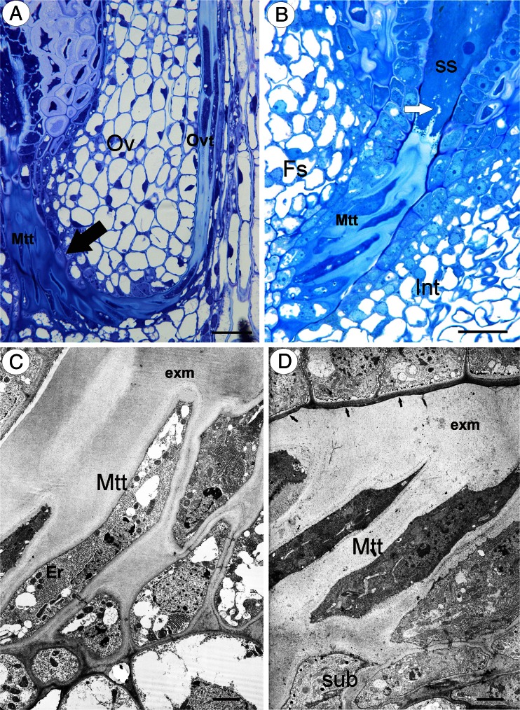
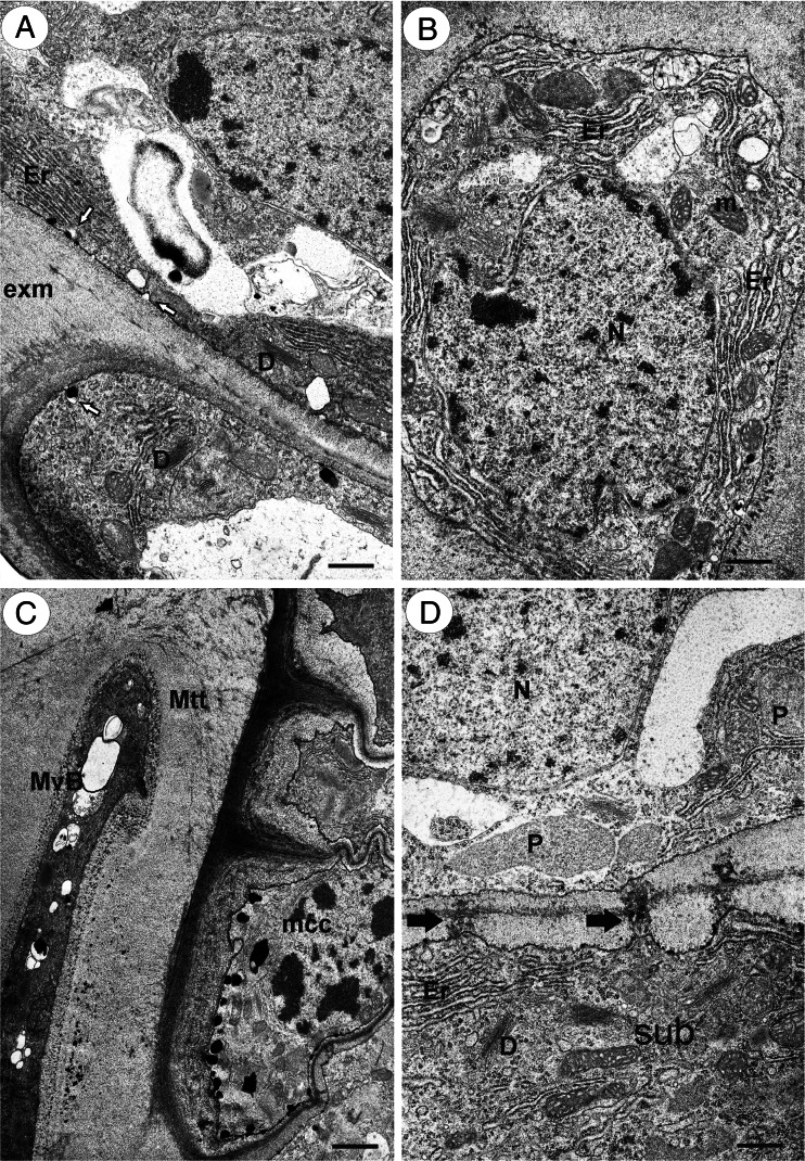
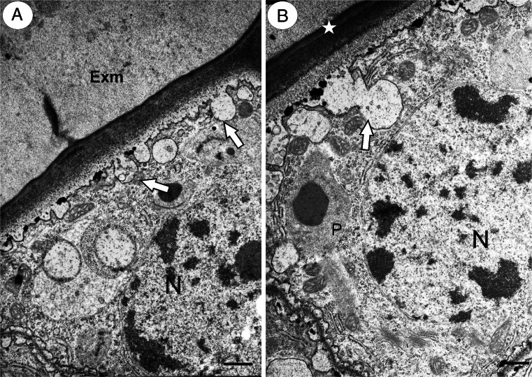
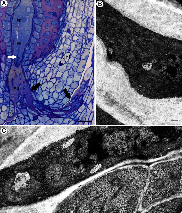
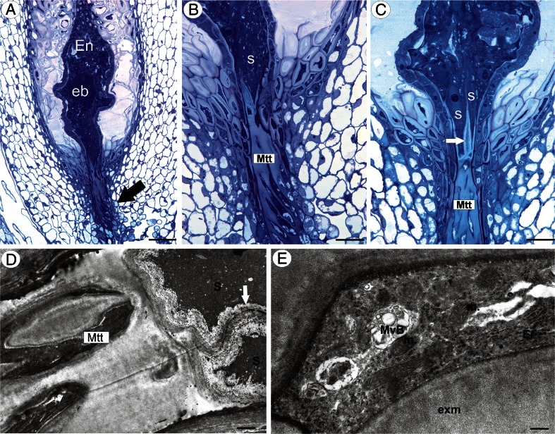
With the exception of the sunflower, little information concerning the micropyle ultrastructure of the family Asteraceae is available. The aim of our study was to compare the micropyle structure in amphimictic and apomictic dandelions. Ultrastructural studies using buds and flowers during anthesis have been done on the micropyle of the sexual and apomictic Taraxacum. In all of the species that were examined, the micropylar canal was completely filled with ovule transmitting tissue and the matrix that was produced by these cells. The ovule transmitting tissue was connected to the ovarian transmitting tissue. The micropyle was asymmetrical because the integument epidermis that forms the transmitting tissue was only on the funicular side. There was a cuticle between the obturator cells and epidermal cells on the other side of integument. The micropylar transmitting tissue cells and theirs matrix reached the synergid apex. The cytoplasm of the transmitting tissue cells was especially rich in rough endoplasmic reticulum (ER), dictyosomes, and mitochondria. No major differences were detected between the micropyle structure of the amphimictic and apomictic species; thus, a structural reduction of obturator does not exist. The ovule transmitting tissue is still active in apomictic dandelions despite the presence of the embryo and endosperm. Differences and similarities between the micropyle structure in the Asteraceae that have been studied to date are discussed.
Figures
Similar articles
-
Immunodetection of some pectic, arabinogalactan proteins and hemicellulose epitopes in the micropylar transmitting tissue of apomictic dandelions (Taraxacum, Asteraceae, Lactuceae).Protoplasma. 2017 Mar;254(2):657-668. doi: 10.1007/s00709-016-0980-0. Epub 2016 May 6. Protoplasma. 2017. PMID: 27154759 Free PMC article.
-
Integument cell differentiation in dandelions (Taraxacum, Asteraceae, Lactuceae) with special attention paid to plasmodesmata.Protoplasma. 2016 Sep;253(5):1365-72. doi: 10.1007/s00709-015-0894-2. Epub 2015 Oct 10. Protoplasma. 2016. PMID: 26454638 Free PMC article.
-
Synergids and filiform apparatus in the sexual and apomictic dandelions from section Palustria (Taraxacum, Asteraceae).Protoplasma. 2014 Jan;251(1):211-7. doi: 10.1007/s00709-013-0539-2. Epub 2013 Aug 24. Protoplasma. 2014. PMID: 23974526 Free PMC article.
-
Style morphology and pollen tube pathway.Plant Reprod. 2017 Dec;30(4):155-170. doi: 10.1007/s00497-017-0312-3. Epub 2017 Nov 7. Plant Reprod. 2017. PMID: 29116403 Review.
-
Paving the Way for Fertilization: The Role of the Transmitting Tract.Int J Mol Sci. 2021 Mar 5;22(5):2603. doi: 10.3390/ijms22052603. Int J Mol Sci. 2021. PMID: 33807566 Free PMC article. Review.
Cited by
-
Immunodetection of some pectic, arabinogalactan proteins and hemicellulose epitopes in the micropylar transmitting tissue of apomictic dandelions (Taraxacum, Asteraceae, Lactuceae).Protoplasma. 2017 Mar;254(2):657-668. doi: 10.1007/s00709-016-0980-0. Epub 2016 May 6. Protoplasma. 2017. PMID: 27154759 Free PMC article.
-
Structural aspects of cypsela and seed development of Trichocline catharinensis (Cabrera): a Brazilian endemic species.Protoplasma. 2019 Nov;256(6):1495-1506. doi: 10.1007/s00709-019-01361-7. Epub 2019 May 29. Protoplasma. 2019. PMID: 31144034
-
Immunodetection of Pectic Epitopes, Arabinogalactan Proteins, and Extensins in Mucilage Cells from the Ovules of Pilosella officinarum Vaill. and Taraxacum officinale Agg. (Asteraceae).Int J Mol Sci. 2020 Dec 17;21(24):9642. doi: 10.3390/ijms21249642. Int J Mol Sci. 2020. PMID: 33348898 Free PMC article.
-
Integument cell differentiation in dandelions (Taraxacum, Asteraceae, Lactuceae) with special attention paid to plasmodesmata.Protoplasma. 2016 Sep;253(5):1365-72. doi: 10.1007/s00709-015-0894-2. Epub 2015 Oct 10. Protoplasma. 2016. PMID: 26454638 Free PMC article.
References
-
- Asker SE, Jerling L. Apomixis in plants. Boca Raton: CRC Press; 1992.
-
- den Nijs JCM. Taraxacum: ploidy levels, hybridization and speciation. The advantage and consequence of combining reproductive systems. Lagascalia. 1997;19:45–56.
-
- den Nijs JCM, Menken SBJ. Relations between breeding system, ploidy level, and taxonomy in some advanced sections of Taraxacum. In: Hind DJN, Beentje HJ, editors. Compositae: systematics. Proceedings of the international Compositae conference. Kew: Royal Botanic Gardens; 1996. pp. 665–677.
-
- Erbar C. Pollen tube transmitting tissue: place of competition of male gametophytes. Int J Plant Sci. 2003;164(suppl):S265–S277. doi: 10.1086/377061. - DOI
-
- Erbar C, Enghofer J. Untersuchungen zum Reproduktionssystem der Wegwarte (Cichorium intybus, Asteraceae): Pollenportionierung, Narbenbelegung und Pollenschlauchkonkurrenz. Bot Jahrb Syst. 2001;123:179–208.
Publication types
MeSH terms
LinkOut - more resources
Full Text Sources
Other Literature Sources
Miscellaneous
