. 2015 Mar 7;282(1802):20142764.
doi: 10.1098/rspb.2014.2764.

Altered retinoic acid signalling underpins dentition evolution

Affiliations

Altered retinoic acid signalling underpins dentition evolution

Yann Gibert et al. Proc Biol Sci. .

Abstract

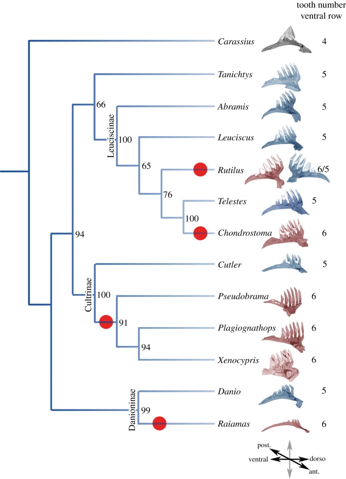
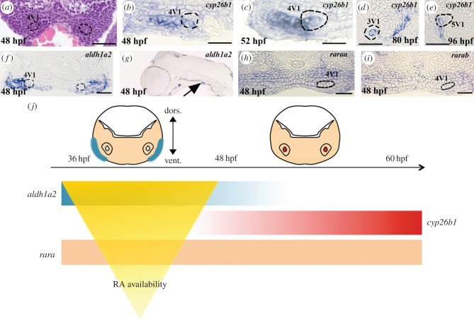
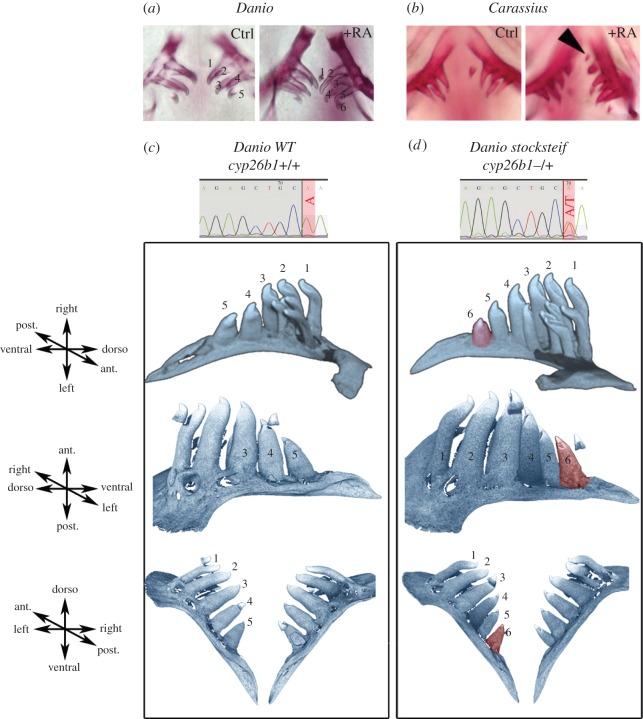
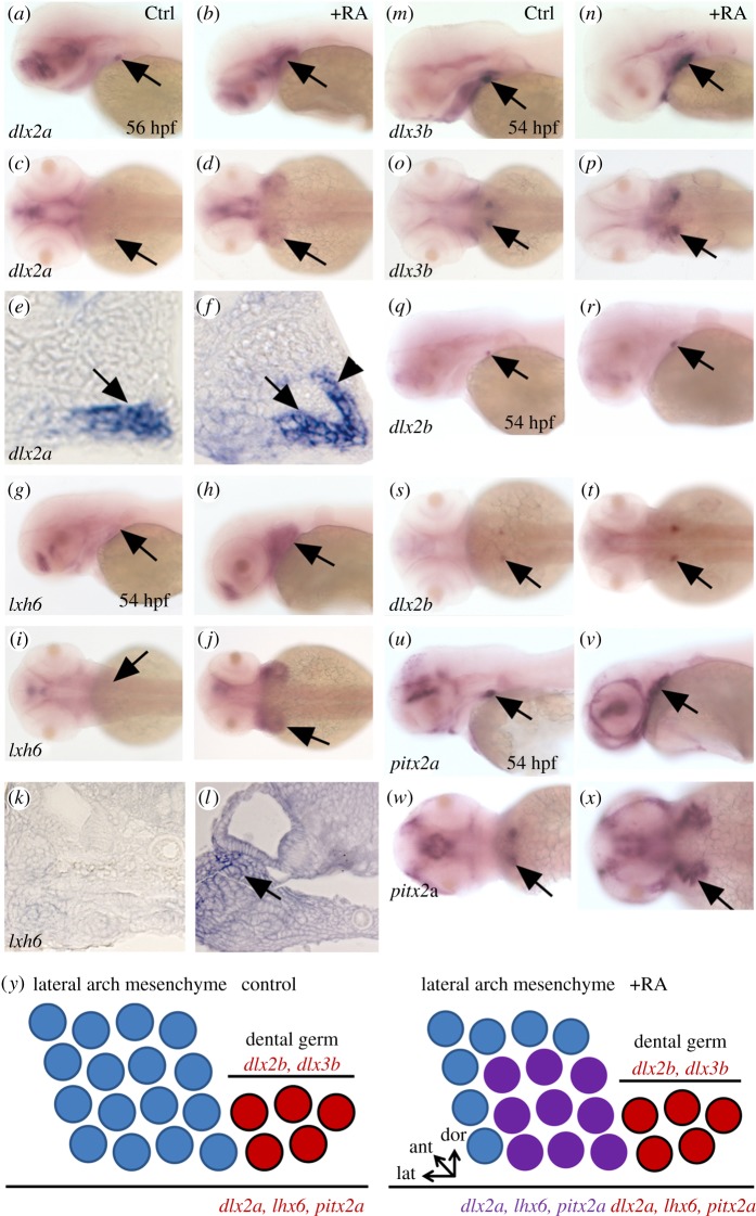
Small variations in signalling pathways have been linked to phenotypic diversity and speciation. In vertebrates, teeth represent a reservoir of adaptive morphological structures that are prone to evolutionary change. Cyprinid fish display an impressive diversity in tooth number, but the signals that generate such diversity are unknown. Here, we show that retinoic acid (RA) availability influences tooth number size in Cyprinids. Heterozygous adult zebrafish heterozygous for the cyp26b1 mutant that encodes an enzyme able to degrade RA possess an extra tooth in the ventral row. Expression analysis of pharyngeal mesenchyme markers such as dlx2a and lhx6 shows lateral, anterior and dorsal expansion of these markers in RA-treated embryos, whereas the expression of the dental epithelium markers dlx2b and dlx3b is unchanged. Our analysis suggests that changes in RA signalling play an important role in the diversification of teeth in Cyprinids. Our work illustrates that through subtle changes in the expression of rate-limiting enzymes, the RA pathway is an active player of tooth evolution in fish.

Keywords: cyprinids; development; evolution; retinoic acid; tooth.


Figures

Figure 1.
Figure 1.
Cyprinidae dentition diversity. Phylogeny of several species of Cyprinidae based on cytb, coi and rag1 markers. Phylogenetic analyses were performed using RaxML, with a GTR + G + I partitioned model, with 1000 bootstraps (values indicated for each branch). For each species, the fifth ceratobranchial bearing pharyngeal teeth is pictured and the number of teeth on the ventral row is indicated. Species with six teeth on the ventral row are red. Red dots indicate the events of tooth gain on the ventral row, based on parsimonious hypotheses. The orientation of the ceratobranchial arch is indicated at the bottom of the tree. Colour code: grey, four teeth; blue, five teeth; red, six teeth in the ventral part of the fifth ceratobranchial arch.
Figure 2.
Figure 2.
Retinoic acid signalling in the tooth-forming region. (a) Transverse sections of the posterior ventral pharynx of a 48 hpf Danio rerio embryo stained with haematoxylin and eosin. The 4V1 tooth germs are marked by dashed lines. (be) Transverse sections of the posterior ventral pharynx of embryos post in situ hybridization embryos stained with cyp26b1 at (b) 48 hpf, (c) 52 hpf, (d) 80 hpf and (e) 96 hpf. The 4V1 tooth bud is marked by dashed lines in (b,c). The 3V1 and 4V2 tooth buds are marked by dashed lines in (d) and (e), respectively. (f) Transverse and (g) longitudinal sections of the posterior ventral pharynx of an embryo post in situ hybridization embryos stained with aldh1a2 at 48 hpf. 4V1 is marked in (f), whereas expression in the ventral posterior pharynx in the vicinity of the forming tooth bud is indicated by an arrow in (g). Note that aldh1a2 expression is excluded from the developing tooth bud in (f). (h) raraa and (i) rarab expression in transverse sections of the posterior ventral pharynx at 48 hpf. The position of the 4V1 tooth bud is indicated. Note that both raraa and rarab are expressed within the tooth bud but also in the entire posterior ventral pharynx. (j) Summary of the temporal and regional expression pattern of RA signalling actors: at 36 hpf, aldh1a2 is expressed in the ventral pharynx until 48 hpf. cyp26b1 expression starts at 50 hpf in the tooth bud mesenchyme. raraa and rarab are expressed through the entire ventral pharynx. The RA availability within the tooth-forming region is temporally sharpened by the expressions of both synthetizing (aldh1a2) and degrading (cyp26b1) enzymes. Scale bars, 25 µm.
Figure 3.
Figure 3.
Retinoic acid controls tooth number. (a) Alizarin red staining of 12 dpf untreated and treated zebrafish (Danio rerio) embryos with 10−8 M of RA from 56 hpf onwards. (b) Alizarin red staining of 14 dpf untreated and treated goldfish (Carassius auratus) embryos with 5 × 10⁻⁸ M of RA from 48 hpf onwards. A black arrowhead indicates the extra tooth induced by RA treatment. (c,d) Computed microtomography scans of (c) wild-type zebrafish adults (cyp26b1+/+) and of (d) stocksteif (sst) heterozygous mutants (cyp26b1+/−). Teeth on the ventral row are numbered and the sixth extra tooth observed in sst mutants is highlighted in red. Chromatograms of genotyping showing the A > T nonsense mutation in heterozygous is provided.
Figure 4.
Figure 4.
Expansion of pharyngeal mesenchyme markers in RA treated embryos. (a–f) expression of dlx2a in the tooth bud of 4V1 and lateral arch mesenchyme marked by an arrow in (a,c,e) control and (b,d,f) RA-treated embryos. Note the lateral, anterior and dorsal expansion of dlx2a expression in RA-treated embryos (arrowhead in (f)). (g–l) Expression of lhx6 in the lateral pharyngeal mesenchyme marked by an arrow in (g,i,k) control and (h,j,l) RA-treated embryos. As for dlx2a, note the lateral, anterior and dorsal expansion of lhx6 expression in RA-treated embryos (arrow in transverse section in (l)). (m–t) Expression of dlx2b and dlx3b in the dental epithelium marked by an arrow in (m,o,q,s) control and (n,p,r,t) RA-treated embryos. RA exposure has only a minor effect on the expression of these dental epithelium markers. (u–x) Expression of pitx2a in the pharyngeal and dental epithelium marked by an arrow in (u,w) control and (v,x) RA-treated embryos. Note the lateral, anterior and dorsal expansion of pitx2a expression in RA treated embryos. (y) Diagram of a longitudinal section of the posterior ventral pharyngeal epithelium and mesenchyme of the 4V1 tooth germ during early morphogenesis, dorsal up, anterior to the left of a control embryo (top) and RA-treated embryo (bottom). In red, dlx2b and dlx3b are restricted to the folding dental epithelium, whereas dlx2a is expressed in both the developing tooth (red) germ and the lateral pharyngeal mesenchyme (purple); lhx6 is expressed in both the dental mesenchyme (red) and in lateral arch mesenchyme (purple); pitx2a is expressed in the dental epithelium (red) and the pharyngeal epithelium (purple). Under RA exposure, expression of dlx2a, lhx6 and pitx2a is expanded in three directions (laterally, dorsally and anteriorly), whereas expression of dlx2b and dlx3b remains confined in the dental epithelium, but their expression domains are slightly enlarged compared with control.

References

    1. Abzhanov A, Protas M, Grant BR, Grant PR, Tabin CJ. 2004. Bmp4 and morphological variation of beaks in Darwin's finches. Science 305, 1462–1465. (10.1126/science.1098095) - DOI - PubMed
    1. Jernvall J, Salazar-Ciudad I. 2007. The economy of tinkering mammalian teeth. Novartis Found. Symp. 284, 207–216; discussion 216–224 (10.1002/9780470319390.ch14) - DOI - PubMed
    1. Stock DW. 2007. Zebrafish dentition in comparative context. J. Exp. Zool. B Mol. Dev. Evol. 308, 523–549. (10.1002/jez.b.21187) - DOI - PubMed
    1. Rhinn M, Dolle P. 2012. Retinoic acid signalling during development. Development 139, 843–858. (10.1242/dev.065938) - DOI - PubMed
    1. Fujiwara S, Kawamura K. 2003. Acquisition of retinoic acid signaling pathway and innovation of the chordate body plan. Zool. Sci. 20, 809–818. (10.2108/zsj.20.809) - DOI - PubMed

Publication types

MeSH terms