Utility of the diffusion-weighted imaging for activity evaluation in Crohn's disease patients underwent magnetic resonance enterography
- PMID: 25653007
- PMCID: PMC4323053
- DOI: 10.1186/s12876-015-0235-0
Utility of the diffusion-weighted imaging for activity evaluation in Crohn's disease patients underwent magnetic resonance enterography
Abstract
Background: Cross-sectional imaging techniques as magnetic resonance enterography (MRE) may offer additional information on transmural inflammation, stricturing and fistulising complications in Crohn's disease (CD). The purpose of our study was to evaluate the diagnostic accuracy of Magnetic Resonance Imaging (MRI) combined with Diffusion-weighted Imaging (DWI) and MRE for determination of inflammation in small bowel CD.
Methods: MR imaging examination was performed with a GE Signa EXCITE 3.0 T MRI scanner. The optimal b value in DWI with a learning cohort of patients was determined. The diagnostic accuracy for active lesions and disease activity were accessed by MRE combined with DWI.
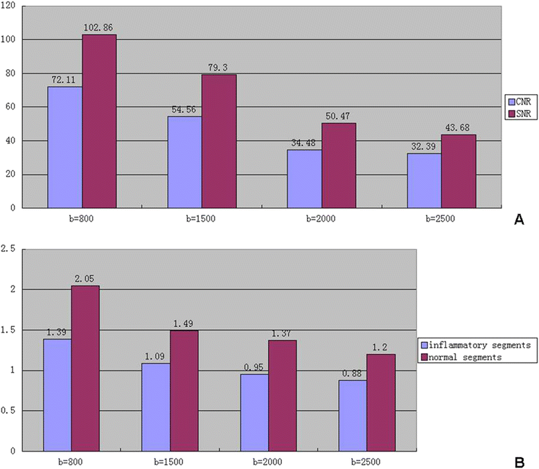
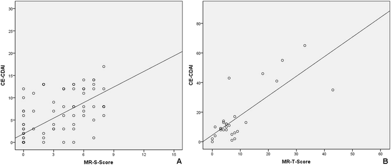
Results: The b value 800 s/mm(2) group showed the highest diagnostic sensitivity (74.19%) for diagnostic assessment of active Crohn's lesions on DWI. MRE combined with DWI showed the highest sensitivity (93.55%), specificity (89.47%) and diagnostic accuracy (92%) compared with MRE or DWI alone. The segmental MR score (MR-score-S) showed a significantly positive correlation with the Capsule Endoscopy Crohn's Disease Activity Index Score (CECDAI-S) (r = 0.717, p < 0.01). The total MR score (MR-score-T) showed significant association with C-reactive protein (CRP) (r = 0.445, p = 0.019) and erythrocyte sedimentation rate (ESR) (r = 0.688, p < 0.01).
Conclusions: MRE combined with DWI improves the diagnostic accuracy for active lesions and correlates the endoscopic disease activity. MRE with DWI could represent a non-invasive tool in assessing active inflammation in CD.
Figures


Similar articles
-
Diffusion-weighted MR enterography for evaluating Crohn's disease: how does it add diagnostically to conventional MR enterography?Inflamm Bowel Dis. 2015 Jan;21(1):101-9. doi: 10.1097/MIB.0000000000000222. Inflamm Bowel Dis. 2015. PMID: 25358063
-
Diffusion-weighted magnetic resonance imaging for detecting and assessing ileal inflammation in Crohn's disease.Aliment Pharmacol Ther. 2013 Mar;37(5):537-45. doi: 10.1111/apt.12201. Epub 2013 Jan 7. Aliment Pharmacol Ther. 2013. PMID: 23289713
-
Accuracy of magnetic resonance enterography in assessing response to therapy and mucosal healing in patients with Crohn's disease.Gastroenterology. 2014 Feb;146(2):374-82.e1. doi: 10.1053/j.gastro.2013.10.055. Epub 2013 Oct 29. Gastroenterology. 2014. PMID: 24177375 Clinical Trial.
-
Cross-sectional imaging modalities in Crohn's disease.Dig Dis. 2013;31(2):199-201. doi: 10.1159/000353692. Epub 2013 Sep 6. Dig Dis. 2013. PMID: 24030225 Review.
-
Magnetic resonance enterography of Crohn's disease.Expert Rev Gastroenterol Hepatol. 2015 Jan;9(1):37-45. doi: 10.1586/17474124.2014.939631. Epub 2014 Sep 3. Expert Rev Gastroenterol Hepatol. 2015. PMID: 25186521 Review.
Cited by
-
Imaging of the small intestine in Crohn's disease: Joint position statement of the Indian Society of Gastroenterology and Indian Radiological and Imaging Association.Indian J Gastroenterol. 2017 Nov;36(6):487-508. doi: 10.1007/s12664-017-0804-y. Epub 2018 Jan 6. Indian J Gastroenterol. 2017. PMID: 29307029
-
Can MR Enterography and Diffusion-Weighted Imaging Predict Disease Activity Assessed by Simple Endoscopic Score for Crohn's Disease?J Belg Soc Radiol. 2019 Jan 18;103(1):10. doi: 10.5334/jbsr.1521. J Belg Soc Radiol. 2019. PMID: 30671568 Free PMC article.
-
Small Bowel Imaging from Stepchild of Roentgenology to MR Enterography: Part I: Guidance in Performing and Observing Normal and Abnormal Imaging Findings.Life (Basel). 2023 Aug 5;13(8):1691. doi: 10.3390/life13081691. Life (Basel). 2023. PMID: 37629548 Free PMC article. Review.
-
Can diffusion weighted imaging be used as an alternative to contrast-enhanced imaging on magnetic resonance enterography for the assessment of active inflammation in Crohn disease?Medicine (Baltimore). 2020 Feb;99(8):e19202. doi: 10.1097/MD.0000000000019202. Medicine (Baltimore). 2020. PMID: 32080107 Free PMC article.
-
Diagnostic Value of Diffusion-Weighted Imaging and Apparent Diffusion Coefficient in Assessment of the Activity of Crohn Disease: 1.5 or 3 T.J Comput Assist Tomogr. 2018 Sep/Oct;42(5):688-696. doi: 10.1097/RCT.0000000000000754. J Comput Assist Tomogr. 2018. PMID: 29958199 Free PMC article.
References
-
- Papi C, Fasci-Spurio F, Rogai F, Settesoldi A, Margagnoni G, Annese V. Mucosal healing in inflammatory bowel disease: treatment efficacy and predictive factors. Digestive Liver Di Off J Italian Soc Gastroenterol Italian Assoc Study Liver. 2013;45(12):978–85. doi: 10.1016/j.dld.2013.07.006. - DOI - PubMed
-
- Panes J, Bouzas R, Chaparro M, Garcia-Sanchez V, Gisbert JP, de GB M, et al. Systematic review: the use of ultrasonography, computed tomography and magnetic resonance imaging for the diagnosis, assessment of activity and abdominal complications of Crohn’s disease. Aliment Pharmacol Ther. 2011;34(2):125–45. doi: 10.1111/j.1365-2036.2011.04710.x. - DOI - PubMed
Publication types
MeSH terms
Substances
LinkOut - more resources
Full Text Sources
Other Literature Sources
Medical
Research Materials
Miscellaneous

