. 2015 Feb 27;43(4):2138-51.
doi: 10.1093/nar/gkv082. Epub 2015 Feb 4.

Fungal Ku prevents permanent cell cycle arrest by suppressing DNA damage signaling at telomeres

Affiliations

Fungal Ku prevents permanent cell cycle arrest by suppressing DNA damage signaling at telomeres

Carmen de Sena-Tomás et al. Nucleic Acids Res. .

Abstract

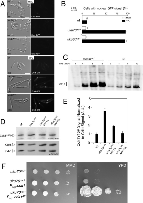
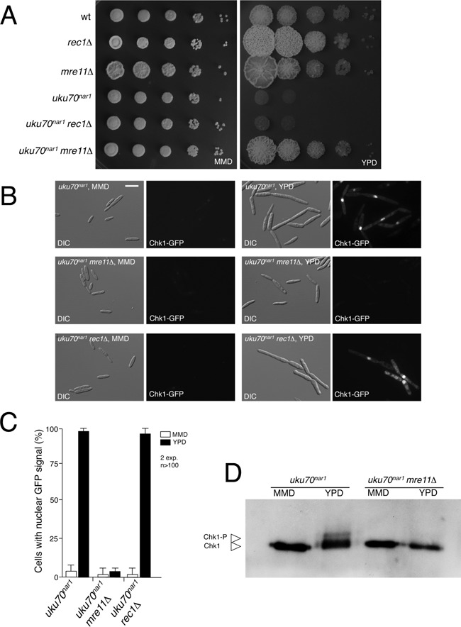
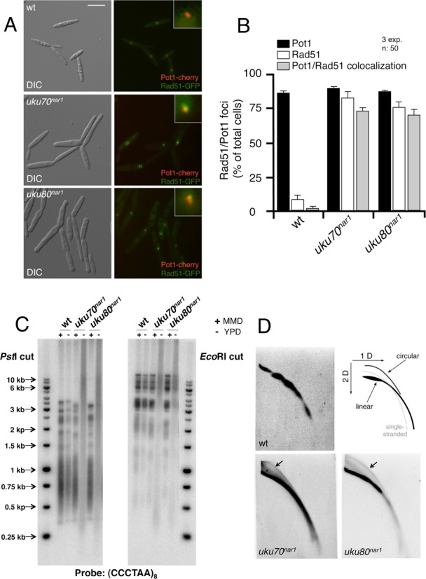
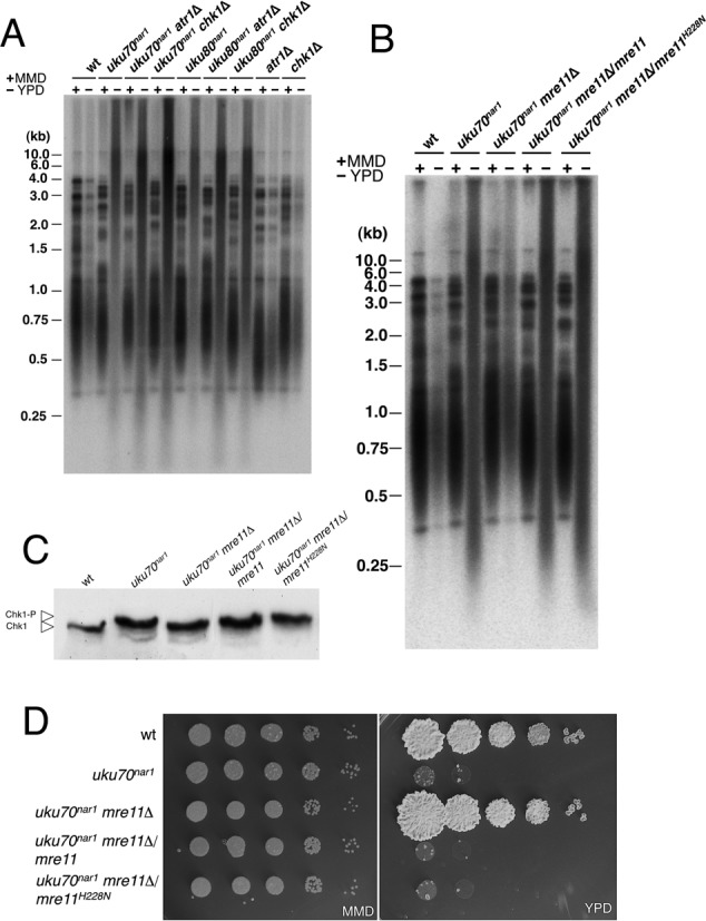
The Ku heterodimer serves in the initial step in repairing DNA double-strand breaks by the non-homologous end-joining pathway. Besides this key function, Ku also plays a role in other cellular processes including telomere maintenance. Inactivation of Ku can lead to DNA repair defects and telomere aberrations. In model organisms where Ku has been studied, inactivation can lead to DNA repair defects and telomere aberrations. In general Ku deficient mutants are viable, but a notable exception to this is human where Ku has been found to be essential. Here we report that similar to the situation in human Ku is required for cell proliferation in the fungus Ustilago maydis. Using conditional strains for Ku expression, we found that cells arrest permanently in G2 phase when Ku expression is turned off. Arrest results from cell cycle checkpoint activation due to persistent signaling via the DNA damage response (DDR). Our results point to the telomeres as the most likely source of the DNA damage signal. Inactivation of the DDR makes the Ku complex dispensable for proliferation in this organism. Our findings suggest that in U. maydis, unprotected telomeres arising from Ku depletion are the source of the signal that activates the DDR leading to cell cycle arrest.


Figures

Figure 1.
Figure 1.
Ku complex is required for proliferation in U. maydis. (A) Growth of conditional strains in solid medium. Serial 10-fold dilutions of FB1 (WT), UCS33 (uku70nar1) and UCS30 (uku80nar1) cultures were spotted on solid rich medium (YPD) and minimal medium with nitrate (MMD). YPD plates were incubated for 2 days and the nitrate plates for 4 days at 28°C. (B) Morphology of wild-type (UMP132), uku70nar1 (UCS34) and uku80nar1 (UCS37) cells incubated for 8 h in permissive (MMD) and restrictive (YPD) conditions. All cells were shown at the same magnification, Bar: 15 μm. Note that the uku70nar1 and uku80nar1 mutants in YPD showed buds substantially more enlarged and elongated, which are characteristic of G2 arrested cells (white stars). Inset showed the nucleus of a representative cell. Nuclear membrane (red) was visualized using Cut11-RFP and DNA (blue) was visualized upon staining with DAPI. (C) FACS analysis of FB1 (WT), UCS33 (uku70nar1) and UCS30 (uku80nar1) cell DNA content after 8 h of incubation in permissive (minimal medium with nitrate, MMD) or restrictive conditions (rich medium, YPD). 1C and 2C indicate haploid and diploid DNA content. The shift to a DNA content higher than 2C observed in conditional cells incubated in YPD was due to mitochondrial DNA staining (31).
Figure 2.
Figure 2.
Disabling the DNA damage response suppressed the requirement of Ku for proliferation of U. maydis cells. (A) Serial 10-fold dilutions of FB1 (WT), UMP122 (chk1Δ), UCS1 (atr1Δ), UCS33 (uku70nar1), UCS35 (uku70nar1 chk1Δ), UCS40 (uku70nar1 atr1Δ), UCS30 (uku80nar1), UCS39 (uku80nar1 chk1Δ) and UCS44 (uku80nar1 atr1Δ) cultures were spotted on solid rich medium (YPD) and minimal medium with nitrate (MMD). YPD plates were incubated for 2 days and the nitrate plates for 4 days at 28°C. (B) Morphology of uku70nar1 (UCS33), uku80nar1 (UCS30), uku70nar1 chk1Δ (UCS35), uku70nar1 atr1Δ (UCS40), uku80nar1 chk1Δ (UCS39) and uku80nar1 atr1Δ (UCS44) cells incubated for 8 h in restrictive conditions (YPD). All cells were shown at the same magnification, Bar: 15 μm. (C) FACS analysis of FB1 (WT), UCS33 (uku70nar1), UCS35 (uku70nar1 chk1Δ), UCS40 (uku70nar1 atr1Δ), UCS30 (uku80nar1), UCS39 (uku80nar1 chk1Δ) and UCS44 (uku80nar1 atr1Δ) after 8 h of incubation in permissive (minimal medium with nitrate, MMD) or restrictive conditions (rich medium, YPD). 1C and 2C indicate haploid and diploid DNA content.
Figure 3.
Figure 3.
Down-regulation of Ku genes triggers the DNA damage response. (A) Cell images of UMP111 (wt), UCS43 (uku70nar1) and UCS42 (uku80nar1) strains carrying a Chk1-3GFP fusion grown for 8 h in permissive (MMD) and restrictive (YPD) conditions. All cells were shown at the same magnification (Bar: 15 μm). (B) Quantification of the cellular response to DNA damage as the percentage of cells carrying a clear nuclear GFP fluorescence signal. (C) In vivo phosphorylation of Chk1 in response to Ku70 protein depletion. UMP124 (wt) and UMP231 (uku70nar1) cells carrying an endogenous Chk1-3MYC allele were incubated in YPD (restrictive conditions) for the indicated time. Protein extracts were immunoprecipitated with a commercial anti-MYC antibody and the immunoprecipitates were subjected to SDS-PAGE and immunoblotting using anti-MYC antibody. Phosphorylated Chk1 migrated as a smear (brackets) most likely because the presence of multiple species of phosphorylated protein. (D) Levels of Cdk1 inhibitory phosphorylation upon Ku70 depletion. Protein extracts from the indicated strains (FB1, wt; UCS33, uku70nar1; UCS35, uku70nar1 chk1Δ; UMP218, uku70nar1 mre11Δ) that were grown in restrictive conditions (YPD) for 8 h were separated by SDS-PAGE. Immunoblots were probed successively with an antibody that recognizes phosphorylated Cdk1 (α-Cdc2-Y15P) and anti-PSTAIRE. (E) Levels of Cdk1 phosphorylation were determined by quantifying the level of antibody signal using a ChemiDoc unit (Bio-Rad). Signal from the phosphopeptide-specific antibodies was normalized to the amount of phosphorylation in the control strain (FB1). Differences in loading of samples were corrected by dividing each phosphopeptide-specific antibody signal by the Cdk1 (α-PSTAIRE) antibody signal. Two independent experiments were used to calculate the mean and s.d. (F) Growth of conditional strains in solid medium. Serial 10-fold dilutions of UCS33 (uku70nar1), UMP221 (uku70nar1 Pscp:cdk1), UMP222 (uku70nar1 Pscp:cdk1AF) cultures were applied to solid rich medium (YPD) and minimal medium with nitrate (MMD). YPD plates were incubated for 2 days at 28°C.
Figure 4.
Figure 4.
The MRN complex is required for the essential role of Ku proteins. (A) Serial 10-fold dilutions of FB1 (WT), UM210 (rec1Δ), UMP219 (mre11Δ), UCS33 (uku70nar1), UMP220 (uku70nar1 rec1Δ), UMP218 (uku70nar1 mre11Δ), cultures were applied to solid rich medium (YPD) and minimal medium with nitrate (MMD). YPD plates were incubated for 2 days and the nitrate plates for 4 days at 28°C. (B) Cell images of UMP111 (wt), UCS43 (uku70nar1), UMP223 (uku70nar1 rec1Δ) and UMP224 (uku70nar1 mre11Δ) carrying a Chk1-GFP fusion grown for 8 h in permissive (MMD) and restrictive (YPD) media. All cells were shown at the same magnification (Bar: 15 μm). (C) The percentages of cells carrying a clear nuclear GFP fluorescence signal in the indicated strains were quantified. (D) In vivo phosphorylation of Chk1. UMP231 (uku70nar1) and UMP228 (uku70nar1 mre11Δ) cells carrying an endogenous Chk1-3MYC allele were incubated in YPD (restrictive conditions) or MMD (permissive conditions) for 8 h. Protein extracts were immunoprecipitated with a commercial anti-MYC antibody and the immunoprecipitates were subjected to SDS-PAGE and immunoblotting using the anti-MYC antibody.
Figure 5.
Figure 5.
Depletion of the Ku proteins produced cells with altered telomeres. (A) Live cell analysis of Rad51-GFP and its co-localization with Pot1-cherry, a telomere marker. Control (wt, UCS57) and Ku conditional (uku70nar1, UCS45; uku80nar1, UCS46) cells were incubated for 8 h in restrictive conditions (YPD), and images were taken with the corresponding set of filters. Pictures showed DIC and merged image from GFP and cherry fluorescence of representative samples. Each inset shows a magnified image of a nucleus. Bar: 15 μm. (B) Quantification of the percentages of cells showing Rad51 and Pot1 foci as well as those with co-localization of Rad51 and Pot1. (C) Southern analysis of telomeric restriction fragments. DNAs from the indicated strains (FB1, wt; UCS33, uku70nar1; UCS30, uku80nar1) grown in restrictive (−,YPD) or permissive (+, MMD) conditions for 18 h was isolated, digested with either PstI or EcoRI and hybridized with a radioactively labeled telomere-specific probe. (D) Ku conditional cells contain elevated levels of extrachromosomal t-circles. Two-dimensional neutral/neutral gel electrophoresis analyses of restriction enzyme-digested genomic DNA from the indicated strains (FB1, wt; UCS33, uku70nar1; UCS30, uku80nar1) grown in restrictive conditions (YPD) for 8 h. After electrophoresis, the DNAs in each gel were transferred to a nitrocellulose memrbane and hybridized with a radioactively labeled telomere-specific probe. A diagram illustrating the expected arcs for linear genomic DNA, open-circular DNA (‘t-circle’) and for single-stranded DNA is shown in the upper right corner. Extrachromosomal t-circles in the uku70nar1 and uku80nar1 blots are marked by arrows.
Figure 6.
Figure 6.
Disabling the DNA damage response does not suppress the formation of altered telomeres. (A) DNAs from the indicated strains (FB1 (WT), UCS33 (uku70nar1), UCS35 (uku70nar1 chk1Δ), UCS40 (uku70nar1 atr1Δ), UCS30 (uku80nar1), UCS39 (uku80nar1 chk1Δ), UCS44 (uku80nar1 atr1Δ), UMP122 (chk1Δ) and UCS1 (atr1Δ)) grown in restrictive (−, YPD) or permissive (+, MMD) conditions for 18 h were isolated, digested with PstI and hybridized with a radioactively labeled telomere-specific probe. (B) DNAs from the indicated strains (FB1 (WT), UCS33 (uku70nar1), UMP218 (uku70nar1 mre11Δ), UMP235 (uku70nar1 mre11Δ/mre11) and UMP236 (uku70nar1 mre11Δ/mre11H228N) grown in restrictive (−, YPD) or permissive (+, MMD) conditions for 18 h were isolated, digested with PstI and hybridized with a radioactively labeled telomere-specific probe. (C) In vivo phosphorylation of Chk1. UMP124 (wt), UMP231 (uku70nar1), UMP228 (uku70nar1 mre11Δ), UMP237 (uku70nar1 mre11Δ/mre11) and UMP238 cells carrying an endogenous Chk1-3MYC allele were incubated in YPD (restrictive conditions) for 8 h. Protein extracts were immunoprecipitated with a commercial anti-MYC antibody and the immunoprecipitates were subjected to SDS-PAGE and immunoblotted with the anti-MYC antibody. (D) Serial 10-fold dilutions of FB1 (WT), UCS33 (uku70nar1), UMP218 (uku70nar1 mre11Δ), UMP235 (uku70nar1 mre11Δ/mre11) and UMP236 (uku70nar1 mre11Δ/mre11H228N) cultures were applied to solid rich medium (YPD) and minimal medium with nitrate (MMD). YPD plates were incubated for 2 days and the nitrate plates for 3 days at 28°C.
Figure 7.
Figure 7.
Mre11 forms foci at telomeres upon Ku depletion. (A) Live cell analysis of Mre11-GFP and its co-localization with Pot1-cherry. UMP230 (wt), UMP231 (uku70nar1) and UMP234 (uku70nar1 chk1Δ) cells were incubated for 8 h in restrictive conditions (YPD) and images were taken with the corresponding set of filters. Pictures showed DIC and merged images from GFP and cherry fluorescence of representative samples. Each inset shows a magnified image of a nucleus. Bar: 15 μm. (B) Quantification of the percentages of cells displaying co-localized Mre11 and Pot1 foci.

Similar articles

Cited by

References

    1. Ciccia A., Elledge S.J. The DNA damage response: making it safe to play with knives. Mol. Cell. 2010;40:179–204. - PMC - PubMed
    1. Deriano L., Roth D.B. Modernizing the nonhomologous end-joining repertoire: alternative and classical NHEJ share the stage. Annu. Rev. Genet. 2013;47:433–455. - PubMed
    1. Panier S., Boulton S.J. Double-strand break repair: 53BP1 comes into focus. Nat. Rev. Mol. Cell Biol. 2014;15:7–18. - PubMed
    1. Lieber M.R. The mechanism of double-strand DNA break repair by the nonhomologous DNA end-joining pathway. Annu Rev. Biochem. 2010;79:181–211. - PMC - PubMed
    1. Downs J.A., Jackson S.P. A means to a DNA end: the many roles of Ku. Nat. Rev. Mol. Cell Biol. 2004;5:367–378. - PubMed

Publication types