Review
. 2015 Jun 15;593(12):2605-15.
doi: 10.1113/jphysiol.2014.287607. Epub 2015 Mar 18.

The KCNQ1 channel - remarkable flexibility in gating allows for functional versatility

Affiliations
Review

The KCNQ1 channel - remarkable flexibility in gating allows for functional versatility

Sara I Liin et al. J Physiol. .

Abstract

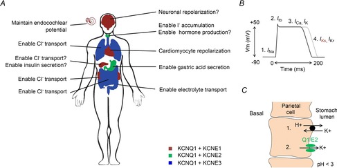
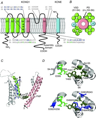
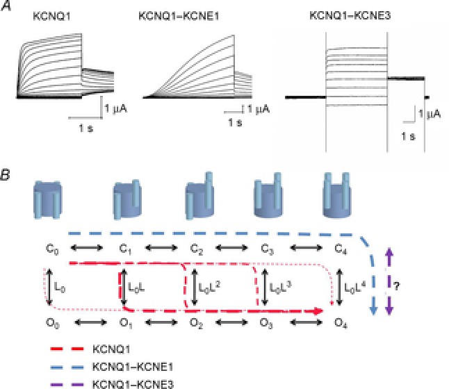
The KCNQ1 channel (also called Kv7.1 or KvLQT1) belongs to the superfamily of voltage-gated K(+) (Kv) channels. KCNQ1 shares several general features with other Kv channels but also displays a fascinating flexibility in terms of the mechanism of channel gating, which allows KCNQ1 to play different physiological roles in different tissues. This flexibility allows KCNQ1 channels to function as voltage-independent channels in epithelial tissues, whereas KCNQ1 function as voltage-activated channels with very slow kinetics in cardiac tissues. This flexibility is in part provided by the association of KCNQ1 with different accessory KCNE β-subunits and different modulators, but also seems like an integral part of KCNQ1 itself. The aim of this review is to describe the main mechanisms underlying KCNQ1 flexibility.


Figures

Figure 1
Figure 1
Suggested role of KCNQ1–KCNE complexes in different tissues A, KCNQ1–KCNE1 is suggested to be expressed in the heart, inner ear, pancreas, kidney and brain and to regulate repolarization of excitable cells, enable transport, and maintain the membrane potential. KCNQ1–KCNE2 is suggested to be expressed in the thyroid gland and stomach and to regulate transport, hormone production, and gastric secretion. KCNQ1–KCNE3 is suggested to be expressed in the intestine, colon and airways and to enable electrolyte transport. B, the action potential in human ventricular cardiomyocytes has a duration of several hundred milliseconds. Cardiomyocyte depolarization (1) is caused by influx of Na+. The transient outward K+ current (Ito) causes the repolarization notch (2) and the plateau is a caused by simultaneous influx of Ca2+ and outflux of K+ (3). Cardiomyocyte repolarization is mainly caused by K+ outflux though the IKs channel and the IKr channel (4). Loss-of-function of the IKs channel tends to impair cardiomyocyte repolarization and prolong the cardiomyocyte action potential duration (grey curve). C, the parietal cells in the gastric glands of stomach epithelium secrete H+ into the stomach lumen. The H+–K+-ATPase that causes stomach acidification (1) requires K+ outflux through KCNQ1–KCNE2 (2) to recycle the K+ transported though the H+–K+-ATPase.
Figure 2
Figure 2
Topology of Kv7.1 and KCNE and model for electro-mechanical coupling A, schematic topology of one KCNQ1 subunit and one KCNE subunit. S1–S4 form the voltage-sensor domain (VSD) and S5–S6 the pore domain (PD). The plus symbols in S4 denote the positive gating charges. Basic residues are shown in blue. Shaker residues suggested to make close contact between voltage-sensor domain and gate are shown in green. B, schematic top view of a tetrameric KCNQ1 channel. Blue spheres marked ‘E’ denote the deduced location of KCNE subunits in the channel complex. C, illustration of model for electro-mechanical coupling in Kv channels. S4 moves up during membrane depolarization (1). This movement pulls the S4–S5 linker (2), which forms bonds with the lower S6. The pulling of S4–S5 moves the lower end of S6 (3) which causes a kink at the PXP motif and opens the gate. Shaker model from Henrion et al. (2012). D, upper panel, molecular details of voltage-sensor domain coupling to the gate via hydrophobic residues in the S4–S5 linker and in the lower part of S6/C-terminus in the Shaker channel. Model from Henrion et al. (2012). Numbering indicates Shaker numbering. Residues in the same subunit are shown in dark green and residues in the neighbouring subunit are shown in light green. D, lower panel, corresponding position of basic KCNQ1 residues. Numbering indicates Shaker numbering/KCNQ1 numbering.
Figure 3
Figure 3
Model for KCNQ1 gating A, representative K+ current families for KCNQ1 (stepping from a holding voltage of –80 mV to test voltages from –80 to +60 mV in 10 mV increments. Tail voltage is –30 mV), KCNQ1–KCNE1 (stepping from a holding voltage of –80 mV to test voltages from –110 to +60 mV in 10 mV increments. Tail voltage is –30 mV), and KCNQ1–KCNE3 (stepping from a holding voltage of –100 mV to test voltages from –100 to +60 mV in 20 mV increments. Tail voltage is –40 mV). B, ten-state model with five closed states (C0–C4) and five open states (O0–O4), where subscript depicts the number of activated S4s. Horizontal transitions are independent activation of S4s and vertical transition is the opening of the gate (with a potential extra concerted movement of all four S4s; Barro-Soria et al. 2014). Suggested transitions for KCNQ1 alone (red dashed lines), KCNQ1–KCNE1 (blue dashed line), and KCNQ1–KCNE3 (magenta dashed line) in response to a depolarization. Even at very negative voltages (all S4s down), KCNQ1 expressed alone displays a significant open probability that increases with more activated S4s (Osteen et al. 2012). In response to a depolarization, most KCNQ1 channels open after one or two S4s have activated (thicker red dashed lines). In contrast, KCNQ1–KCNE1 only opens after all four S4s have activated (all four S4s up; Barro-Soria et al. 2014), whereas in KCNQ1–KCNE3 the S4s are always activated independently of voltage and channel opening is relatively voltage independent (Nakajo & Kubo, ; Rocheleau & Kobertz, 2008).

References

    1. Abbott GW. Biology of the KCNQ1 potassium channel. New J Sci. 2014;2014:237431.
    1. Anantharam A, Markowitz SM. Abbott GW. Pharmacogenetic considerations in diseases of cardiac ion channels. J Pharmacol Exp Ther. 2003;307:831–838. - PubMed
    1. Angelo K, Jespersen T, Grunnet M, Nielsen MS, Klaerke DA. Olesen SP. KCNE5 induces time- and voltage-dependent modulation of the KCNQ1 current. Biophys J. 2002;83:1997–2006. - PMC - PubMed
    1. Barhanin J, Lesage F, Guillemare E, Fink M, Lazdunski M. Romey G. KVLQT1 and IsK (minK) proteins associate to form the IKs cardiac potassium current. Nature. 1996;384:78–80. - PubMed
    1. Barrett KE. Keely SJ. Chloride secretion by the intestinal epithelium: molecular basis and regulatory aspects. Annu Rev Physiol. 2000;62:535–572. - PubMed

Publication types

MeSH terms

Substances

LinkOut - more resources