. 2015 Feb 4;7(273):273ra14.
doi: 10.1126/scitranslmed.3009951.

Local iontophoretic administration of cytotoxic therapies to solid tumors

Affiliations

Local iontophoretic administration of cytotoxic therapies to solid tumors

James D Byrne et al. Sci Transl Med. .

Abstract

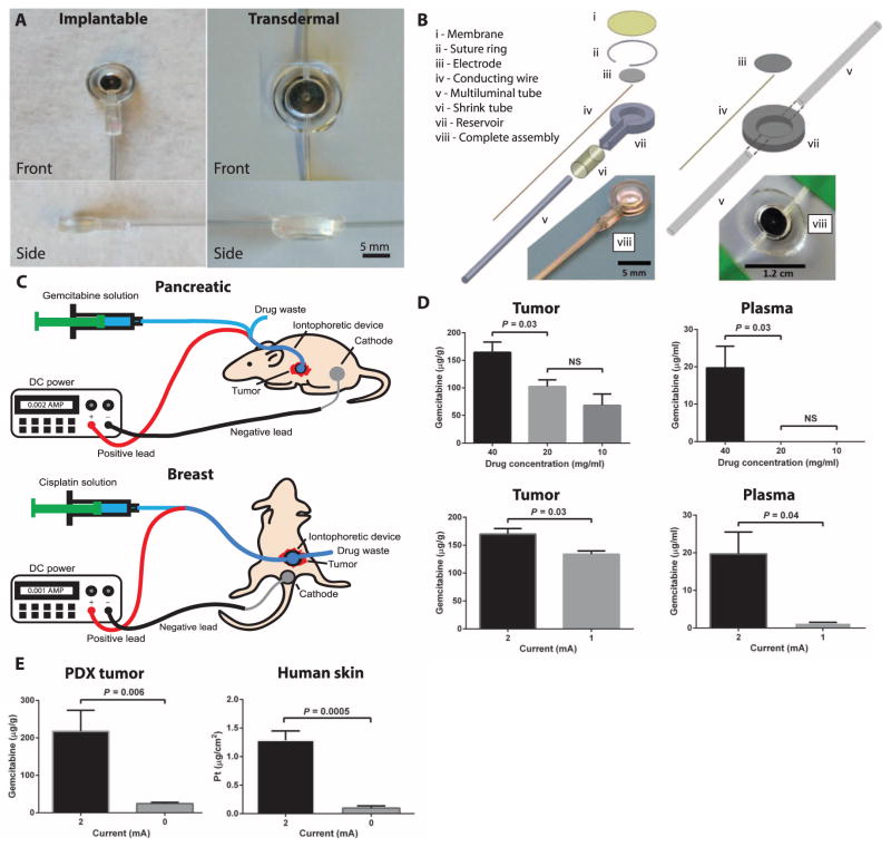
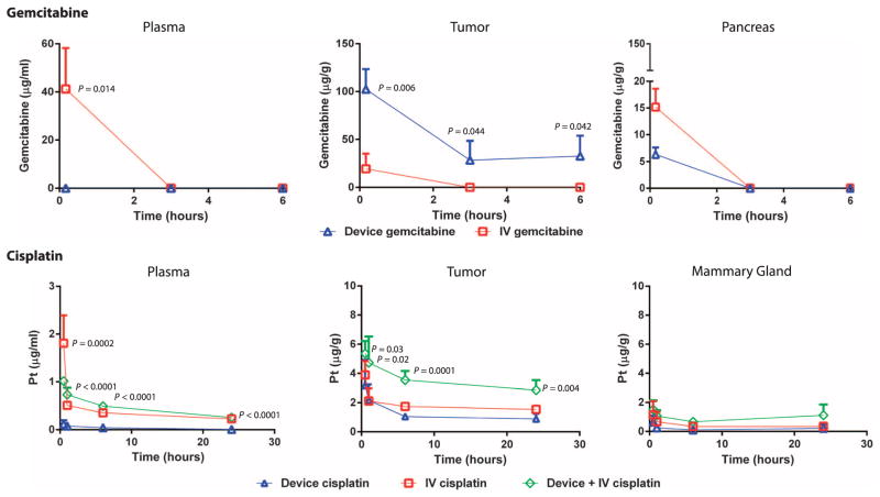
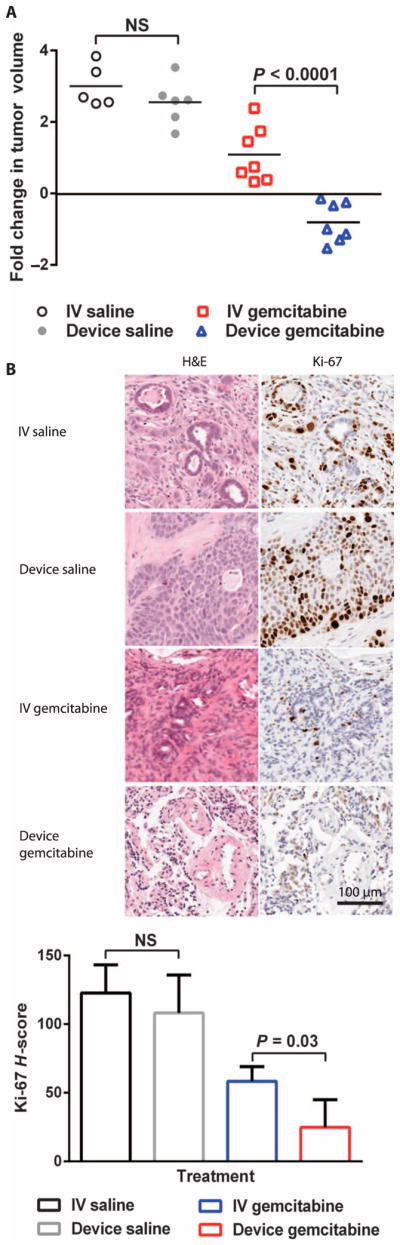
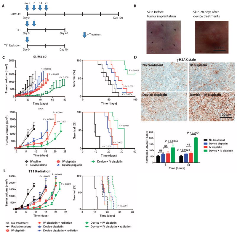
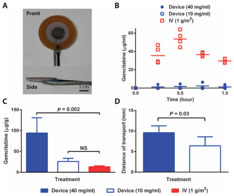
Parenteral and oral routes have been the traditional methods of administering cytotoxic agents to cancer patients. Unfortunately, the maximum potential effect of these cytotoxic agents has been limited because of systemic toxicity and poor tumor perfusion. In an attempt to improve the efficacy of cytotoxic agents while mitigating their side effects, we have developed modalities for the localized iontophoretic delivery of cytotoxic agents. These iontophoretic devices were designed to be implanted proximal to the tumor with external control of power and drug flow. Three distinct orthotopic mouse models of cancer and a canine model were evaluated for device efficacy and toxicity. Orthotopic patient-derived pancreatic cancer xenografts treated biweekly with gemcitabine via the device for 7 weeks experienced a mean log2 fold change in tumor volume of -0.8 compared to a mean log2 fold change in tumor volume of 1.1 for intravenous (IV) gemcitabine, 3.0 for IV saline, and 2.6 for device saline groups. The weekly coadministration of systemic cisplatin therapy and transdermal device cisplatin therapy significantly increased tumor growth inhibition and doubled the survival in two aggressive orthotopic models of breast cancer. The addition of radiotherapy to this treatment further extended survival. Device delivery of gemcitabine in dogs resulted in more than 7-fold difference in local drug concentrations and 25-fold lower systemic drug levels than the IV treatment. Overall, these devices have potential paradigm shifting implications for the treatment of pancreatic, breast, and other solid tumors.


Conflict of interest statement

Competing interests: J.D.B., M.E.N., J.J.Y., and J.M.D. have filed one patent related to the device PCT/US2010/025416. All other authors declare that they have no competing interests or patents to disclose.

Figures

Fig. 1
Fig. 1. Iontophoretic devices used for the delivery of cytotoxic agents to solid tumors
(A) Front and side images of the implantable and transdermal devices. (B) Device components and assemblies. (C) Device treatment setups in the pancreatic (implanted device) and breast (transdermal device) cancer models where the drug is supplied to the device, using a syringe pump and electrical current via a DC power supply. Positive and negative leads connect to the device and counter electrode, respectively. (D) Device parameters of drug concentration (at constant current and time) and applied current (at constant concentration and time) were evaluated in mice with patient-derived pancreatic cancer xenografts. Data are means ± SD (n = 5). P values were determined by one-way ANOVA with unpaired t test. (E) Role of current on drug transport in ex vivo tumor and human skin tissue. Gemcitabine transport through PDX tumor tissue was evaluated by applying a current of 2 or 0 mA for 10 min and comparing drug transport into tumor. Data are means ± SD (n = 6). Cisplatin transport into human skin was evaluated by applying a current of 1 or 0 mA for 25 min and comparing drug transport into and through the skin. Data are means ± SD (n = 5). P values were determined by unpaired t test. NS, not significant.
Fig. 2
Fig. 2. PK of gemcitabine and cisplatin delivered by iontophoretic devices in mouse models of human pancreatic and breast cancers
Iontophoretic device delivery was compared with IV delivery. PK of gemcitabine (20 mg/ml) delivered by device compared to IV was evaluated in an orthotopic PDX model of pancreatic cancer. Mice were administered a single treatment of gemcitabine through the device. Organs were collected from each animal at various times, and total gemcitabine concentrations were analyzed. Data are means ± SD (n = 3 to 5 animals per group). The limit of gemcitabine quantitation was 1 μg/ml. P values were determined by unpaired t test. PK of cisplatin delivered by device compared to IV and device + IV was evaluated in SUM149 orthotopic xenografts of breast cancer. Mice were administered a single treatment of cisplatin. Organs were collected from each animal at various times, and total platinum concentrations were analyzed. Data are means ± SD (n = 5 animals per group). The limit of platinum quantitation was 5 ng/ml. P values were determined by one-way ANOVA with unpaired t test comparing device cisplatin and device + IV cisplatin.
Fig. 3
Fig. 3. Therapeutic effect of gemcitabine delivered iontophoretically in a pancreatic cancer PDX model
(A) Efficacy of device gemcitabine, IV gemcitabine, device saline, and IV saline in PDX mice treated twice per week for 7 weeks. Data are fold change in tumor volume (log2) (n = 7 for IV and device gemcitabine, n = 5 to 6 for IV and device saline). (B) Histological staining of representative tumors in (A) for Ki-67. Ki-67 staining was quantified according to H-score. P values were determined by one-way ANOVA with unpaired t test.
Fig. 4
Fig. 4. Therapeutic effect of cisplatin delivered iontophoretically in mouse tumor xenograft and syngeneic models of breast cancer
(A) Treatment schedule according to mouse model. (B) Efficacy and survival of animals treated with device cisplatin, IV cisplatin (5 mg/kg), device + IV cisplatin (5 mg/kg), device saline, and IV saline. SUM149 tumor xenografts were treated once per week for a total of four doses (n = 8 to 9 per treatment group). T11 syngeneic tumors were treated once per week for a total of two doses (n = 9 per treatment group). The study endpoint was time to tumor progression to 2.0 cm in one dimension. Volume data are means ± SD. (C) Representative images of murine skin before and after 4 weeks of transdermal device treatment. (D) γH2AX staining of tumors harvested from SUM149 xenografts 24 hours after a single treatment. γH2AX staining was quantified according to H-score. P values were determined by one-way ANOVA with unpaired t test. (E) Efficacy and survival of animals with T11 syngeneic tumors after a single treatment of radiation (dose), device cisplatin, device cisplatin + radiation, IV cisplatin (dose), IV cisplatin (dose) + radiation, device + IV cisplatin, or device + IV cisplatin + radiation. Data are mean tumor volumes ± SEM (n = 8 per treatment group). P values for tumor growth inhibition were determined by one-way ANOVA with unpaired t test; P values for survival determined by log-rank test.
Fig. 5
Fig. 5. Evaluation of single device treatments in dogs
(A) Device to be implanted directly onto the canine pancreas. (B) Plasma PK of gemcitabine during the single device (10 or 40 mg/ml) or IV (1 g/m2) treatment. (C) Organs were removed 1 hour after the initiation of treatment, and gemcitabine content was quantified in the pancreas of dogs after the administration of a single treatment. (D) Distance of gemcitabine transport away from the device and into the pancreatic tissue. Data are means ± SD (n = 5). P values were determined by Wilcoxon rank sum tests.

References

    1. Goodman LS, Wintrobe MM, Dameshek W, Goodman MJ, Gilman MA, McLennan MT. Nitrogen mustard therapy; use of methyl-bis (β-chloroethyl) amine hydrochloride and tris (β-chloroethyl) amine hydrochloride for Hodgkin’s disease, lymphosarcoma, leukemia and certain allied and miscellaneous disorders. J Am Med Assoc. 1946;132:126–132. - PubMed
    1. Neoptolemos JP, Stocken DD, Friess H, Bassi C, Dunn JA, Hickey H, Beger H, Fernandez-Cruz L, Dervenis C, Lacaine F, Falconi M, Pederzoli P, Pap A, Spooner D, Kerr DJ, Büchler; MW European Study Group for Pancreatic Cancer. A randomized trial of chemoradiotherapy and chemotherapy after resection of pancreatic cancer. N Engl J Med. 2004;350:1200–1210. - PubMed
    1. Sauer R, Becker H, Hohenberger W, Rödel C, Wittekind C, Fietkau R, Martus P, Tschmelitsch J, Hager E, Hess CF, Karstens JH, Liersch T, Schmidberger H, Raab; R German Rectal Cancer Study Group. Preoperative versus postoperative chemoradiotherapy for rectal cancer. N Engl J Med. 2004;351:1731–1740. - PubMed
    1. Cunningham D, Allum WH, Stenning SP, Thompson JN, Van de Velde CJ, Nicolson M, Scarffe JH, Lofts FJ, Falk SJ, Iveson TJ, Smith DB, Langley RE, Verma M, Weeden S, Chua; YJ MAGIC Trial Participants. Perioperative chemotherapy versus surgery alone for resectable gastroesophageal cancer. N Engl J Med. 2006;355:11–20. - PubMed
    1. Cunningham D, Starling N, Rao S, Iveson T, Nicolson M, Coxon F, Middleton G, Daniel F, Oates J, Norman; AR Upper Gastrointestinal Clinical Studies Group of the National Cancer Research Institute of the United Kingdom. Capecitabine and oxaliplatin for advanced esophagogastric cancer. N Engl J Med. 2008;358:36–46. - PubMed

Publication types

MeSH terms