. 2015 Feb 4;35(5):2133-45.
doi: 10.1523/JNEUROSCI.4098-14.2015.

B-lymphocyte-mediated delayed cognitive impairment following stroke

Affiliations

B-lymphocyte-mediated delayed cognitive impairment following stroke

Kristian P Doyle et al. J Neurosci. .

Abstract

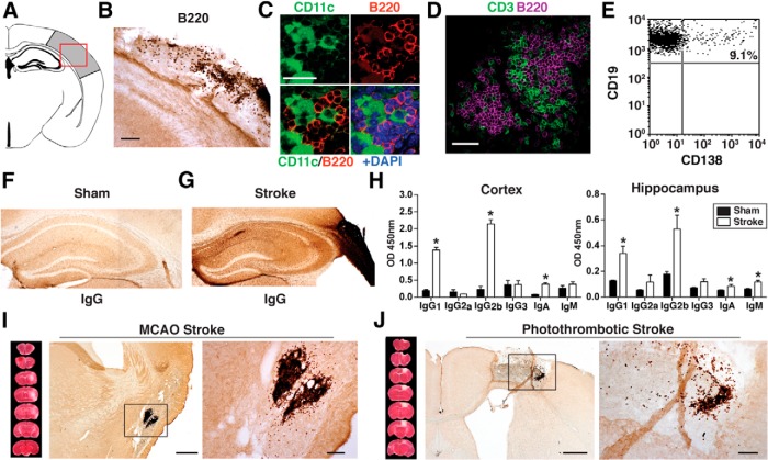
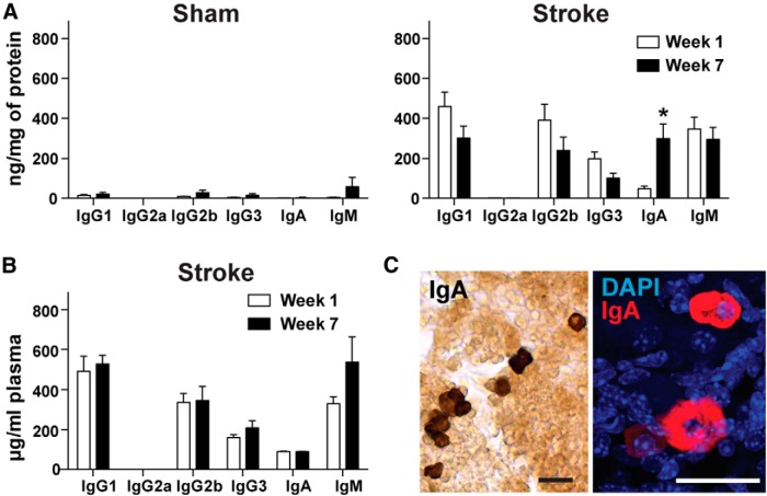
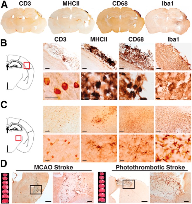
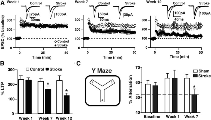
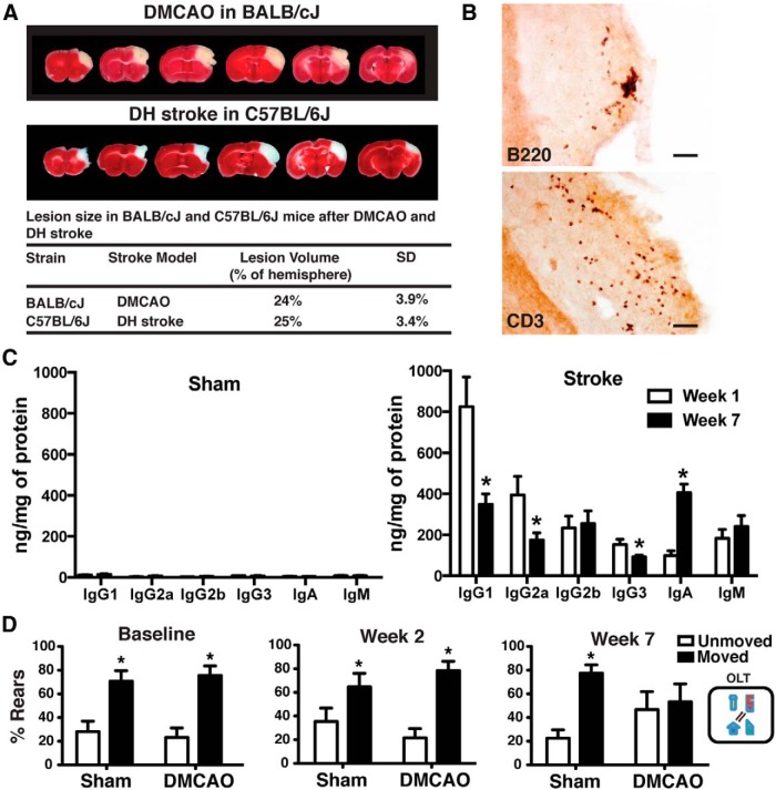
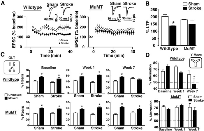
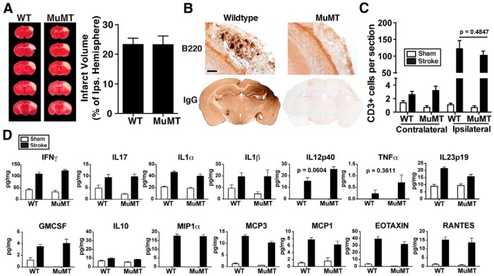
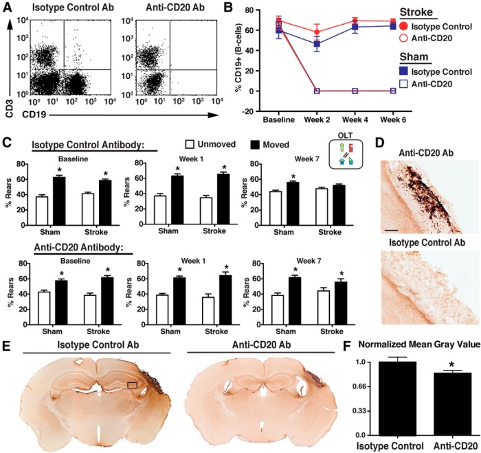
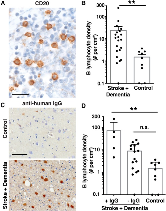
Each year, 10 million people worldwide survive the neurologic injury associated with a stroke. Importantly, stroke survivors have more than twice the risk of subsequently developing dementia compared with people who have never had a stroke. The link between stroke and the later development of dementia is not understood. There are reports of oligoclonal bands in the CSF of stroke patients, suggesting that in some people a B-lymphocyte response to stroke may occur in the CNS. Therefore, we tested the hypothesis that a B-lymphocyte response to stroke could contribute to the onset of dementia. We discovered that, in mouse models, activated B-lymphocytes infiltrate infarcted tissue in the weeks after stroke. B-lymphocytes undergo isotype switching, and IgM, IgG, and IgA antibodies are found in the neuropil adjacent to the lesion. Concurrently, mice develop delayed deficits in LTP and cognition. Genetic deficiency, and the pharmacologic ablation of B-lymphocytes using an anti-CD20 antibody, prevents the appearance of delayed cognitive deficits. Furthermore, immunostaining of human postmortem tissue revealed that a B-lymphocyte response to stroke also occurs in the brain of some people with stroke and dementia. These data suggest that some stroke patients may develop a B-lymphocyte response to stroke that contributes to dementia, and is potentially treatable with FDA-approved drugs that target B cells.

Keywords: B-lymphocyte; dementia; immunology; stroke.


Conflict of interest statement

The authors declare no competing financial interests.

Figures

Figure 1.
Figure 1.
A B-lymphocyte response to stroke occurs in C57BL/6J mice. A, Schematic diagram of a mouse coronal brain section following DH stroke, with the stroke core shaded in gray and a red box to indicate the region immunostained in B–D, and used for flow in E. B, Immunostaining for B220 (B-lymphocytes) shows that B cells are present in the lesion 7 weeks following DH stroke in C57BL/6J mice. Scale bar, 250 μm. C, Fluorescent immunostaining for the B-lymphocyte marker B220 (red) and the dendritic cell marker CD11c (green) shows that B cells are still present in the lesion 12 weeks following stroke and are juxtaposed to cells immunostaining for CD11c. Scale bar, 25 μm. Images are representative of 10 mice that underwent stroke. D, Fluorescent immunostaining for the B-lymphocyte marker B220 (magenta) and the T-cell marker CD3 (green) reveals that B cells and T cells are compartmentalized 7 weeks following DH stroke. Scale bar, 100 μm. E, Flow cytometry revealed that 9% of CD19+ B-lymphocytes present in the stroke lesion were CD19+ CD138+ plasma cells (n = 10 pooled stroke lesions). F, G, Representative images of IgG immunostaining in the hippocampus 7 weeks following stroke or sham surgery (n = 10). H, Determination of antibody isotypes present in the cortex and hippocampus 7 weeks following stroke or sham surgery (stroke n = 7; sham n = 3). *p < 0.05, compared with sham. Error bars indicate SD. I, J, Immunostaining for the B-cell marker B220 4 weeks after stroke in two additional stroke models performed in C57BL/6J mice: MCAO (I) and photothrombotic stroke (PT stroke) (J). Scale bars, 500 and 100 μm (lower and higher magnifications, respectively).
Figure 2.
Figure 2.
There is a delayed appearance of IgA in the brain but not the blood between 1 and 7 weeks after stroke. A, Multiplex immunoassay revealed that the amount of IgA present in the ipsilateral hemisphere significantly increased between week 1 and week 7 after stroke (n = 4–7 per group). *p < 0.05, compared with 1 week (two-way ANOVA). Error bars indicate mean ± SEM. B, There was no difference in immunoglobulin content in the plasma at week 1 and week 7 after stroke. There was no difference between stroke and sham plasma at week 1 and 7 after stroke (data not shown). C, IgA immunostaining (left: DAB, right: fluorescence) revealed the presence of IgA-producing plasma cells in the stroke lesion 7 weeks following stroke (n = 7). Scale bars, 75 μm.
Figure 3.
Figure 3.
The B-lymphocyte response to stroke is a component of a larger adaptive immune response to stroke. A, Whole-brain sections showing the locations of T cells (CD3+), MHCII expression, and activated macrophages/microglia (CD68+ and Iba1+) 7 weeks after stroke. Staining is evident both in the lesion and locations where there is axonal degeneration. Images are representative of 10 mice. B, C, Progressively higher-magnification images of CD3, MHCII, CD68, and Iba1 staining in the lesion (B) and in an area of axonal degeneration (C). Equivalent findings were observed in two additional independent experiments. D, Equivalent T-cell responses after stroke were also independently observed with CD3 immunostaining in two additional stroke models: MCAO and photothrombotic stroke. This demonstrates that an adaptive immune response to stroke is not model-dependent in C57BL/6J mice. Scale bars: B, C, 250 μm, 10 μm; D, 500 μm, 100 μm (lower and higher magnifications, respectively).
Figure 4.
Figure 4.
Mice develop a deficit in LTP in the hippocampus between weeks 1 and 7 after stroke, and a delayed cognitive deficit on the Y maze. A, B, Whole-cell EPSCs were recorded from CA1 pyramidal neurons in acute hippocampal slices prepared from C57BL/6J mice that had undergone stroke or sham surgery 1, 7, and 12 weeks earlier (n = 6–9 per group). The time course (A) and magnitude (B) of LTP are illustrated and demonstrate an impairment at 7 and 12 weeks, but not 1 week, following stroke. *p < 0.05, compared with controls. C, C57BL/6J mice developed a delayed cognitive deficit on the Y maze, a test of short-term memory, between weeks 1 and 7 following stroke (n = 10 per group). *p < 0.05, compared with sham week 7. Error bars indicate SD.
Figure 5.
Figure 5.
A B-lymphocyte response to stroke and delayed cognitive dysfunction also occurs in BALB/CJ mice. A, Lesion size and location are equivalent following DMCAO in BALB/CJ mice to lesion size and location following DH stroke in C57BL/6J mice. B, Immunostaining for the B- and T-lymphocyte markers B220 and CD3 in the stroke lesion 7 weeks after stroke. Scale bars, 100 μm. C, Quantification of immunoglobulins in the ipsilateral hemisphere in BALB/CJ mice 1 and 7 weeks after stroke (n = 10 per group). *p < 0.05, compared with 1 week (two-way ANOVA). Error bars indicate mean ± SEM. D, BALB/CJ mice also develop a delayed cognitive deficit on the OLT between weeks 1 and 7 after stroke (n = 10 per group). *p < 0.05, moved versus unmoved objects.
Figure 6.
Figure 6.
B-cell-deficient muMT mice do not develop either LTP or cognitive deficits in the weeks after stroke. A, B, Whole-cell EPSCs were recorded from CA1 pyramidal neurons in acute hippocampal slices prepared from WT and muMT mice that had undergone DH stroke or sham surgery 12 weeks earlier (n = 6–9 per group). The time course (A) and magnitude (B) of LTP demonstrated that muMT mice that had undergone DH stroke did not develop an LTP impairment 12 weeks following stroke. *p < 0.05, compared with WT sham control. There was no difference between sham groups (p = 0.85). LTP plot represents 0–40 min after LTP. Values in the bar graph were taken at 35 min after LTP. C, muMT mice also did not develop a cognitive deficit on the OLT test 7 weeks after DH stroke (n = 10 per group). *p < 0.05, compared with unmoved objects. D, WT, but not muMT, mice developed a cognitive deficit on the Y maze 7 weeks after DH stroke (n = 10 per group). *p < 0.05, compared with sham.
Figure 7.
Figure 7.
Lesion size and the T-lymphocyte response to stroke are equivalent in WT and muMT mice. A, B-cell-deficient muMT mice and wild-type (WT) C57BL/6J mice had equivalent infarct volumes 72 h after DH stroke (n = 5 per group). B, muMT mice did not have B cell (B220+) or IgG immunostaining in the lesion 7 weeks after DH stroke (n = 10). Scale bar, 125 μm. C, The number of T cells in the brain was equivalent in WT and muMT mice 7 weeks after stroke. D, Levels of proinflammatory cytokines and chemokines in the lesion were similarly elevated in both WT and muMT mice compared with corresponding cortical tissue from sham mice 7 weeks after DH stroke (sham, n = 4 per group; DH stroke, n = 7 per group). There was no difference between WT and muMT mice by two-way ANOVA. The p value before correcting for multiple comparisons is shown for IL12p40 and TNFα, which were not different statistically.
Figure 8.
Figure 8.
B-cell depletion with an anti-CD20 antibody beginning 5 d after stroke ameliorates the development of delayed cognitive deficits. A, Representative scatter plot showing complete B-cell ablation 7 d following injection with anti-CD20 antibody. CD3, T-cell marker; CD19, B-cell marker. B, Flow cytometry showed that B-cell ablation was sustained throughout the experiment in the mice treated with the anti-CD20 antibody. C, Isotype control-treated, but not anti-CD20 antibody-treated, mice developed a cognitive deficit on the OLT test. Data are from 7 weeks after DH stroke (n = 10 per group). *p < 0.05, compared with unmoved objects. D, Anti-CD20 antibody-treated mice did not have B-cell (B220+) infiltration in the lesion 7 weeks after stroke. Scale bar, 125 μm. E, Anti-CD20 antibody-treated mice exhibited less IgG infiltration in the lesion and hippocampus 7 weeks after stroke. Black box represents region where grayscale was quantified in IgG-stained sections, demonstrating less immunostaining in anti-CD20-treated mice (F). n = 8–10 per group. *p < 0.05.
Figure 9.
Figure 9.
Evidence of a B-cell response in the brain after stroke in humans with concurrent dementia. A, CD20 immunostaining from an ischemic infarct that had a density of 240 B-lymphocytes/cm2. Scale bar, 25 μm. B, Quantification of B-cell infiltration in stroked brain tissue and age-matched control tissue. **p = 0.0021 (Mann–Whitney test). C, IgG immunostaining from 1 of the 2 control subjects that had any cellular staining (top) and from a representative section from stroke survivors with dementia that were scored as IgG+ for IgG immunoreactivity in the neuropil. Scale bar, 100 μm. D, Graph of B-cell densities in subjects with stroke and dementia with and without IgG immunoreactivity in the neuropil, compared with control subjects. **p < 0.01 (Kruskal-Wallis with Dunn's multiple-comparison post hoc test). n.s., Not significant. More detailed information on B-cell densities, IgG immunoreactivity, and associated clinical information is provided in Table 2.

Comment in

  • B cells: Meddling with the mind.
    Bordon Y. Bordon Y. Nat Rev Immunol. 2015 Mar;15(3):135. doi: 10.1038/nri3826. Epub 2015 Feb 20. Nat Rev Immunol. 2015. PMID: 25698677 No abstract available.
  • Stroke: Meddling with the mind.
    Bordon Y. Bordon Y. Nat Rev Drug Discov. 2015 Mar;14(3):166. doi: 10.1038/nrd4561. Nat Rev Drug Discov. 2015. PMID: 25722242 No abstract available.

References

    1. Ankeny DP, Popovich PG. B cells and autoantibodies: complex roles in CNS injury. Trends Immunol. 2010;31:332–338. doi: 10.1016/j.it.2010.06.006. - DOI - PMC - PubMed
    1. Ankeny DP, Lucin KM, Sanders VM, McGaughy VM, Popovich PG. Spinal cord injury triggers systemic autoimmunity: evidence for chronic B lymphocyte activation and lupus-like autoantibody synthesis. J Neurochem. 2006;99:1073–1087. doi: 10.1111/j.1471-4159.2006.04147.x. - DOI - PubMed
    1. Ankeny DP, Guan Z, Popovich PG. B cells produce pathogenic antibodies and impair recovery after spinal cord injury in mice. J Clin Invest. 2009;119:2990–2999. doi: 10.1172/JCI39780. - DOI - PMC - PubMed
    1. Arac A, Brownell SE, Rothbard JB, Chen C, Ko RM, Pereira MP, Albers GW, Steinman L, Steinberg GK. Systemic augmentation of alphaB-crystallin provides therapeutic benefit twelve hours post-stroke onset via immune modulation. Proc Natl Acad Sci U S A. 2011;108:13287–13292. doi: 10.1073/pnas.1107368108. - DOI - PMC - PubMed
    1. Barba R, Martínez-Espinosa S, Rodríguez-García E, Pondal M, Vivancos J, Del Ser T. Poststroke dementia: clinical features and risk factors. Stroke. 2000;31:1494–1501. doi: 10.1161/01.STR.31.7.1494. - DOI - PubMed

Publication types

Substances